The Muscular System
Every voluntary thing you do — a smile, a step, a breath, a signature — is a muscle pulling on a bone or on your skin. In the last chapters you met the microscopic machinery inside a muscle fiber; now we zoom out to the whole body and ask a different set of questions. How do more than 600 skeletal muscles cooperate to produce smooth, controlled movement when each one can only pull? Why do their names read like Latin riddles — and how can you crack those riddles to know a muscle you have never memorized? This chapter is a guided tour of the body's muscles, region by region, from the tiny muscles that shape a facial expression to the powerhouses that launch you off the ground.
By the end of this chapter you can
- Explain why skeletal muscles can only pull, and how that forces them to work in opposing pairs across a joint.
- Define a muscle's origin and insertion, and assign the roles of agonist, antagonist, synergist, and fixator in a given movement.
- Describe how bones and joints act as levers, and explain the speed-versus-force trade-off of a third-class lever.
- Use the six naming clues to predict a muscle's location, shape, size, fiber direction, number of heads, or action from its name alone.
- Distinguish the muscles of facial expression from the muscles of mastication, and name the cranial nerve that drives each group.
- Identify the major muscles of the trunk — the breathing muscles, the abdominal wall, and the deep back — and state what each does.
- Name the principal muscles that move the shoulder, arm, and forearm, including the rotator cuff and the classic flexor/extensor pairs.
- Name the principal muscles that move the hip, knee, and ankle, including the quadriceps, hamstrings, and calf.
Section 1How muscles work together
A muscle can only pull
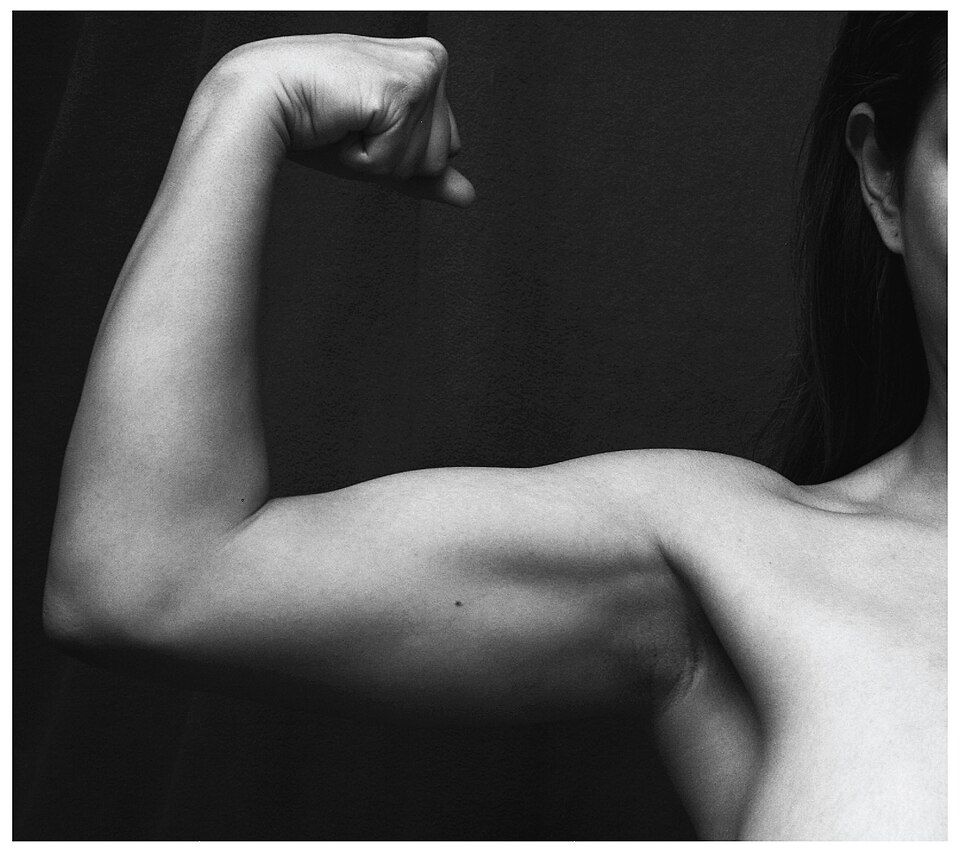
Here is the single fact that everything in this section is built on: a muscle can only pull, never push. When a muscle contracts, its fibers shorten and draw its two ends toward each other — that is the only trick it has. It cannot lengthen itself to shove a bone away. So how does your forearm both bend up and straighten back down? The answer is that muscles are almost never soloists. They are arranged in opposing teams on either side of a joint, and to reverse a movement the body simply relaxes one team and contracts the other.
To describe that pull precisely, we name a muscle's two ends. The origin is the attachment on the bone that stays relatively still — usually the more proximal, heavier bone. The insertion is the attachment on the bone that actually moves — usually the more distal one. Between them is the thick, contractile middle called the belly. When the muscle shortens, the insertion is dragged toward the origin. Muscles attach to bone through tough, cord-like tendons, or through broad, flat sheets called aponeuroses.
Four roles in every movement
Because muscles cooperate, any single motion is really a small committee at work, and each member has a job. Learn these four roles and you can analyze any movement in the body:
| Role | What it does | Example (bending the elbow) |
|---|---|---|
| Agonist (prime mover) | The main muscle producing the movement | Biceps brachii flexes the elbow |
| Antagonist | Opposes the agonist; relaxes and stretches as it acts | Triceps brachii relaxes to allow flexion |
| Synergist | Assists the agonist or steadies the joint | Brachialis helps the biceps flex |
| Fixator | Stabilizes the origin so the pull is efficient | Shoulder muscles hold the scapula still |
Notice that the same muscle switches roles depending on the movement. When you bend your elbow, the biceps is the agonist and the triceps is the antagonist; when you straighten it, they swap jobs entirely. There is no such thing as a “flexor muscle” in the abstract — a muscle is a flexor or an extensor only for a particular joint in a particular motion.
Bones are levers, joints are fulcrums
Muscles do not move bones by brute force alone; they work through levers. A lever is a rigid bar that pivots on a fixed point, and the body is full of them: the bone is the bar, the joint is the pivot (the fulcrum), the muscle's pull is the effort, and the weight being moved is the load. The way these three points are arranged decides whether a joint favors power or speed and range.
Most joints in the body are third-class levers, where the effort is applied between the fulcrum and the load — exactly the setup at your elbow, where the biceps inserts just past the joint while the load sits far out at the hand. This arrangement is mechanically “inefficient” for lifting heavy weight, but it buys something more useful: a small, quick muscle shortening produces a large, fast sweep of the hand. Your body traded raw force for speed and reach, which is why you can hurl a ball but strain to curl a heavy dumbbell.
Bend and straighten the elbow in the interactive and watch the biceps and triceps trade the agonist and antagonist roles.
- A muscle can only pull; opposing muscles across a joint reverse each other's actions.
- The origin is the fixed (usually proximal) attachment; the insertion is the moving (usually distal) one; the insertion is drawn toward the origin.
- Every movement uses an agonist, antagonist, synergist, and fixator, and muscles swap these roles depending on the motion.
- Bones act as levers; most body joints are third-class levers that favor speed and range over force.
Section 2How muscles are named
Names are descriptions in disguise
The first time you see words like extensor carpi radialis longus, muscle names feel like a wall of Latin memorization. They are not. Anatomists named muscles the way sailors named knots — by describing them. Almost every muscle name is a compact description of where the muscle is, what it looks like, how big it is, which way its fibers run, how many heads it has, or what it does. Once you know the vocabulary, a name you have never seen becomes a set of instructions for finding and understanding the muscle.
The six clues
There are six kinds of information a name can carry, and many muscles pack several clues into one name:
| Clue | Tells you | Example words & muscles |
|---|---|---|
| Location | The body region or nearby bone | temporalis (temporal bone), tibialis anterior (front of tibia) |
| Shape | The muscle's outline | deltoid (triangle), trapezius (trapezoid), rhomboid |
| Size | Relative size or length | maximus/minimus, longus/brevis, major/minor |
| Fiber direction | How the fibers run relative to the midline | rectus (straight), transversus (across), oblique (angled) |
| Number of heads | How many origins the muscle has | biceps (two), triceps (three), quadriceps (four) |
| Action | The movement it produces | flexor, extensor, adductor, levator, masseter (chewer) |
Watch the clues stack. Extensor digitorum longus unpacks to “the long muscle that extends the digits” — action, then body part, then size. Rectus femoris is “the straight muscle of the femur.” Even a monster like flexor carpi radialis is just “the muscle that flexes the wrist (carpus) along the radial side.” You do not have to memorize it as one blob; you read it like a sentence.
Tap each of the six clues in the interactive to see how a name reveals a muscle.
- Muscle names are descriptions, not arbitrary labels — decode them and you can reason about muscles you never memorized.
- The six clues: location, shape, size, fiber direction, number of heads, and action.
- Many names combine several clues; read them left to right like a sentence.
Section 3Muscles of the head and neck
The muscles that make a face
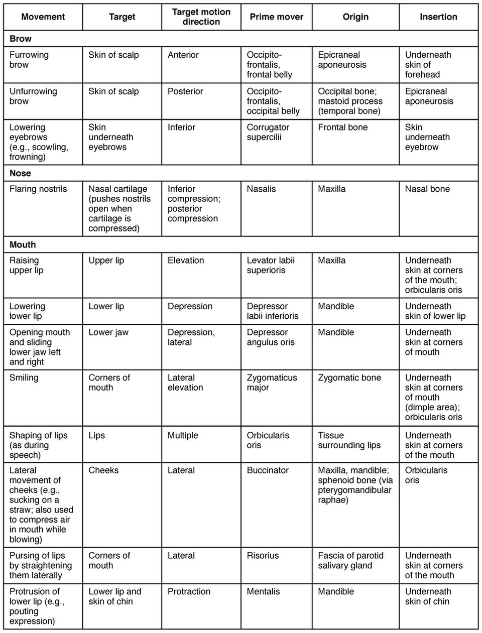
The muscles of facial expression are unlike almost any others in the body, because most of them do not move a bone at all — they insert into the skin. When they contract, they tug the skin of the face into the countless expressions that carry human emotion: a smile, a frown, a raised eyebrow, a wink. This whole group is driven by a single nerve, the facial nerve (cranial nerve VII). Key players include the orbicularis oris, the ring around the mouth that purses your lips to whistle or kiss; the orbicularis oculi, the ring around the eye that closes it to blink and squint; the buccinator in the cheek, which flattens the cheek to blow out a candle and holds food against the teeth while you chew; the zygomaticus, which pulls the corner of the mouth up to smile; and the occipitofrontalis, which raises the eyebrows and wrinkles the forehead.
The muscles that chew
The second job up here is mastication — chewing. Unlike the facial muscles, the muscles of mastication do move a bone: they close and grind the mandible (lower jaw) at the temporomandibular joint. This group answers to a different nerve, the trigeminal nerve (cranial nerve V). The masseter runs from the cheekbone to the angle of the jaw and is, for its size, one of the strongest muscles in the body — you can feel it bulge when you clench your teeth. The fan-shaped temporalis spreads over the side of the skull and helps close and retract the jaw, while the deep pterygoid muscles produce the side-to-side grinding motion.
| Group | Job | Inserts on | Nerve | Members |
|---|---|---|---|---|
| Facial expression | Move the skin of the face | Skin | Facial (CN VII) | Orbicularis oris & oculi, buccinator, zygomaticus, occipitofrontalis |
| Mastication | Move the mandible to chew | Mandible (bone) | Trigeminal (CN V) | Masseter, temporalis, pterygoids |
| Neck | Move and support the head | Skull & girdle | Accessory / cervical | Sternocleidomastoid, trapezius |
Down in the neck, two large muscles move the head itself. The paired sternocleidomastoid muscles flex the neck when they work together and rotate the head to the opposite side when one works alone. The broad trapezius extends the head and stabilizes the shoulder girdle behind it.
Tap each muscle in the interactive to light it up and see whether it makes an expression or chews.
- Muscles of facial expression insert into skin and are driven by the facial nerve (CN VII).
- Muscles of mastication move the mandible to chew and are driven by the trigeminal nerve (CN V); the masseter is the strongest for its size.
- The sternocleidomastoid flexes the neck and rotates the head to the opposite side.
Section 4Muscles of the trunk
The muscles that breathe
The trunk muscles do the body's quiet, essential work — and none is more essential than breathing. The diaphragm is the primary muscle of respiration: a broad dome of muscle that seals off the bottom of the chest. When it contracts it flattens, enlarging the thoracic cavity, dropping the pressure inside, and pulling air into the lungs. Relax it and the dome springs back up, pushing air out. Helping it are the external intercostals, the muscles between the ribs that lift the rib cage during inhalation; the internal intercostals pull the ribs down for forceful exhalation, such as when you cough or blow out a candle.
The abdominal wall and the back
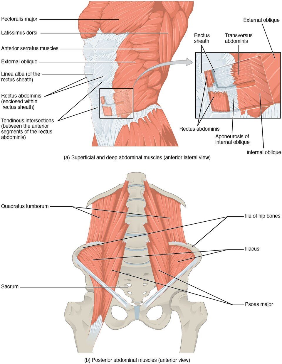
The front and sides of the belly are wrapped by four flat muscles layered like plywood, with their fibers running in different directions for strength. The rectus abdominis — the “six-pack” — runs straight up the midline and flexes the vertebral column (the motion of a sit-up). Over the sides run the external oblique, the internal oblique, and, deepest of all, the transversus abdominis, whose fibers wrap horizontally like a corset. Together this wall compresses the abdomen, bends and twists the trunk, protects the soft organs behind it, and boosts the pressure needed for coughing, childbirth, and moving the bowels.
Along the back, the erector spinae — a set of three muscle columns running the length of the spine — do the opposite of the abdominals: they extend the vertebral column to straighten you up from a bend and hold you upright against gravity all day long.
| Region | Muscles | Main action |
|---|---|---|
| Breathing | Diaphragm, external & internal intercostals | Change chest volume to move air in and out |
| Abdominal wall | Rectus abdominis, external & internal obliques, transversus abdominis | Flex/rotate trunk, compress and protect abdomen |
| Deep back | Erector spinae group | Extend the spine and maintain upright posture |
| Pelvic floor | Levator ani and companions | Support pelvic organs; control continence |
Tap each trunk muscle in the interactive to see whether it breathes, bends, or protects.
- The diaphragm is the primary breathing muscle; contracting it flattens the dome and pulls air in.
- Four layered abdominal muscles — rectus abdominis plus the two obliques and transversus abdominis — flex and rotate the trunk and protect the viscera.
- The erector spinae extends the spine and holds posture; the diaphragm runs on the phrenic nerve (C3–C5).
Section 5Muscles of the upper limb
Anchoring and moving the shoulder
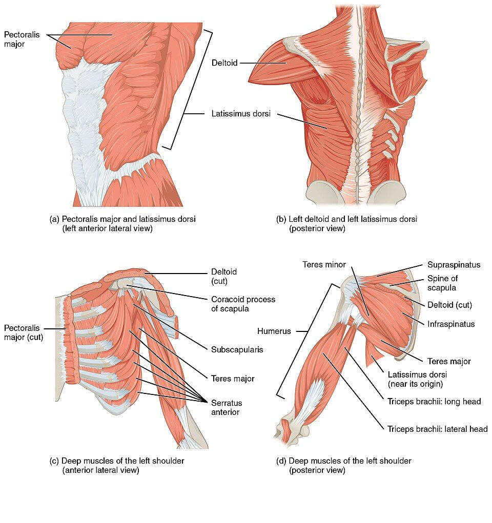
The upper limb is built for reach and dexterity, and it starts at the shoulder — the most mobile, and therefore least stable, joint in the body. Several large muscles anchor the shoulder blade and swing the arm. The trapezius and serratus anterior hold and rotate the scapula so it can serve as a moving base. The fan-shaped pectoralis major on the chest pulls the arm forward and across the body, while the broad latissimus dorsi on the back pulls it down and behind — the muscle of a swimmer's stroke. Capping the shoulder is the deltoid, the triangular muscle that abducts the arm, lifting it out to the side.
Wrapped deep around the joint itself is the rotator cuff, a set of four small muscles — supraspinatus, infraspinatus, teres minor, and subscapularis (the “SITS” group). They do not generate big movements; their job is to hold the ball of the humerus snugly in its shallow socket while the larger muscles move the arm.
The arm and forearm
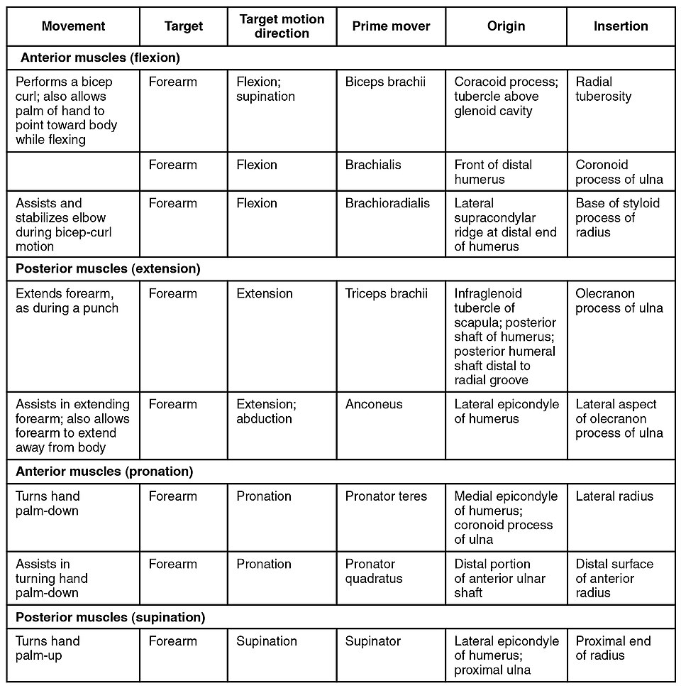
The arm proper (shoulder to elbow) hosts the classic agonist/antagonist pair. On the front, the biceps brachii flexes the elbow and turns the palm up (supination), aided beneath it by the brachialis. On the back, the triceps brachii — the only major muscle there — extends the elbow to straighten the arm. Cross the elbow into the forearm and you find two opposing compartments: a group of flexors on the palm side that curl the wrist and fingers, and a group of extensors on the back side that straighten them. Their long tendons run down into the hand, which is why so much of the power for gripping actually comes from muscle bellies up in the forearm.
| Muscle | Location | Main action |
|---|---|---|
| Deltoid | Cap of the shoulder | Abducts the arm |
| Pectoralis major | Anterior chest | Flexes and adducts the arm |
| Latissimus dorsi | Broad of the back | Extends and adducts the arm |
| Rotator cuff (SITS) | Deep around the shoulder | Stabilizes the humeral head |
| Biceps brachii | Anterior arm | Flexes elbow, supinates forearm |
| Triceps brachii | Posterior arm | Extends the elbow |
Tap each muscle in the interactive to find the shoulder, arm, and forearm movers and their pairs.
- The deltoid abducts the arm; pectoralis major and latissimus dorsi pull it forward and back.
- The rotator cuff (SITS) stabilizes the loose shoulder joint; the supraspinatus tears most often.
- At the elbow, biceps brachii flexes and supinates while triceps brachii extends; forearm flexors and extensors move the wrist and fingers by long tendons.
Section 6Muscles of the lower limb
The hip and the buttock
The lower limb carries the whole weight of the body and drives every step, so it holds the largest, strongest muscles you own. At the hip, the gluteus maximus — the bulk of the buttock and the single largest muscle in the body — powerfully extends the hip to drive you up from a squat, up stairs, and into a run. Beneath and to the side sit the gluteus medius and gluteus minimus, which abduct the thigh and, crucially, steady the pelvis every time you stand on one leg to take a step. On the front, the deep iliopsoas is the chief hip flexor, lifting the thigh toward the chest.
The thigh and the leg
The thigh is organized into opposing groups. On the front, the quadriceps femoris — four muscles (rectus femoris and the three vastus muscles) sharing one tendon over the kneecap — extend the knee to straighten the leg for a kick or a step. On the back, the three hamstrings (biceps femoris, semitendinosus, semimembranosus) do the reverse: they flex the knee and help extend the hip. On the inner thigh, the adductors squeeze the legs together.
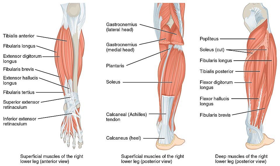
Below the knee, the calf provides the push-off. The two-headed gastrocnemius and the deeper soleus join into the Achilles (calcaneal) tendon and plantarflex the ankle — they point the toes and lift the heel, launching you into every step and jump. Opposing them on the shin, the tibialis anterior dorsiflexes the ankle, pulling the toes up to clear the ground as your foot swings forward.
| Muscle / group | Location | Main action |
|---|---|---|
| Gluteus maximus | Buttock | Extends the hip (running, stairs) |
| Gluteus medius & minimus | Lateral hip | Abduct thigh; stabilize pelvis when walking |
| Iliopsoas | Deep anterior hip | Flexes the hip |
| Quadriceps femoris | Anterior thigh | Extends the knee |
| Hamstrings | Posterior thigh | Flex the knee, extend the hip |
| Gastrocnemius & soleus | Calf | Plantarflex the ankle (push off) |
| Tibialis anterior | Anterior leg (shin) | Dorsiflexes the ankle |
Tap each muscle in the interactive to trace the movers of the hip, knee, and ankle.
- The gluteus maximus extends the hip; the gluteus medius/minimus abduct the thigh and stabilize the pelvis in walking.
- The quadriceps extend the knee and the hamstrings flex it — a front/back antagonist pair; the iliopsoas flexes the hip.
- The gastrocnemius and soleus plantarflex the ankle through the Achilles tendon; the tibialis anterior dorsiflexes it.
The chapter in one breath
- A skeletal muscle can only pull, so muscles work in opposing pairs across joints, drawing the insertion toward the fixed origin.
- Every movement has an agonist, antagonist, synergist, and fixator, and bones act as levers — most of them third-class levers built for speed and range.
- Muscle names are descriptions coded from six clues: location, shape, size, fiber direction, number of heads, and action.
- The head carries muscles of facial expression (insert into skin, CN VII) and muscles of mastication (move the mandible, CN V).
- The trunk holds the diaphragm and intercostals for breathing, the four-layer abdominal wall, and the erector spinae for posture.
- The upper limb is moved by the deltoid, pectoralis major, latissimus dorsi, the stabilizing rotator cuff (SITS), and the biceps/triceps pair.
- The lower limb's powerhouses are the glutes, the quadriceps and hamstrings, and the calf muscles that plantarflex through the Achilles tendon.
ReferenceWords worth knowing
A quick reference for the terms in this chapter — skim it now, or circle back whenever a word trips you up.
- Abduction
- Movement of a limb away from the midline of the body (the deltoid abducts the arm).
- Adduction
- Movement of a limb toward the midline of the body (the thigh adductors squeeze the legs together).
- Agonist (prime mover)
- The muscle chiefly responsible for producing a particular movement.
- Antagonist
- The muscle that opposes an agonist, relaxing and lengthening while the agonist contracts.
- Aponeurosis
- A broad, flat sheet of connective tissue that attaches a muscle to bone or to another muscle.
- Belly
- The thick, contractile middle portion of a muscle between its origin and insertion.
- Diaphragm
- The dome-shaped primary muscle of breathing that separates the thoracic and abdominal cavities; powered by the phrenic nerve.
- Erector spinae
- The group of deep back muscles that extend the vertebral column and maintain upright posture.
- Fixator
- A muscle that stabilizes the origin (or a nearby joint) so a prime mover can act efficiently.
- Insertion
- The attachment of a muscle on the bone that moves — usually the more distal bone.
- Intercostal muscles
- The muscles between the ribs; externals lift the rib cage to inhale, internals pull it down for forced exhalation.
- Lever
- A rigid bar (a bone) that pivots on a fulcrum (a joint) to move a load; most body levers are third-class.
- Masseter
- A powerful muscle of mastication running from the cheekbone to the angle of the jaw; closes the mandible.
- Mastication
- Chewing — the grinding of food produced by muscles that move the mandible, supplied by the trigeminal nerve (CN V).
- Muscles of facial expression
- Muscles that insert into the skin of the face to create expressions; supplied by the facial nerve (CN VII).
- Origin
- The attachment of a muscle on the bone that stays relatively fixed — usually the more proximal bone.
- Quadriceps femoris
- The four-headed anterior thigh muscle group that extends the knee.
- Rotator cuff
- Four muscles (supraspinatus, infraspinatus, teres minor, subscapularis — “SITS”) that stabilize the shoulder joint.
- Sternocleidomastoid
- A neck muscle connecting the sternum, clavicle, and mastoid process; flexes the neck and rotates the head.
- Synergist
- A muscle that assists an agonist or stabilizes a joint during a movement.
- Tendon
- A tough, cord-like band of dense connective tissue that attaches a muscle to a bone.