Anatomy of the Nervous System
Tucked inside the vault of your skull sits the most complicated object we know of in the universe — about three pounds of tissue that lets you read this sentence, remember your childhood, and pull your hand off a hot stove before you even feel the burn. This chapter is a guided tour of that hardware. We will walk the wrinkled surface of the cerebrum, drop down through its inner regions to the life-support brainstem, float in the cerebrospinal fluid that cushions it all, peel back the three protective meninges, follow the spinal cord down its bony canal, and finish by counting off the twelve cranial nerves that leave the brain directly. By the end you will be able to hold a whole brain in your mind's eye and name what you are looking at.
By the end of this chapter you can
- Identify the four lobes of the cerebrum, name the skull bone each lies under, and match each lobe to its main jobs.
- Locate the primary motor and primary somatosensory cortices and the central sulcus that separates them.
- Divide the brain into its four major regions — cerebrum, diencephalon, brainstem, and cerebellum — and state what each does.
- Trace the flow of cerebrospinal fluid through the four ventricles and out into the subarachnoid space, and explain its three functions.
- Name the three meninges in order, describe the spaces between them, and connect each to a clinical emergency.
- Read a cross-section of the spinal cord — gray horns, white columns, dorsal and ventral roots — and explain the one-way traffic rule.
- List the twelve cranial nerves in order, classify each as sensory, motor, or mixed, and give its main function.
Section 1The cerebral lobes
A wrinkled sheet of gray matter
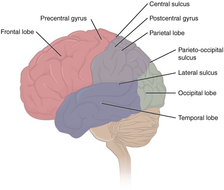
The part of the brain you picture when you hear the word — that pale, deeply folded dome — is the cerebrum, the largest region and the seat of everything we call thought. Its outer shell, only a few millimeters thick, is the cerebral cortex: a sheet of gray matter packed with billions of neuron cell bodies. Underneath the cortex lies white matter, the myelinated axons that wire one region to another. The famous wrinkles are not decoration. Each ridge is a gyrus and each groove a sulcus (a deep one is a fissure), and folding the sheet this way lets an enormous area of cortex fit inside a skull — roughly two-thirds of it is hidden down in the sulci. More cortex means more processing power, which is why the brains of intelligent animals are so heavily folded.
Four lobes, four skull bones
The cortex of each cerebral hemisphere is divided into four lobes, and nature made them easy to name: each lobe is called after the skull bone lying directly over it. Two landmark grooves mark the borders. The central sulcus runs down across the top of the brain and separates the frontal lobe in front from the parietal lobe behind it. The deep lateral sulcus (Sylvian fissure) runs along the side and marks off the temporal lobe below. At the very back sits the occipital lobe. A fifth region, the insula, is buried deep inside the lateral sulcus and is often counted as a hidden fifth lobe.
The two hemispheres are not identical twins working in lockstep — they show lateralization, a division of labor in which one side tends to lead on certain tasks (language usually on the left, spatial and musical processing often on the right). They stay in constant conversation through a thick bridge of white matter called the corpus callosum, which carries hundreds of millions of axons from one hemisphere to the other.
Who does what
Each lobe has its own specialties, and a handful of cortical areas are worth memorizing because damage to them produces classic, testable deficits. The strip of cortex just in front of the central sulcus — the precentral gyrus — is the primary motor cortex, which issues the commands for voluntary movement. The strip just behind the central sulcus — the postcentral gyrus — is the primary somatosensory cortex, which receives touch, pressure, and temperature from the whole body. In each of these strips the body is mapped out upside-down and out of proportion, with huge territory given to the hands and face.
| Lobe | Sits under | Main jobs |
|---|---|---|
| Frontal | Frontal bone | Voluntary movement (primary motor cortex), planning, judgment, personality, and speech production (Broca’s area) |
| Parietal | Parietal bone | Touch, pressure, pain, and temperature (primary somatosensory cortex); spatial awareness |
| Temporal | Temporal bone | Hearing, smell, memory, and language comprehension (Wernicke’s area) |
| Occipital | Occipital bone | Vision — the primary visual cortex |
Tap through the lobes and watch each one light up in the interactive below.
- The cerebral cortex is a folded sheet of gray matter; gyri (ridges) and sulci (grooves) pack more surface into the skull.
- Four lobes — frontal, parietal, temporal, occipital — each named for the overlying skull bone.
- The central sulcus separates the motor cortex (precentral, in front) from the somatosensory cortex (postcentral, behind).
- The corpus callosum connects the two hemispheres, which show lateralization of function.
Section 2The major brain regions
Slice it down the middle
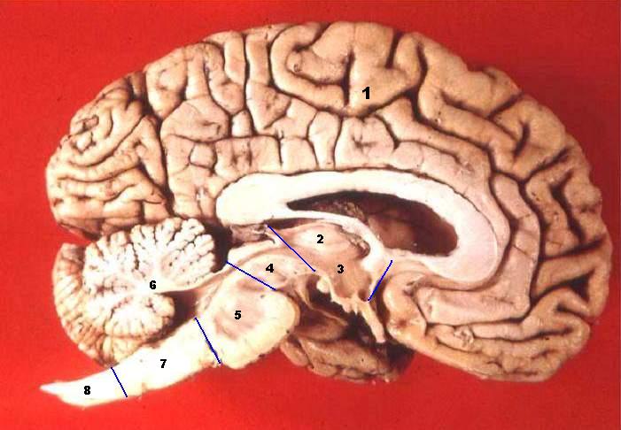
The lobes are only the outer story. To see how the whole brain is built, imagine cutting it exactly down the midline — a midsagittal section — and looking at the cut face. Four major regions appear, stacked from the thinking machinery on top to the life-support wiring below: the cerebrum, the diencephalon, the brainstem, and the cerebellum. As a rough rule, the higher a structure sits, the more “human” and deliberate its job; the lower it sits, the more ancient and automatic. You can lose a good deal of cerebrum and still live, but a tiny stroke in the brainstem can be fatal.
The diencephalon: the brain’s relay and thermostat
Buried beneath the cerebrum, wrapped around the third ventricle, is the diencephalon, a small but critical hub with two stars. The thalamus is the grand central station of the brain: nearly every sensory signal on its way to the cortex — sight, sound, touch, taste — stops here first to be sorted and forwarded. (Smell is the one sense that skips it.) Below and in front sits the hypothalamus, a structure the size of an almond that runs an astonishing amount of homeostasis: body temperature, hunger and thirst, the sleep–wake cycle, and the whole hormonal output of the pituitary gland dangling beneath it. The hypothalamus is where the nervous system and the endocrine system shake hands.
The brainstem and the cerebellum
The brainstem connects the brain to the spinal cord and, region for region, is the most vital tissue you own. From top to bottom it is the midbrain, the pons, and the medulla oblongata. The medulla contains the reflex centers that set your heart rate, blood pressure, and breathing rhythm — jobs you never think about but cannot live without. Every one of the twelve cranial nerves except the first two attaches to the brainstem. Behind the brainstem, tucked under the occipital lobe, is the cerebellum (“little brain”), a densely folded structure that fine-tunes movement, coordinates balance, and keeps your motions smooth. It does not start a movement; it edits one, which is why cerebellar damage makes a person clumsy and unsteady rather than paralyzed.
| Region | Key parts | Main job |
|---|---|---|
| Cerebrum | Two hemispheres, cortex, corpus callosum | Thought, sensation, voluntary movement, memory, language |
| Diencephalon | Thalamus, hypothalamus, epithalamus | Sensory relay (thalamus); homeostasis and hormones (hypothalamus) |
| Brainstem | Midbrain, pons, medulla oblongata | Vital reflexes (heartbeat, breathing), cranial-nerve hub, brain–cord highway |
| Cerebellum | Two hemispheres, folia, vermis | Coordination, balance, smooth and precise movement |
Cut the brain down the middle and tap each region in the interactive below.
- Four major regions: cerebrum, diencephalon, brainstem, cerebellum — higher means more deliberate, lower means more automatic.
- The thalamus relays almost all sensory input to the cortex; the hypothalamus runs homeostasis and the pituitary.
- The brainstem (midbrain, pons, medulla) controls vital reflexes and anchors most cranial nerves.
- The cerebellum coordinates and smooths movement and balance — it edits motion, it does not command it.
Section 3Ventricles & cerebrospinal fluid
A brain that floats
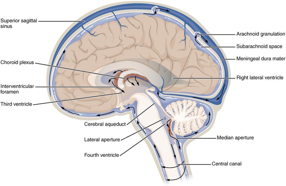
The brain is not packed solid. Running through its core is a set of connected hollow chambers, the ventricles, and the whole organ is bathed inside and out in a clear liquid called cerebrospinal fluid, or CSF. There are four ventricles: a pair of large, C-shaped lateral ventricles (one in each cerebral hemisphere), a slit-like third ventricle in the middle of the diencephalon, and a diamond-shaped fourth ventricle between the brainstem and the cerebellum. CSF is produced by the choroid plexus, a lacework of capillaries lining the ventricles that filters plasma from the blood at a rate of about 500 mL a day, keeping the total volume around 150 mL by constantly draining and replacing it several times over.
Following the flow
CSF is not a stagnant pool; it circulates on a one-way path from the top of the ventricles down and out around the brain and cord. Trace it once and it sticks: fluid made in the lateral ventricles passes through the interventricular foramina into the third ventricle, then down the narrow cerebral aqueduct into the fourth ventricle. From there it escapes through three small openings into the subarachnoid space that surrounds the entire brain and spinal cord, giving the whole central nervous system a liquid cushion. Finally, it is reabsorbed into the blood through cauliflower-like arachnoid granulations that poke up into the large veins draining the brain, completing the loop back into the circulation.
| Step | Structure | What happens |
|---|---|---|
| 1 | Choroid plexus (lateral ventricles) | CSF is made from filtered blood plasma |
| 2 | Interventricular foramina | Fluid passes into the third ventricle |
| 3 | Cerebral aqueduct | Fluid descends to the fourth ventricle |
| 4 | Apertures → subarachnoid space | Fluid bathes the brain and spinal cord |
| 5 | Arachnoid granulations | Fluid is reabsorbed into venous blood |
Why the fluid matters
CSF does three jobs. First, buoyancy: the brain effectively floats, so that an organ weighing about 1,400 g presses down with an effective weight of only around 50 g — without this, the brain would crush its own base under its weight. Second, protection: the liquid layer absorbs shocks, letting the brain bob rather than slam against the skull during a jolt. Third, chemical stability: CSF carries nutrients in and washes wastes out, and it helps maintain the precise ionic environment neurons need to fire.
Watch the fluid circulate and tap each chamber in the interactive below.
- Four ventricles — two lateral, the third, and the fourth — are hollow chambers filled with CSF.
- CSF is made by the choroid plexus and flows lateral → third → fourth ventricle → subarachnoid space → venous blood.
- CSF provides buoyancy, shock protection, and chemical stability for the brain and cord.
- Blocked circulation causes hydrocephalus — swollen ventricles and rising pressure.
Section 4The meninges
Three coats around the cord
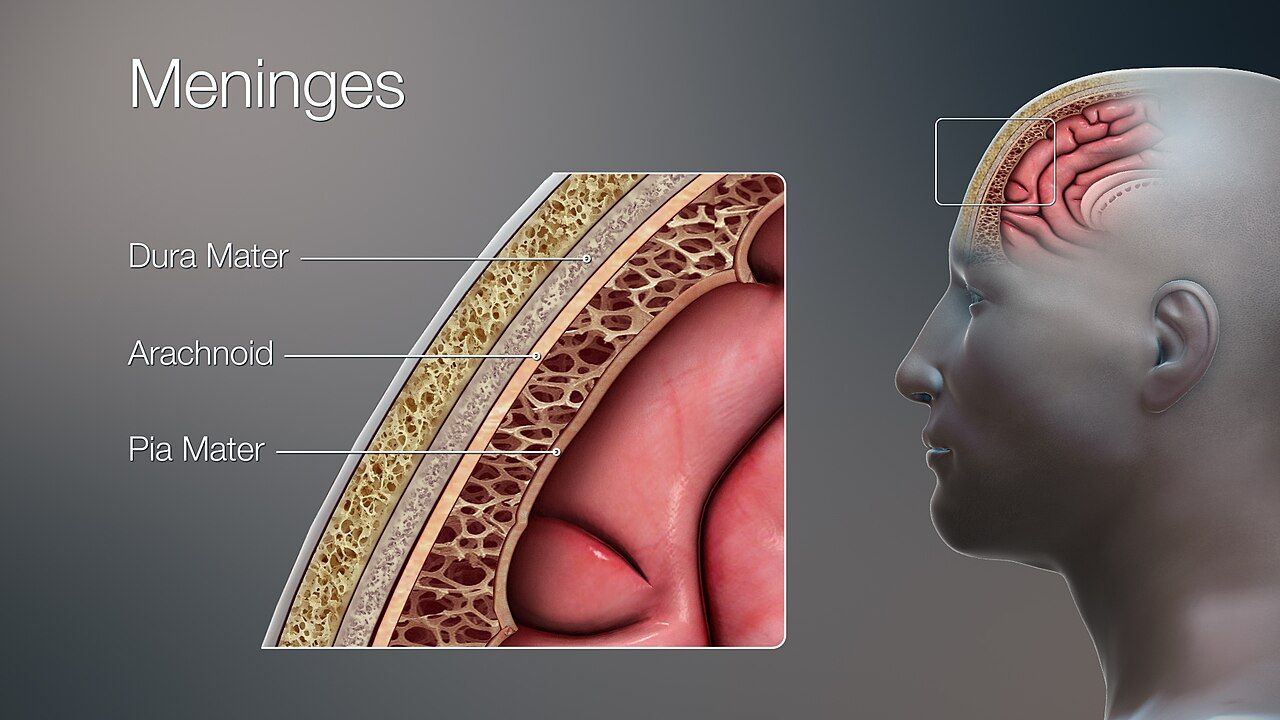
The brain and spinal cord are far too precious to leave bare against bone, so they are wrapped in three connective-tissue membranes called the meninges. From the outside in, they are the dura mater, the arachnoid mater, and the pia mater — a sequence worth memorizing in order, because the spaces between them are where the important clinical drama happens. The Latin names describe them: dura mater means “tough mother,” and pia mater means “tender mother.”
From tough to tender
The dura mater is the outermost layer: thick, leathery, and strong. Around the brain it has two sublayers, and where they separate they form the dural venous sinuses that drain blood from the brain, and inward folds like the falx cerebri that dip between the hemispheres to hold the brain steady. Beneath the dura lies the arachnoid mater, a delicate, web-like layer — its name comes from the Greek for spider, because fine threads called arachnoid trabeculae span the gap beneath it. That gap, the subarachnoid space, is filled with the CSF from the last section. Finally, the pia mater is a microscopically thin, richly vascular film that clings to every gyrus and sulcus of the brain like shrink-wrap, following the surface into its every fold.
| Layer | Character | Space / feature |
|---|---|---|
| Dura mater | Tough, leathery outer coat | Forms dural venous sinuses; epidural space lies outside it |
| Arachnoid mater | Thin, web-like middle layer | Subdural space above, subarachnoid space (CSF) below |
| Pia mater | Delicate, vascular inner film | Adheres directly to the brain and cord surface |
Peel back the layers one at a time in the interactive below.
- Three meninges, outside in: dura mater (tough), arachnoid mater (web-like), pia mater (delicate).
- The subarachnoid space, beneath the arachnoid, holds circulating CSF.
- The pia mater follows every fold of the brain; the dura forms venous sinuses and stabilizing folds like the falx cerebri.
- The spaces define the bleeds: epidural (arterial, lens-shaped) vs subdural (venous, crescent-shaped); infection of the meninges is meningitis.
Section 5The spinal cord
A cable and a switchboard
The spinal cord is the great communication cable of the body, running from the base of the brainstem down through the protective canal of the vertebral column to about the level of the first or second lumbar vertebra (L1–L2). Below that point it frays into a spray of nerve roots called the cauda equina (“horse’s tail”). The cord does two things at once: it carries traffic up and down between the body and the brain, and it acts as its own local switchboard, handling reflexes without waiting for the brain to weigh in.
Reading a slice
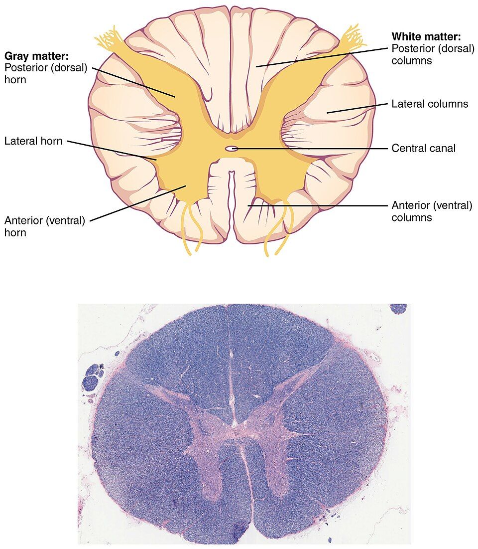
Cut the cord across and the anatomy is beautifully organized. In the center is a butterfly- or H-shaped region of gray matter — neuron cell bodies — surrounded by white matter, the myelinated tracts. This is the reverse of the brain, where gray is on the outside. The gray matter has wings called horns: the dorsal (posterior) horns handle incoming sensory information, the ventral (anterior) horns send out motor commands, and small lateral horns in the trunk region house autonomic neurons. A tiny central canal runs down the middle, filled with CSF.
One-way traffic at the roots
Where each spinal nerve joins the cord it splits into two roots, and they obey a strict traffic rule. The dorsal root carries sensory signals into the cord, and it bears a visible bulge, the dorsal root ganglion, where the sensory neuron cell bodies sit. The ventral root carries motor signals out to muscles and glands. Just beyond the cord the two roots merge into a single mixed spinal nerve — and there are 31 pairs of them. The white matter between is organized into columns of ascending (sensory) and descending (motor) tracts, the express lanes carrying information the length of the cord.
| Structure | Type | Function |
|---|---|---|
| Dorsal (posterior) horn | Gray matter | Receives sensory input |
| Ventral (anterior) horn | Gray matter | Sends motor output to muscles |
| Dorsal root (with ganglion) | Sensory | Carries sensation into the cord |
| Ventral root | Motor | Carries commands out of the cord |
| White columns / tracts | White matter | Ascending (sensory) and descending (motor) highways |
Slice the cord and tap each part in the interactive below.
- The cord runs to about L1–L2, then continues as the nerve roots of the cauda equina; 31 pairs of spinal nerves branch off.
- In cross-section, central gray matter (horns) is surrounded by white matter (tracts) — the opposite arrangement to the brain.
- Dorsal horns/roots = sensory in; ventral horns/roots = motor out (the Bell–Magendie rule).
- The dorsal root ganglion holds sensory cell bodies; a lumbar puncture is done below the cord for safety.
Section 6The cranial nerves
Twelve nerves straight from the brain
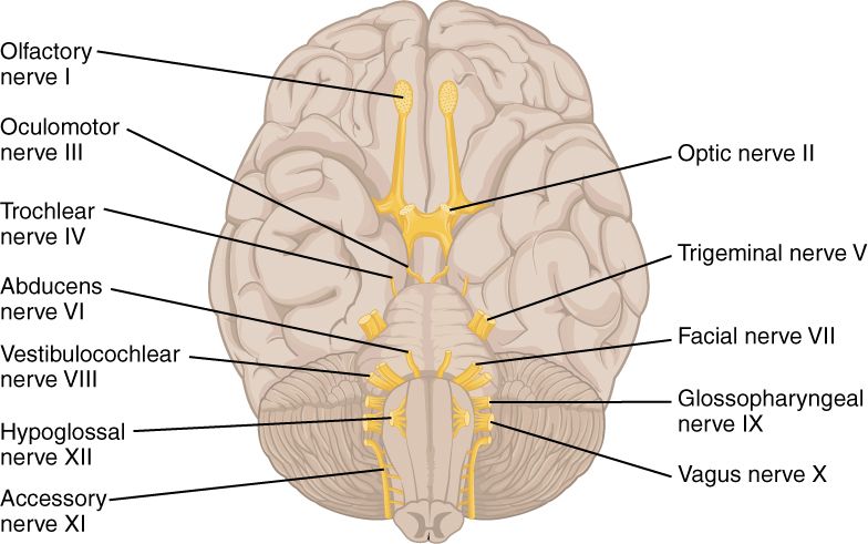
Most nerves branch off the spinal cord, but a special set of twelve pairs emerges directly from the brain itself: the cranial nerves. They are numbered with Roman numerals I through XII, from front to back, in the order their roots appear along the underside of the brain. The first two — olfactory and optic — grow out of the cerebrum and diencephalon; the remaining ten sprout from the brainstem. Together they handle the special senses of the head, move the eyes and face and tongue, and — in the case of one wandering nerve — reach all the way down into the chest and abdomen.
Sensory, motor, or both
Each cranial nerve is classified by the kind of traffic it carries. Some are purely sensory (they bring information in), some are purely motor (they send commands out), and some are mixed, doing both. A time-honored mnemonic captures the pattern — “Some Say Marry Money, But My Brother Says Big Brains Matter Most” — where the first letter of each word (S, S, M, M, B, M, B, S, B, B, M, M) tells you whether nerve I through XII is Sensory, Motor, or Both. For the names themselves, generations of students have used “On Old Olympus’ Towering Tops, A Finn And German Viewed Some Hops.”
| # | Name | Type | Main function |
|---|---|---|---|
| I | Olfactory | Sensory | Smell |
| II | Optic | Sensory | Vision |
| III | Oculomotor | Motor | Most eye movements; pupil constriction; raises the eyelid |
| IV | Trochlear | Motor | Moves the eye (superior oblique muscle) |
| V | Trigeminal | Both | Facial sensation; chewing muscles |
| VI | Abducens | Motor | Moves the eye laterally (lateral rectus) |
| VII | Facial | Both | Facial expression; taste (front tongue); tears and saliva |
| VIII | Vestibulocochlear | Sensory | Hearing and balance |
| IX | Glossopharyngeal | Both | Taste (back tongue); swallowing; saliva |
| X | Vagus | Both | Parasympathetic control of heart, lungs, and gut |
| XI | Accessory | Motor | Moves the neck and shoulders (sternocleidomastoid, trapezius) |
| XII | Hypoglossal | Motor | Moves the tongue |
The wandering nerve
One cranial nerve deserves a special note. The vagus nerve (X) is the odd one out: while every other cranial nerve stays in the head and neck, the vagus — its name literally means “wanderer” — travels down into the thorax and abdomen. It is the great parasympathetic highway, slowing the heart, driving digestion, and monitoring the internal organs. When you hear about the “rest-and-digest” response, you are hearing about the vagus at work.
Number off each nerve and tap it in the interactive below.
- Twelve pairs of cranial nerves, numbered I–XII front to back; all but the first two arise from the brainstem.
- Each is sensory, motor, or mixed — the mnemonic “Some Say Marry Money…” gives the pattern.
- Several move the eye (III, IV, VI); others carry special senses (I smell, II vision, VIII hearing/balance).
- The vagus (X) is the only cranial nerve to leave the head and neck, carrying most parasympathetic output to the organs.
The chapter in one breath
- The cerebrum is a folded sheet of cortical gray matter split into four lobes — frontal, parietal, temporal, occipital — each under its namesake skull bone.
- The central sulcus divides the motor cortex (front) from the somatosensory cortex (back); the corpus callosum links the hemispheres.
- Four brain regions stack from thinking to survival: cerebrum → diencephalon → brainstem → cerebellum, with the medulla running vital reflexes.
- Cerebrospinal fluid made by the choroid plexus flows through four ventricles into the subarachnoid space, giving the brain buoyancy, protection, and chemical stability.
- Three meninges — dura, arachnoid, pia — wrap the CNS; the spaces between them define epidural, subdural, and subarachnoid bleeds.
- The spinal cord holds central gray horns inside white tracts; sensation enters the dorsal root, motor leaves the ventral root.
- Twelve cranial nerves (I–XII) serve the head and neck — each sensory, motor, or mixed — and the vagus alone reaches the chest and abdomen.
ReferenceWords worth knowing
A quick reference for the terms in this chapter — skim it now, or circle back whenever a word trips you up.
- Arachnoid mater
- The web-like middle meningeal layer; beneath it, the subarachnoid space holds circulating cerebrospinal fluid.
- Brainstem
- The stalk connecting brain to spinal cord — midbrain, pons, and medulla oblongata — housing vital reflex centers and most cranial nerves.
- Central sulcus
- The groove separating the frontal lobe (and its motor cortex) from the parietal lobe (and its somatosensory cortex).
- Cerebellum
- The “little brain” behind the brainstem that coordinates balance and smooths voluntary movement.
- Cerebral cortex
- The thin outer sheet of gray matter covering the cerebrum, where higher thought, sensation, and voluntary movement are processed.
- Cerebrospinal fluid (CSF)
- The clear liquid made by the choroid plexus that fills the ventricles and subarachnoid space, cushioning and nourishing the CNS.
- Choroid plexus
- The capillary network lining the ventricles that filters blood plasma to produce cerebrospinal fluid.
- Corpus callosum
- The thick band of white matter carrying axons between the two cerebral hemispheres.
- Cranial nerves
- The twelve pairs of nerves (I–XII) emerging directly from the brain to serve the head, neck, and (via the vagus) the internal organs.
- Diencephalon
- The central brain region containing the thalamus and hypothalamus, wrapped around the third ventricle.
- Dorsal root ganglion
- The swelling on a spinal nerve’s dorsal root that houses the cell bodies of sensory neurons.
- Dura mater
- The tough, leathery outermost meningeal layer; it forms the dural venous sinuses and folds like the falx cerebri.
- Gyrus
- A raised ridge on the surface of the cerebral cortex (plural, gyri).
- Hypothalamus
- The diencephalon structure that regulates temperature, hunger, thirst, sleep, and hormone release, linking the nervous and endocrine systems.
- Medulla oblongata
- The lowest part of the brainstem, containing the reflex centers for heart rate, blood pressure, and breathing.
- Meninges
- The three connective-tissue membranes — dura, arachnoid, and pia mater — that wrap the brain and spinal cord.
- Pia mater
- The delicate, vascular innermost meningeal layer that clings directly to the surface of the brain and cord.
- Sulcus
- A shallow groove between the ridges of the cerebral cortex (plural, sulci); a deep one is a fissure.
- Thalamus
- The paired diencephalon relay station that sorts and forwards nearly all sensory input on its way to the cortex.
- Vagus nerve
- Cranial nerve X, the only cranial nerve to extend beyond the head and neck, carrying most parasympathetic output to the heart, lungs, and gut.
- Ventricles
- The four interconnected fluid-filled chambers within the brain — two lateral, third, and fourth — that make and circulate CSF.