Development & Inheritance
Every human life begins as a single cell no bigger than the period at the end of this sentence — and yet folded inside it is a complete instruction book for building a person. This chapter follows that cell forward: the dramatic meeting of sperm and egg, the first week of frantic cell division and burrowing into the uterus, the moment three germ layers appear and quietly divide up the job of making every organ, and the long stretch of fetal growth that ends in birth. Then we turn the story around and ask how the instruction book itself is passed on — the rules of inheritance that let us predict, with a simple square grid, the odds that a child will have brown eyes or a particular disease. It is the most compressed miracle in all of physiology, and it follows rules you can learn.
By the end of this chapter you can
- Describe the steps of fertilization, from the sperm crossing the egg's outer coats to the fusion that forms a zygote, and explain how the egg blocks fertilization by more than one sperm.
- Trace the first week of development through cleavage, the morula, and the blastocyst, and describe how and where implantation occurs.
- Explain gastrulation and name the three primary germ layers along with the major tissues each one gives rise to.
- Distinguish the embryonic period from the fetal period and outline the milestones of the three trimesters.
- Describe the structure and functions of the placenta, and list the three stages of labor.
- Define allele, genotype, and phenotype, and contrast dominant with recessive inheritance.
- Use a Punnett square to predict the genotype and phenotype ratios of offspring from a given cross.
- Recognize why two unaffected parents can have a child with a recessive condition.
Section 1Fertilization
A race with one finisher
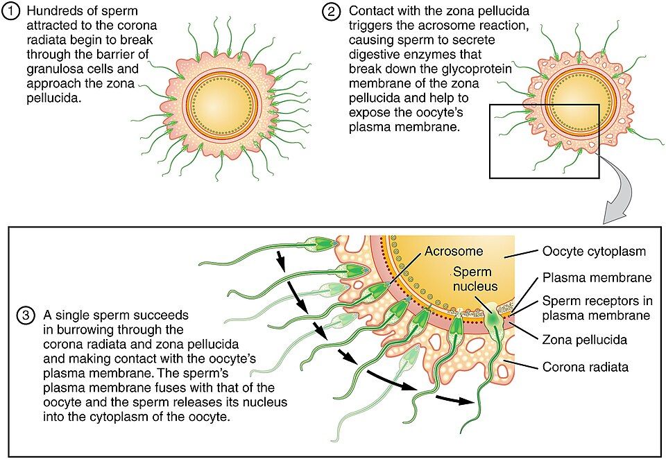
Fertilization is the fusion of a sperm and an oocyte (egg) to create a new, genetically unique cell. It usually happens not in the uterus but high up in the ampulla of the uterine tube, within roughly a day of ovulation, because the ovulated egg is only fertile for about 12 to 24 hours and sperm survive at most a few days. Hundreds of millions of sperm are deposited, but the journey is brutal: most are lost to the acidic vagina, the cervical mucus, and simply taking wrong turns. Only a few hundred reach the egg. Along the way the survivors undergo capacitation — a final chemical maturation in the female tract that makes their membranes ready to fuse. The egg they find is not naked. It is wrapped in a halo of follicle cells called the corona radiata and, beneath that, a thick glycoprotein shell called the zona pellucida.
Breaking in: the acrosome reaction
To reach the egg's plasma membrane, a sperm must get through both coats. Sitting like a cap over the sperm's head is the acrosome, a lysosome-like sac full of digestive enzymes. When the sperm contacts the zona pellucida, the acrosome ruptures and spills those enzymes — the acrosome reaction — digesting a narrow tunnel through the shell. Because it takes the combined effort of many sperm chipping away at the corona and zona, fertilization is very much a team effort with a single winner. The first sperm to reach the oocyte's plasma membrane binds to receptors there, and the two plasma membranes fuse. The sperm's nucleus, carrying its 23 chromosomes, slides into the egg's cytoplasm.
| Barrier | What it is | How the sperm passes |
|---|---|---|
| Corona radiata | Outer halo of granulosa (follicle) cells | Many sperm push and enzymatically loosen the cells |
| Zona pellucida | Thick glycoprotein shell around the egg | The acrosome reaction digests a path through it |
| Oocyte plasma membrane | The egg cell's own surface | Sperm receptors bind; the two membranes fuse |
One sperm only: the blocks to polyspermy
Fertilization by two sperm — polyspermy — would give the embryo the wrong number of chromosomes and is lethal, so the egg has two safeguards that snap shut the instant one sperm fuses. The fast block is an immediate change in the electrical charge across the egg membrane that stops further sperm from binding. The slow block, or cortical reaction, follows seconds later: tiny vesicles just under the egg's surface release their contents into the zona pellucida, chemically hardening it into an impenetrable wall. Only now does the egg finish its own final division (meiosis II). The two sets of 23 chromosomes — one from each parent — come together, restoring the full human count of 46, and the fertilized egg is now a single diploid cell called the zygote. Its sex was decided at this exact moment, by whether the winning sperm carried an X or a Y.
Step through the meeting of sperm and egg in the interactive below.
- Fertilization normally occurs in the ampulla of the uterine tube; the egg is fertile for only about 12–24 hours.
- The sperm crosses the corona radiata and zona pellucida, using the acrosome reaction, then fuses with the oocyte membrane.
- A fast block (membrane charge) and a slow block (cortical reaction hardening the zona) prevent polyspermy.
- Union of the two nuclei restores 46 chromosomes, forming a diploid zygote whose sex is set by the sperm.
Section 2The first week: cleavage & implantation
Cleavage: dividing without growing
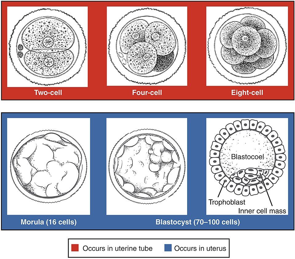
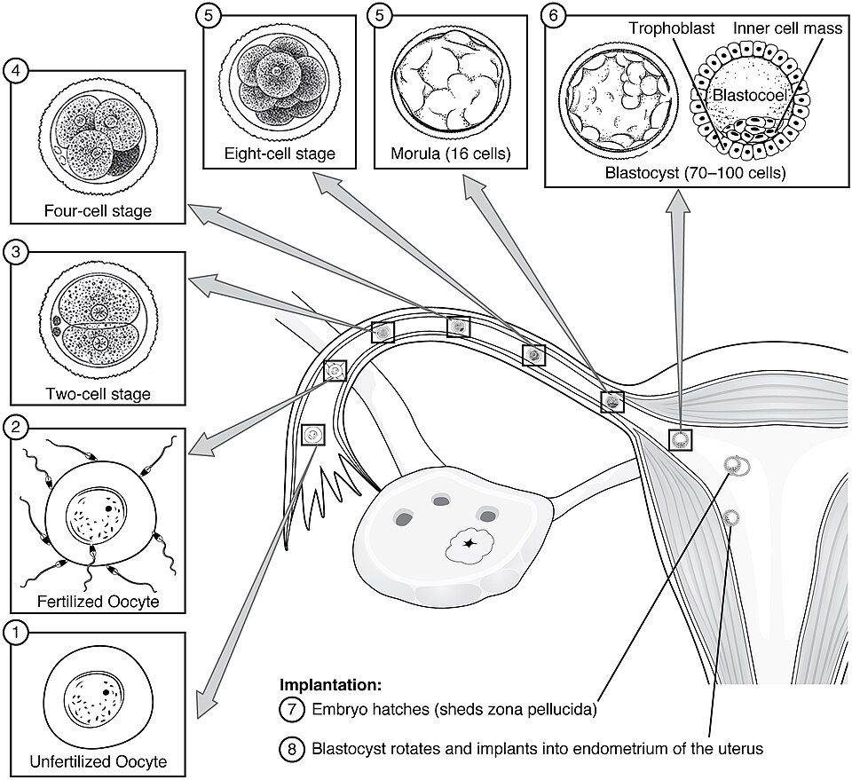
Within about a day, the zygote begins a series of rapid mitotic divisions called cleavage. Here is the surprising part: the whole ball does not get bigger. Each division splits the existing cytoplasm into smaller and smaller cells, so the cluster stays roughly the size of the original egg while the number of cells climbs — 2, then 4, then 8, then 16. These early cells are called blastomeres. All of this happens while the tiny embryo is still drifting down the uterine tube, swept along by cilia and muscular waves toward the uterus. By about day 4 it has become a solid mulberry-shaped ball of roughly 16 cells known as the morula (Latin for "little mulberry").
From morula to blastocyst
Around day 5, fluid seeps into the center of the morula and hollows it out into a fluid-filled sphere called the blastocyst. This is the first time the embryo's cells commit to different fates, and it splits into two populations you must know. The outer shell of cells, the trophoblast, will become the embryo's contribution to the placenta and membranes — it is destined to be support tissue, not the baby. Clustered against one inner wall is the inner cell mass (embryoblast), the small group of cells that will actually become the embryo itself. The fluid space between them is the blastocoel. In these few days the embryo has quietly answered its first big question: which cells build the child, and which cells build the life-support system.
| Day (approx.) | Stage | Where |
|---|---|---|
| 0 | Zygote (fertilized egg) | Ampulla of uterine tube |
| 1–3 | Cleavage: 2-, 4-, 8-cell stages | Traveling down the uterine tube |
| 4 | Morula (~16 cells) | Entering the uterus |
| 5 | Blastocyst (trophoblast + inner cell mass) | Free in the uterine cavity |
| 6–10 | Implantation into the endometrium | Uterine wall |
Implantation and the pregnancy signal
To implant, the blastocyst must first "hatch" — break free of the leftover zona pellucida — so its sticky trophoblast can contact the uterine lining. Around day 6 or 7, the trophoblast adheres to the endometrium and begins to burrow in, its cells secreting enzymes that erode into the lining until the embryo is fully embedded, usually by day 10. The invading trophoblast immediately starts producing human chorionic gonadotropin (hCG), a hormone that signals the ovary's corpus luteum to keep making progesterone so the endometrium is not shed. That single hormone is what keeps the pregnancy from ending in a normal period — and it is exactly what a pregnancy test detects.
Follow the zygote's first-week journey from division to implantation below.
- Cleavage divides the zygote into more, smaller cells without the cluster growing in size, producing the morula.
- The blastocyst splits into the outer trophoblast (future placenta) and the inner cell mass (future embryo), around a fluid blastocoel.
- Implantation into the endometrium occurs about days 6–10 and requires the blastocyst to hatch from the zona pellucida.
- The trophoblast secretes hCG, which sustains the pregnancy and is the target of pregnancy tests.
Section 3The germ layers
Gastrulation: one sheet becomes three
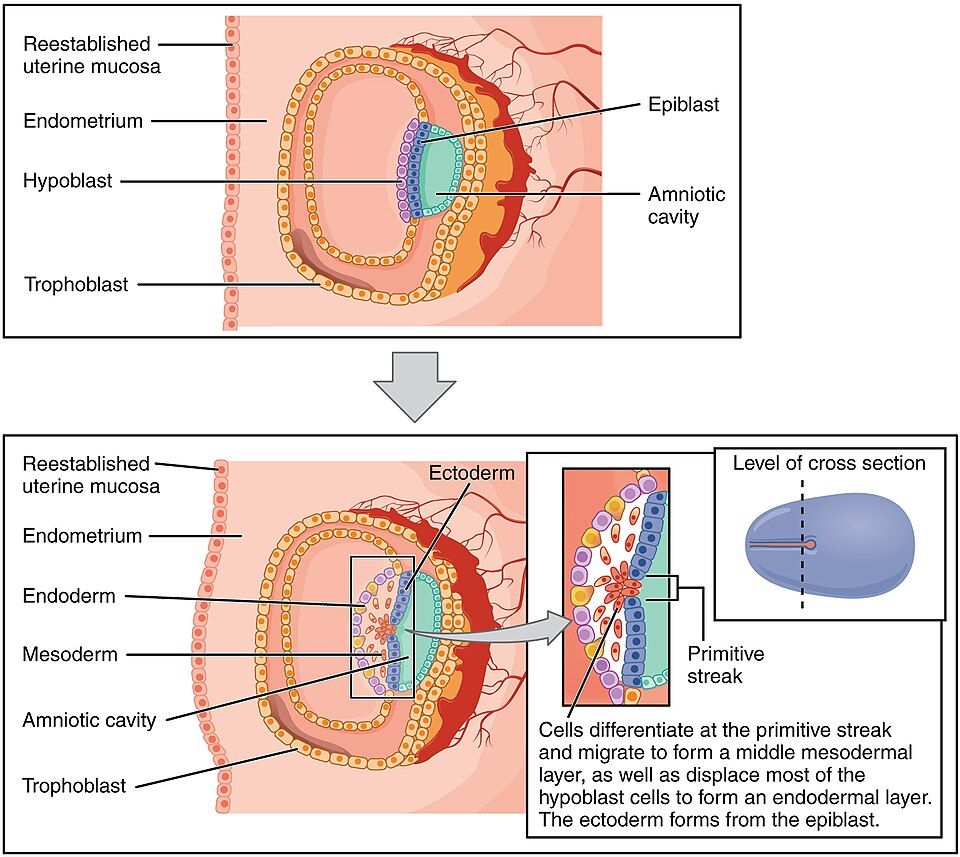
Around the third week, the inner cell mass performs one of the most important moves in all of development: gastrulation. A groove called the primitive streak appears down the middle of the flat embryonic disc, and cells from the upper layer (the epiblast) stream toward it and dive underneath, rearranging themselves into three primary germ layers. Every one of the more than 200 cell types in your body traces its lineage back to one of these three sheets. Getting these layers laid down — in the right order, in the right place — is the foundation for building every organ that follows.
Three layers, three destinies
The three germ layers divide up the entire body between them. The outer layer, the ectoderm, becomes what faces the world and what senses it — the epidermis of the skin and the entire nervous system. The middle layer, the mesoderm, becomes the body's bulk and machinery — muscle, bone, blood, heart, blood vessels, and kidneys. The inner layer, the endoderm, becomes the linings of the hollow tubes and the glands that grow off them — the epithelium of the digestive and respiratory tracts, plus the liver and pancreas. A simple way to hold it: ectoderm is the outside and the wiring, endoderm is the innermost linings, and mesoderm is everything in the middle.
| Germ layer | Position | Major derivatives |
|---|---|---|
| Ectoderm | Outer | Epidermis, hair, nails; entire nervous system; parts of the eye |
| Mesoderm | Middle | Muscle; bone and cartilage; blood and heart; blood vessels; kidneys; dermis |
| Endoderm | Inner | Epithelial lining of the GI and respiratory tracts; liver; pancreas |
Folding into a body: organogenesis
With the three layers in place, organogenesis — the actual building of organs — begins and continues through the eighth week. The flat disc folds into a tube-within-a-tube shape, and each layer starts sculpting its assigned structures. One of the earliest and most critical events is the folding of ectoderm into the neural tube, the forerunner of the brain and spinal cord. If that tube fails to close completely, the result is a neural tube defect such as spina bifida. Because the neural tube closes so early — often before a person even knows they are pregnant — adequate folic acid at the time of conception dramatically lowers the risk.
Tap each of the three layers to see what it becomes in the interactive below.
- Gastrulation (week 3) forms three primary germ layers at the primitive streak.
- Ectoderm → skin and nervous system; mesoderm → muscle, bone, blood, heart, kidney; endoderm → gut and respiratory linings, liver, pancreas.
- Organogenesis builds the organs through week 8; early neural tube closure depends on adequate folic acid.
Section 4Embryo to fetus: the timeline
The embryonic period versus the fetal period
Development is divided at the end of week 8 into two named periods. From fertilization through week 8 is the embryonic period, when the germ layers form and every major organ is first laid down. From week 9 until birth is the fetal period, which is mostly about growth, maturation, and refinement of organs that already exist. The distinction matters clinically: because organs are being built during the embryonic weeks, that is exactly when they are most vulnerable to damage. A teratogen — a drug, infection, or other agent that disrupts development — does its worst harm during weeks 3 to 8, when structures are forming. The same exposure later in the fetal period may cause little or no structural malformation.
Three trimesters
Counting from fertilization, a full-term pregnancy lasts about 38 weeks (about 40 weeks if counted from the last menstrual period, the way clinicians usually date it). That span is divided into three roughly equal trimesters. The first trimester is where the action is: implantation, gastrulation, organ formation, and the transition from embryo to fetus — by its end the fetus is recognizably human but only about the size of a plum. The second trimester is a stretch of rapid growth and movement; the mother usually feels the first fetal movements ("quickening"), and the fetus develops toward the edge of viability — about 23 to 24 weeks of gestation as clinicians date it from the last menstrual period, which is roughly 21 to 22 weeks after fertilization. (Watch the dating convention whenever a week number is quoted: clinical practice counts from the LMP, embryology counts from fertilization, and the two differ by two weeks.) The third trimester is dominated by weight gain and final maturation, especially of the lungs and brain, preparing the fetus to survive outside the uterus.
| Trimester | Weeks (from fertilization) | Headline events |
|---|---|---|
| First | 1–12 | Implantation, germ layers, organ formation; embryo becomes fetus at week 9 |
| Second | 13–27 | Rapid growth; first movements felt; approaches viability (~21–22 weeks after fertilization; ~23–24 weeks by LMP dating) |
| Third | 28–38 | Major weight gain; lung and brain maturation; readiness for birth |
Tap through the stages of the earliest journey in the interactive below.
- The embryonic period (weeks 1–8) builds organs; the fetal period (week 9 to birth) grows and matures them.
- Teratogens cause the most structural harm during weeks 3–8, when organs are forming.
- Pregnancy is about 38 weeks from fertilization, split into three trimesters; viability begins around 23–24 weeks by LMP dating (~21–22 weeks after fertilization).
Section 5Pregnancy & birth
The placenta: a shared border organ
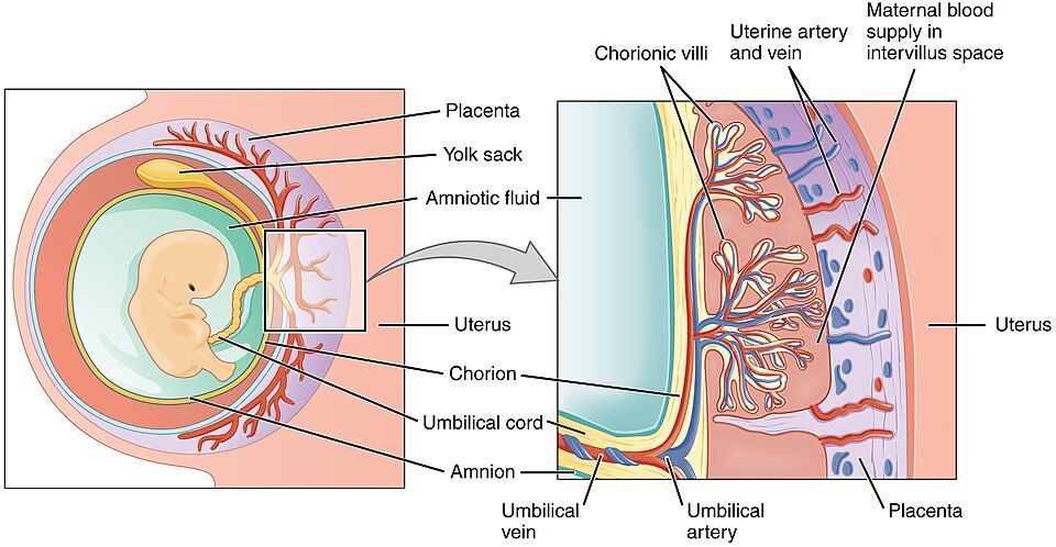
The placenta is a temporary organ built jointly from the embryo's trophoblast-derived tissue and the mother's endometrium, and it is the lifeline for the entire pregnancy. Finger-like chorionic villi from the fetal side project into pools of maternal blood, creating an enormous surface for exchange. Across that thin barrier, O₂ and nutrients pass from mother to fetus while CO₂ and wastes pass the other way — yet, crucially, the two bloodstreams normally never mix. The placenta also becomes an endocrine powerhouse, taking over from the corpus luteum to secrete the progesterone and estrogen that maintain the pregnancy. The fetus is tethered to it by the umbilical cord, and floats within the fluid-filled amnion that cushions it.
A body remade for pregnancy
Sustaining a fetus reshapes nearly every maternal system. Blood volume rises by roughly 30–50 percent to supply the placenta; the heart pumps more each minute; the growing uterus crowds the diaphragm and bladder, changing breathing and increasing urinary frequency. Hormones loosen ligaments (including those of the pelvis, in preparation for delivery) and shift metabolism to prioritize fetal fuel. These are normal, coordinated adaptations — but they also explain why pregnancy stresses conditions like the heart, and why blood pressure and glucose are watched so closely.
Labor: birth in three stages
Labor is triggered by a shift in the hormonal balance near term, and once it begins it is driven by a textbook positive feedback loop: the fetal head pushes on the cervix, which triggers release of oxytocin, which strengthens uterine contractions, which push the head harder against the cervix — a self-amplifying cycle that only ends when the baby is delivered. Labor unfolds in three stages.
| Stage | Name | What happens |
|---|---|---|
| 1 | Dilation | The longest stage: contractions thin (efface) and open the cervix to full dilation (~10 cm) |
| 2 | Expulsion | Strong contractions and maternal pushing deliver the baby through the birth canal |
| 3 | Placental | The uterus contracts to expel the placenta (the "afterbirth"), then clamps down to limit bleeding |
Step through the placenta and the three stages of birth in the interactive below.
- The placenta exchanges O₂, CO₂, nutrients, and wastes without mixing maternal and fetal blood, and secretes pregnancy hormones.
- Pregnancy drives large adaptations — a 30–50% rise in blood volume, higher cardiac output, and displaced abdominal organs.
- Labor is driven by oxytocin positive feedback and proceeds in three stages: dilation → expulsion → placental.
Section 6Inheritance
Two copies of every gene
Because you inherit one set of chromosomes from each parent, you carry two copies of nearly every gene — one maternal, one paternal. The alternative versions of a gene are called alleles. For a gene with two alleles, an individual may be homozygous (two identical alleles) or heterozygous (two different ones). The pair of alleles a person carries is their genotype; the observable trait that results — eye color, blood type, whether a disease appears — is their phenotype. The whole logic of inheritance is a bridge between these two: how the genotype you cannot see produces the phenotype you can.
Dominant and recessive
When the two alleles differ, they do not always contribute equally. A dominant allele masks the effect of a recessive one, so a single copy is enough to show its phenotype; a recessive allele only shows its phenotype when both copies are recessive. By convention the dominant allele gets a capital letter and the recessive a lowercase. Using the interactive's example, where B = brown eyes (dominant) and b = blue (recessive): genotypes BB and Bb both give brown eyes, and only bb gives blue. This is why a recessive trait can seem to "skip" generations — it can be carried silently for years by heterozygous carriers and only reappears when two carriers each pass the recessive allele to the same child.
| Genotype | Name | Phenotype (B = brown, b = blue) |
|---|---|---|
| BB | Homozygous dominant | Brown eyes |
| Bb | Heterozygous (carrier) | Brown eyes |
| bb | Homozygous recessive | Blue eyes |
Predicting the odds: the Punnett square
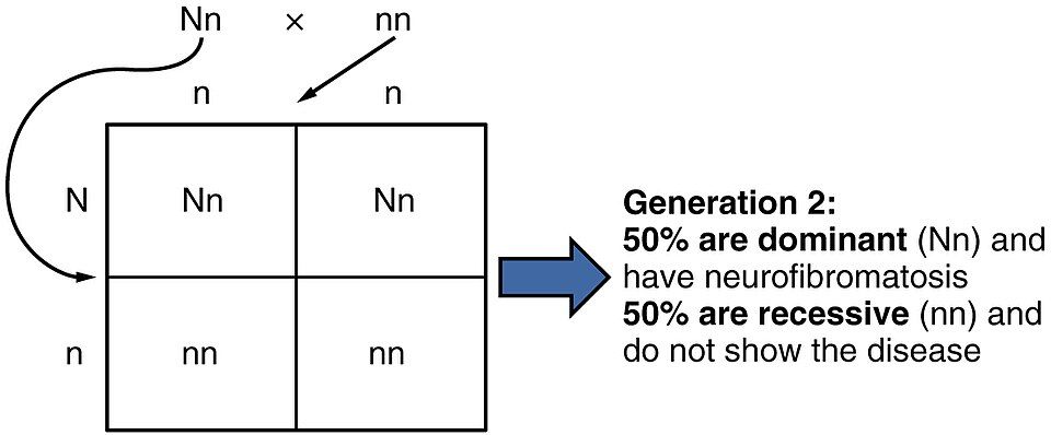
A Punnett square is a simple grid that predicts the probability of each offspring genotype. You list one parent's possible alleles across the top, the other parent's down the side, and fill each box by combining them. Cross two brown-eyed carriers (Bb × Bb) and the four boxes come out 1 BB : 2 Bb : 1 bb — a 3:1 ratio of brown to blue, meaning each child has a 25 percent chance of blue eyes even though both parents are brown-eyed. It is essential to read these as probabilities, not guarantees: a 1-in-4 chance applies fresh to every pregnancy, exactly like flipping a coin, so four children could easily be all brown or include two blue. Not every trait follows this clean dominant/recessive pattern — some alleles are codominant (both show, as in AB blood type), and genes on the X chromosome produce distinctive sex-linked patterns — but the two-allele Punnett square is the foundation everything else builds on.
Set the two parents and watch the ratios change in the interactive below.
- You carry two alleles of most genes; genotype is the allele pair, phenotype is the visible trait.
- A dominant allele shows with one copy; a recessive allele shows only when both copies are recessive.
- A Punnett square predicts offspring ratios (e.g., Bb × Bb → 3:1); results are probabilities that reset each pregnancy.
- Two heterozygous carriers can produce an affected child, explaining recessive diseases that skip generations.
The chapter in one breath
- Fertilization in the uterine tube fuses sperm and egg into a diploid zygote; fast and slow blocks stop polyspermy, and the sperm sets the child's sex.
- Cleavage divides the zygote into a morula and then a blastocyst (trophoblast + inner cell mass), which implants in the endometrium and secretes hCG.
- Gastrulation forms three germ layers: ectoderm (skin, nerves), mesoderm (muscle, bone, blood, kidney), and endoderm (gut and respiratory linings, liver, pancreas).
- The embryonic period (weeks 1–8) builds organs and is most vulnerable to teratogens; the fetal period grows them across three trimesters (~38 weeks total).
- The placenta exchanges gases and nutrients without mixing bloodstreams and secretes pregnancy hormones; labor is oxytocin positive feedback in three stages (dilation, expulsion, placental).
- Genotype (allele pair) produces phenotype (visible trait); dominant alleles mask recessive ones.
- A Punnett square predicts offspring probabilities, and two carriers can have a child with a recessive condition.
ReferenceWords worth knowing
A quick reference for the terms in this chapter — skim it now, or circle back whenever a word trips you up.
- Allele
- One of the alternative versions of a gene; a person carries two alleles of most genes, one from each parent.
- Blastocyst
- The early, fluid-filled hollow ball of cells that implants in the uterus, made of an outer trophoblast and an inner cell mass.
- Cleavage
- The series of rapid mitotic divisions after fertilization that increase cell number without increasing the embryo's overall size.
- Dominant allele
- An allele that shows its phenotype when only one copy is present, masking any recessive allele (written with a capital letter).
- Ectoderm
- The outer germ layer, giving rise to the epidermis and the entire nervous system.
- Endoderm
- The inner germ layer, giving rise to the linings of the digestive and respiratory tracts and to the liver and pancreas.
- Fertilization
- The fusion of a sperm and an oocyte to form a diploid zygote, normally in the ampulla of the uterine tube.
- Gastrulation
- The reorganization of the embryo (around week 3) that forms the three primary germ layers at the primitive streak.
- Genotype
- The specific pair of alleles an individual carries for a gene.
- Germ layer
- One of the three primary cell sheets — ectoderm, mesoderm, endoderm — from which all tissues develop.
- Heterozygous
- Having two different alleles of a gene (for example, Bb).
- Homozygous
- Having two identical alleles of a gene (for example, BB or bb).
- Human chorionic gonadotropin (hCG)
- The hormone secreted by the trophoblast that sustains the corpus luteum and pregnancy; the target of pregnancy tests.
- Implantation
- The embedding of the blastocyst into the uterine endometrium, occurring about days 6–10 after fertilization.
- Mesoderm
- The middle germ layer, giving rise to muscle, bone, blood, the heart and vessels, and the kidneys.
- Morula
- The solid, mulberry-shaped ball of about 16 cells produced by cleavage before the blastocyst forms.
- Phenotype
- The observable trait that results from a genotype (and its interaction with the environment).
- Placenta
- The temporary organ, built from fetal and maternal tissue, that exchanges gases, nutrients, and wastes and secretes pregnancy hormones.
- Punnett square
- A grid that predicts the probability of each offspring genotype from a given cross.
- Recessive allele
- An allele whose phenotype appears only when both copies are recessive (written with a lowercase letter).
- Trophoblast
- The outer cell layer of the blastocyst that forms the embryo's contribution to the placenta and secretes hCG.
- Zygote
- The single diploid cell formed at fertilization, carrying the full set of 46 chromosomes.