The Tissue Level of Organization
In the last chapter we zoomed in on a single cell. Now we take one step back up the ladder. Cells rarely work alone. They gather into tissues, which are teams of similar cells that share a job. Together, they do things no single cell could do. The amazing part is how few kinds there are. Your whole body, and every organ in it, is built from just four primary tissues. This chapter introduces all four. You will see how each looks under the microscope, where it lives, and what it does. The chapter ends with a story every tissue must tell sooner or later: how it heals when it gets hurt.
By the end of this chapter you can
- Define tissue. Name the four primary tissue types and give the general job of each.
- List the features all epithelial tissue shares. Name any epithelium by its layering and cell shape.
- Describe the basic plan of all connective tissue: cells spread out in an extracellular matrix of fibers and ground substance. Name its main types.
- Tell the three muscle tissues apart by stripes, by whether you control them, and by cell shape.
- Draw a neuron and label its parts. Show how nervous tissue carries a signal in one direction.
- List the four stages of tissue repair. Compare regeneration with fibrosis (scarring).
- Explain how the four tissues join to build organs and body membranes.
Section 1The four tissue types
A team of cells
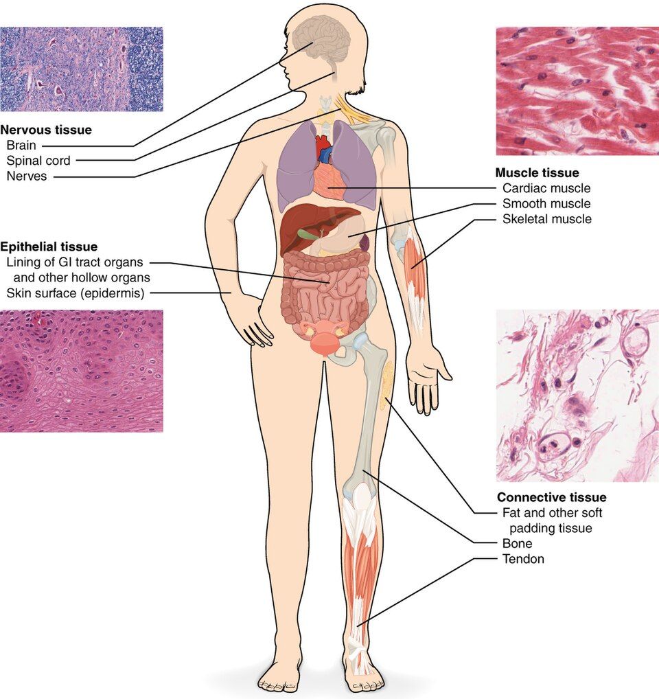
A tissue is a group of similar cells that work together to do a shared job. That is the whole definition. But the word "together" matters a lot. The cells in a tissue are not just neighbors that look alike. They work as a team. They are held in place by links between them, or by material they make around themselves. And each tissue is built for one kind of job.
The study of tissues under the microscope is called histology. It is really anatomy at very high zoom. Instead of asking what an organ looks like to the eye, we ask what it is made of.
Four families do all the work
Here is what makes this subject easy to learn. No matter how complex an organ looks, it is built from only four primary tissue types. Learn these four, and you have the words to describe any part of the body. Each family meets a different basic need. A body must cover and line its surfaces. It must bind and support its parts. It must move. And it must communicate.
| Tissue | What it does | Where you find it |
|---|---|---|
| Epithelial | Covers surfaces, lines spaces, and forms glands. It protects, absorbs, and releases substances. | Skin surface, gut lining, kidney tubes, gland cells |
| Connective | Binds, supports, protects, stores, and carries things. It is the body's packing material. | Bone, fat, tendon, cartilage, blood |
| Muscle | Squeezes (contracts) to make movement and force. It also makes heat. | Skeletal muscles, heart wall, walls of hollow organs |
| Nervous | Senses, sorts out, and quickly sends electrical signals | Brain, spinal cord, nerves in the rest of the body |
Notice how different the four are in how much room the cells give each other. Epithelial cells are packed so tightly that there is almost no space between them. That is what makes them a good barrier. Connective tissue is the opposite. Its cells are few and far apart. They float in a sea of non-living material that they make. This one contrast is one of the fastest ways to tell the first two tissues apart on a slide. Look for crowded cells or cells spread out in a matrix.
From tissues to organs and membranes
An organ is two or more tissue types layered together. The whole organ can do something none of its tissues could do alone. The stomach is the clearest example. It has an epithelial lining that makes acid and mucus. It has a connective tissue layer that carries its blood vessels and nerves. It has smooth muscle that churns food. And it has nervous tissue that times the churning. That is four tissues in one organ.
The simplest of these teams is a body membrane. This is a thin sheet that covers or lines a surface. There are only four kinds.
The cutaneous membrane is the skin itself. It is stratified squamous epithelium on top of dense irregular connective tissue. It is the only membrane that is dry. A mucous membrane lines every space that opens to the outside. This includes the airway, the gut, and the urinary and reproductive tracts. It pairs an epithelium with a loose connective layer under it, called the lamina propria.
A serous membrane lines the closed body spaces in the front of the trunk and wraps the organs inside them. Examples are the pleura, pericardium, and peritoneum. It joins a simple squamous epithelium called mesothelium to a thin areolar layer. It makes a watery fluid so organs can slide past each other. The synovial membrane is the odd one out. It lines the spaces inside joints. It is made of connective tissue only, with no epithelium at all.
Meet all four tissues side by side in the interactive below.
- A tissue is a group of similar cells doing a shared job. The study of tissues is histology.
- The body has four primary tissues: epithelial (cover and line), connective (bind and support), muscle (move), and nervous (communicate).
- Epithelial cells are tightly packed. Connective cells are spread out in a matrix. This is a quick way to tell them apart.
- Tissues layer together to build organs and the four body membranes. These are cutaneous (skin, dry), mucous (lines spaces open to the outside), serous (lines closed spaces in the front of the trunk), and synovial (joint spaces, connective tissue only).
Section 2Naming an epithelium
What every epithelium has in common
Epithelial tissue covers every open surface of the body. It lines every space, tube, and duct. Think of your outer skin, the inside of your gut, your airways, and your blood vessels. Because it sits at every border, it has a set of features built for that role.
Its cells are tightly packed, with very little material between them. So almost nothing gets across without going through a cell. Every epithelium has polarity, which means it has a top and a bottom. The apical surface is the top. It faces the open space and often has tiny fingers (microvilli) or hairs (cilia). The basal surface is the bottom. It is anchored to a thin basement membrane that glues it to the connective tissue below.
Epithelium is avascular. This means it has no blood vessels of its own. It gets its food by diffusion from the tissue underneath. Yet it has plenty of nerves. It also regenerates, or regrows, quickly. It has to, because being a surface means cells are always wearing away.
The two-word naming rule
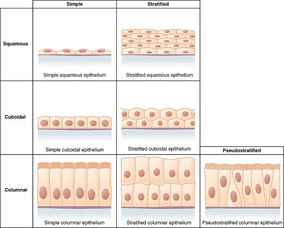
Here is the big reward of this section. Every epithelium is named with the same simple formula. Once you learn the rule, you can name a tissue you have never seen. The name has two parts. First comes the number of layers. Then comes the shape of the cells at the surface.
- Layers. One layer of cells is simple. Two or more stacked layers is stratified.
- Shape. Flat cells, like fish scales, are squamous. Cells shaped like cubes are cuboidal. Tall cells shaped like columns are columnar.
Snap the two halves together and you have the name. One layer of flat cells is simple squamous. Many stacked layers of flat cells make stratified squamous. One row of tall cells is simple columnar.
The build even tells you the job. Thin simple epithelia are best where things must cross quickly. Simple squamous lines the air sacs of the lungs and the walls of capillaries. Oxygen and nutrients slip across it in an instant. Thick stratified epithelia are built for protection. Stratified squamous forms the surface of your skin and lines your mouth and esophagus. You can afford to lose and rebuild these layers.
| Epithelium | Build | Best-known job and place |
|---|---|---|
| Simple squamous | One layer of flat cells | Fast diffusion: lung air sacs, lining of capillaries and vessels (endothelium) |
| Simple cuboidal | One layer of cube cells | Releasing and absorbing: kidney tubes, gland ducts |
| Simple columnar | One layer of tall cells | Absorbing and releasing: lining of the stomach and intestines |
| Stratified squamous | Many stacked flat layers | Protection from rubbing and scraping: skin surface, mouth, esophagus, vagina |
| Pseudostratified columnar | One layer that looks layered | Lining of the airways, with cilia that sweep mucus upward |
| Transitional | Stretchy layers that change shape | Bladder and ureters: stretches as they fill |
Two rows in that table are the famous exceptions. Pseudostratified columnar epithelium is really just one layer. Every cell touches the basement membrane. But the nuclei sit at different heights, so it looks stacked. That is why it is called "pseudo" (false) stratified. Transitional epithelium is the shape-shifter of the urinary tract. Its cells slide over each other and flatten as the bladder fills. Then they bunch back up as it empties.
Build an epithelium's name box by box and watch it appear in the interactive.
- Epithelia are tightly packed and polar (apical top, basal bottom). They sit on a basement membrane, are avascular, and regenerate fast.
- Name any epithelium by layers (simple = one, stratified = many) + cell shape (squamous, cuboidal, columnar).
- Simple epithelia are for absorbing, releasing, and diffusion. Stratified epithelia are for protection. Pseudostratified and transitional are the special cases.
Section 3Connective tissue
One recipe, many results
Connective tissue is the most common tissue in the body. It is also the most varied. At first it can seem like a random mix. Bone, fat, tendon, cartilage, and even blood all belong to it. What links them is one shared plan.
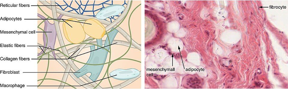
In epithelium, the cells are everything. Connective tissue is different. It is mostly extracellular matrix. This is non-living material that the cells make and then sit inside. The cells are few and spread far apart. The feel of the tissue comes almost entirely from the matrix around them.
This is the key idea of the whole section: change the matrix and you change the tissue. Make the matrix a firm, rubbery gel, and you get cartilage. Harden it with calcium salts, and you get bone. Make it liquid, and you get blood. Adipose tissue, or fat, is the useful exception. Its cells store fat inside themselves. Each cell swells up with one big droplet. So the matrix gets squeezed down to almost nothing. Fill the cells instead of the matrix, and you get fat.
The matrix: fibers and ground substance
The extracellular matrix always has two parts. The first part is protein fibers. There are three kinds. Collagen fibers are strong, like ropes, and resist pulling. Elastic fibers stretch and snap back into shape, like rubber bands. Reticular fibers are thin and form a fine mesh that holds things up.
The second part is the ground substance. It fills the space between the fibers and cells. Depending on the tissue, it can be watery, like jelly, or rock hard.
The cells often come in two kinds, named by a simple rule. A name that ends in -blast is a young, busy builder cell that makes matrix. Examples are the fibroblast, osteoblast, and chondroblast. A name that ends in -cyte is a grown-up cell that keeps the matrix in good shape. Examples are the fibrocyte, osteocyte, and chondrocyte.
| Connective tissue | Matrix | Job and place |
|---|---|---|
| Loose (areolar) | Soft and gel-like, with loosely woven fibers | All-purpose packing and cushion; found under epithelia everywhere |
| Adipose (fat) | Cells so full of fat that there is little matrix | Stores energy, holds in heat, pads organs |
| Dense regular | Tightly packed collagen, all lined up in the same direction | Strong pull in one direction: tendons and ligaments |
| Cartilage | Firm, rubbery gel | Bendable support: nose, ears, joint surfaces, discs in the spine |
| Bone (osseous) | Collagen hardened with calcium salts | Stiff support and protection; stores minerals |
| Blood | Liquid matrix (plasma) | Carries cells, gases, nutrients, and wastes |
Read that table from top to bottom. You can watch one idea stretch across every level of stiffness. It goes from a soft cushion, to a fat pad, to a tough cable, to a rubbery shock absorber, to solid mineral, and all the way to a flowing liquid. It is the same recipe with very different results. What changes is what the cells put into the matrix.
Rebuild the matrix in the interactive and watch one tissue turn into the next.
- All connective tissue follows one recipe: spread-out cells in an extracellular matrix made of fibers + ground substance.
- There are three fiber types: collagen (strength), elastic (stretch), and reticular (mesh support). -blast cells build the matrix. -cyte cells keep it up.
- Changing the matrix gives the whole family: loose, adipose, dense, cartilage, bone, and blood.
Section 4Muscle tissue
Cells built to shorten
Muscle tissue has one skill that no other tissue has. It can contract. That means it can actively shorten to pull on something. Its cells are packed with two protein threads, actin and myosin. These threads slide past each other to shorten the cell. Everything about a muscle cell's shape helps with that pull.
Sometimes these threads line up in neat, repeating rows. Then they catch the light in bands, and the tissue looks striated, or striped. When the threads are less orderly, the tissue looks smooth. Contraction does more than move the body. It also makes much of your body heat. That is why shivering warms you up.
The three muscle tissues
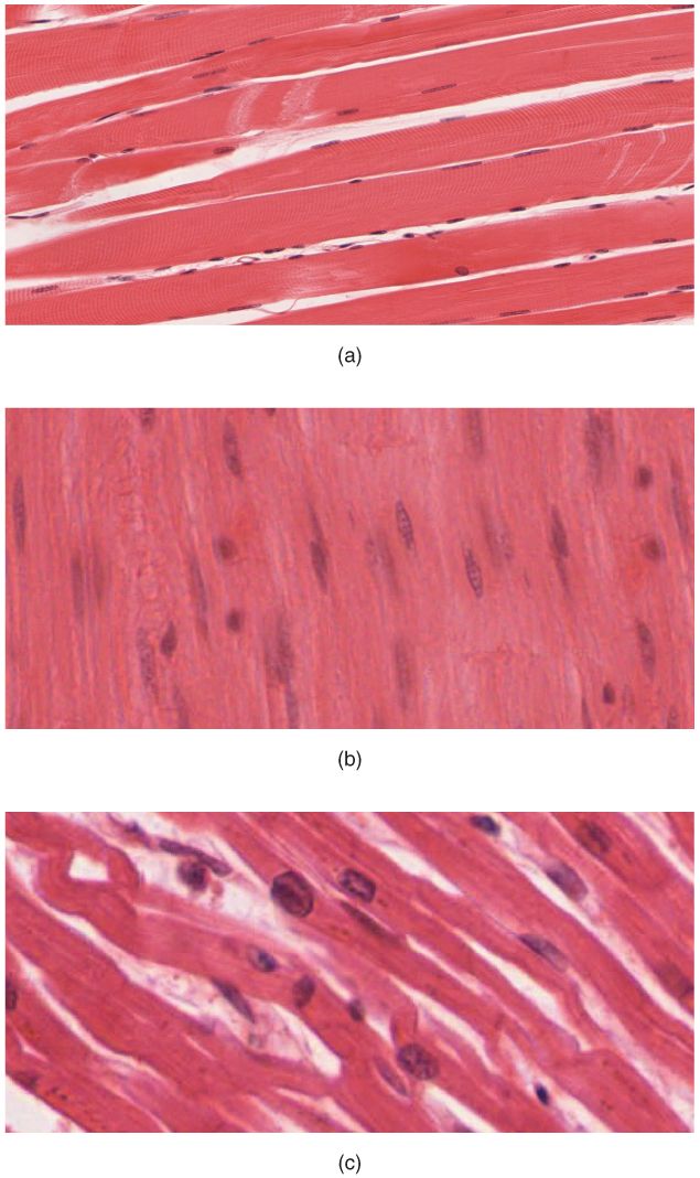
There are exactly three muscle tissues. You can tell them apart with three questions. Is it striped? Can you control it on purpose? What shape are its cells?
Skeletal muscle is the meat that moves your bones. It is striated and voluntary, which means you control it on purpose. It is made of long, tube-shaped fibers, each with many nuclei.
Cardiac muscle is found only in the wall of the heart. It is striated but involuntary, so it works without you thinking about it. Its short, branching cells are joined end to end by intercalated discs. These let the whole heart squeeze as one team.
Smooth muscle lines the walls of hollow organs like the gut, blood vessels, and bladder. It is non-striated and involuntary. It is made of small, tapered cells shaped like spindles, thick in the middle and pointed at the ends. They squeeze those organs slowly and without tiring.
| Feature | Skeletal | Cardiac | Smooth |
|---|---|---|---|
| Striations (stripes) | Striated | Striated | Non-striated |
| Control | Voluntary | Involuntary | Involuntary |
| Cell shape | Long tubes | Short, branched | Tapered spindles |
| Nuclei per cell | Many | One (or two) | One |
| Where | Attached to bones | Heart wall only | Walls of hollow organs |
Here is a handy shortcut. The two striated muscles, skeletal and cardiac, are the fast, strong ones. The one smooth muscle is the slow, steady one. And only one of the three, skeletal, does what you tell it. The heart and the gut run on their own.
Watch each of the three muscles contract in the interactive below.
- Muscle tissue contracts as actin and myosin threads slide. Neat rows of threads make it look striated.
- Skeletal is striated and voluntary, with long fibers that have many nuclei. Cardiac is striated and involuntary, with branched cells joined by intercalated discs. Smooth is non-striated and involuntary, with spindle cells.
- Tell them apart with three questions: striped? voluntary? cell shape?
Section 5Nervous tissue
The neuron: a cell shaped like a message
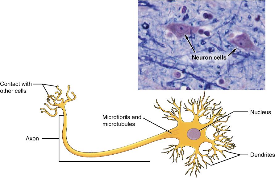
Nervous tissue senses changes, makes decisions, and sends electrical signals. It does this faster than any other tissue in the body. Its star cell is the neuron, or nerve cell. The neuron's odd shape is really a map of how a signal flows through it. The signal goes in one end and out the other.
Signals arrive at the bushy dendrites. These are short, branching arms that pick up messages from other cells. The signals flow into the cell body (or soma). The cell body holds the nucleus and keeps the cell running. If the incoming signal is strong enough, the neuron fires an electrical pulse. The pulse races away down the axon, one long fiber that carries the message to the next cell.
Many axons are wrapped in a fatty myelin sheath. It works like the plastic coating on a wire. It lets the signal jump along much faster. The traffic only goes one way: dendrite → cell body → axon, every time.
| Part | What it is | Its job in the signal |
|---|---|---|
| Dendrites | Short, branching arms | Receive incoming signals from other cells |
| Cell body (soma) | Holds the nucleus and organelles | Integrates, or adds up, the input; keeps the cell alive |
| Axon | One long fiber | Sends the electrical pulse toward the next cell |
| Myelin sheath | Fatty wrap on the axon, like the coating on a wire | Speeds the signal along the axon |
| Axon terminals | Branched tips of the axon | Pass the signal to the next cell at a synapse, the meeting point |
Neuroglia: the support crew
Neurons get the fame. But there are at least as many neuroglia, also called glial cells. These are the support cells of nervous tissue. Careful counts show about one glial cell for each neuron across the whole brain. Glia outnumber neurons in the cerebral cortex, the brain's wrinkled outer layer. Neurons far outnumber glia in the cerebellum, at the back of the brain.
Glia do not carry the signal themselves. Instead, they feed neurons and wrap their axons in myelin. They hold the tissue together and defend it from germs. Two features define what nervous tissue can do. Excitability is the ability to respond to a trigger by making an electrical signal. Conductivity is the ability to carry that signal from one place to another. Together, they make this tissue the body's high-speed communication network.
Tap a part of the neuron and watch a signal fire in the interactive.
- The neuron carries a one-way signal. Dendrites receive → the cell body adds it up → the axon sends it on.
- The fatty myelin sheath wraps the axon and speeds the signal. Neuroglia support and protect neurons.
- Nervous tissue is defined by excitability and conductivity. Grown-up neurons rarely divide, so damage to the brain and spinal cord (the CNS) often heals poorly.
Section 6Tissue repair
Two ways to fix a wound
When a tissue is hurt, the body can fix it in one of two ways. The better result is regeneration. The damaged cells are replaced with new cells of the very same type. The tissue goes back to normal, as if the injury never happened.
The backup plan is fibrosis. Here the gap is filled with thick, tough scar tissue instead of the original cells. Scar tissue plugs the hole and makes the area strong again. But it cannot do the original job. Scar in the heart cannot beat. Scar in the skin has no hair or sweat glands.
Which path a tissue takes depends mostly on how well its cells can divide. Tissues whose cells divide fast, like skin and gut, regenerate very well. Tissues whose cells barely divide, like heart muscle and neurons, tend to scar.
The healing playbook
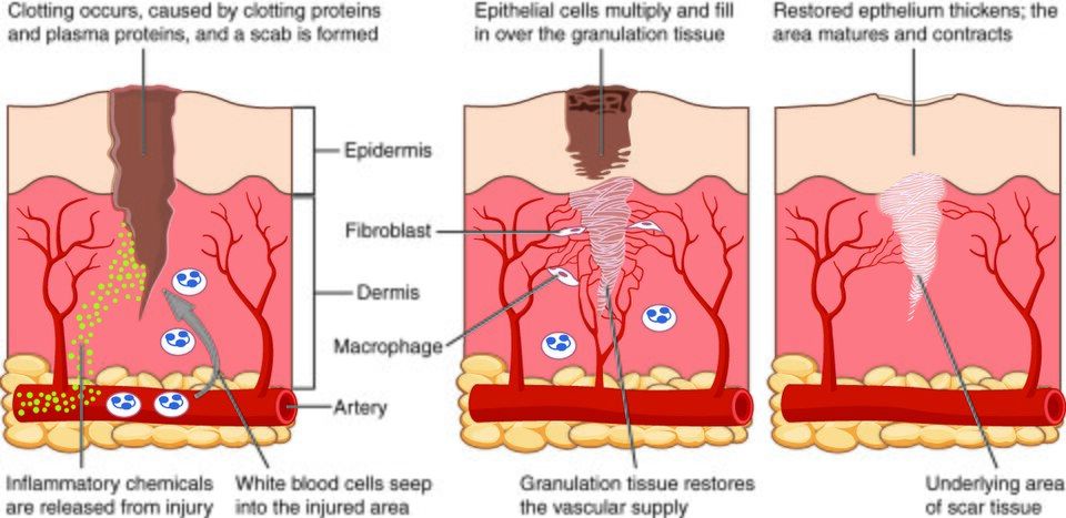
For any tissue, the body runs the same four-step repair plan, in order. Think of it as a building project on a rush schedule. First stop the flooding. Then clear the site. Then rebuild the frame. Then finish the surfaces.
| Stage | What happens | Purpose |
|---|---|---|
| 1. Hemostasis | Blood clots seal the broken vessels, and a scab forms | Stop the bleeding |
| 2. Inflammation | Vessels widen, and immune cells arrive to clear away junk and germs | Clean up the wound |
| 3. Proliferation | New tiny blood vessels and collagen build pink granulation tissue. Epithelium spreads across the top. | Rebuild the tissue |
| 4. Remodeling | Collagen is rearranged and made stronger. The scar matures and shrinks. | Make the area strong again over weeks to months |
The signs you can see in stage two are redness, heat, swelling, and pain. These are the classic marks of inflammation. They feel like the problem, but they are really the body doing its job. The vessels widen and leak. That causes the redness and swelling. It also brings in the clotting factors, germ fighters, and cleanup crews the wound needs. Inflammation is not the injury. It is the repair getting started.
Step through a healing wound from clot to scar in the interactive below.
- Healing happens by regeneration (the same cells come back) or fibrosis (scar tissue fills the gap). Which one depends on how well the cells divide.
- The repair steps always go hemostasis → inflammation → proliferation → remodeling.
- Inflammation (redness, heat, swelling, pain) is the repair at work, not the injury itself.
The chapter in one breath
- A tissue is a group of similar cells that share a job. The whole body is built from just four of them.
- Epithelial tissue covers and lines. It is tightly packed, polar, and avascular. It is named by layers (simple or stratified) + cell shape (squamous, cuboidal, or columnar).
- Connective tissue binds and supports. Its cells are spread out in an extracellular matrix of fibers and ground substance. Change the matrix and you get cartilage, bone, or blood. Adipose is the exception. It fills its cells with fat instead.
- Muscle tissue contracts. Skeletal is striated and voluntary. Cardiac is striated and involuntary, with intercalated discs. Smooth is non-striated and involuntary.
- Nervous tissue communicates. The neuron carries a one-way signal from dendrites → cell body → axon. Neuroglia support it.
- Hurt tissue heals by regeneration or fibrosis. It always goes through hemostasis → inflammation → proliferation → remodeling.
- How well a tissue heals comes down mostly to its blood supply and how easily its cells divide.
ReferenceWords worth knowing
Here are the key words from this chapter. Skim them now, or come back any time a word trips you up.
- Apical surface
- The free top surface of an epithelial cell. It faces the open space, cavity, or lumen, and often has microvilli or cilia.
- Areolar tissue
- The most common loose connective tissue. It is soft, all-purpose packing that lies under epithelia and cushions organs.
- Axon
- The one long fiber of a neuron. It carries the electrical signal away from the cell body toward the next cell.
- Basement membrane
- A thin layer that anchors the bottom (basal) surface of an epithelium to the connective tissue below it.
- Cardiac muscle
- Striated, involuntary muscle found only in the heart wall. Its branched cells are joined by intercalated discs, so the heart squeezes as one.
- Collagen fiber
- The strong, rope-like protein fiber of connective tissue. It resists pulling.
- Connective tissue
- The varied tissue that binds, supports, protects, and carries things. It is made of spread-out cells in an extracellular matrix.
- Dendrite
- A short, branching arm of a neuron. It picks up signals from other cells.
- Epithelial tissue
- Tightly packed tissue that covers body surfaces, lines spaces and tubes, and forms glands.
- Extracellular matrix
- The non-living material, fibers plus ground substance, that connective tissue cells make and sit inside.
- Fibrosis
- Repair by laying down tough scar tissue in place of the original cells. It brings back strength but not full function.
- Ground substance
- The part of the extracellular matrix that fills the space between cells and fibers. It can be watery, like jelly, or solid.
- Histology
- The study of tissues under the microscope. It is anatomy at high zoom.
- Muscle tissue
- Tissue built to contract using actin and myosin threads. It makes movement, force, and heat.
- Nervous tissue
- Tissue built to sense, sort out, and quickly send electrical signals. It is made of neurons and neuroglia.
- Neuroglia
- The support cells of nervous tissue. They feed, wrap, hold together, and protect neurons.
- Neuron
- The signal-carrying cell of nervous tissue. It has dendrites, a cell body, and an axon.
- Regeneration
- Repair in which damaged cells are replaced by new cells of the same type. Normal structure and function come back.
- Simple epithelium
- An epithelium one cell layer thick. It is suited to diffusion, absorbing, and releasing substances.
- Squamous
- Describes flat, scale-like epithelial cells. They are ideal where substances must cross quickly.
- Stratified epithelium
- An epithelium with two or more stacked layers of cells. It is built to protect against rubbing and scraping.
- Tissue
- A group of similar cells that work together to do a shared job.