Vital Signs
Vital signs are the only part of the assessment that produces a number every patient, every shift, in every setting — and because they are numbers, they are the first thing anyone deteriorating shows you. Hours before a patient looks septic, the respiratory rate has already climbed. Hours before a patient bleeds out visibly, the pulse pressure has already narrowed and the pulse has quickened. This chapter teaches you to produce numbers that can be trusted: the correct route, the correct cuff, the correct count, the correct position, the correct amount of time. It then teaches you to read them — against the exact adult ranges, against the lifespan variations that make a heart rate of 140 normal in an infant and an emergency in an adult, and against the patient’s own baseline, which almost always matters more than the textbook. It ends with the values that mean stop, call now, and the words you will use to call.
By the end of this chapter you can
- State adult ranges for temperature, pulse, respiration, blood pressure and SpO2 to the exact number, and adjust each for infants, children, pregnancy and older adults.
- Sequence the four assessment techniques — inspection, palpation, percussion, auscultation — and state the abdominal exception and why it exists.
- Select and perform the correct temperature route, and name the contraindications to oral and rectal measurement.
- Assess a pulse for rate, rhythm and amplitude, grade it on the 0 to 4+ scale, and measure an apical–radial pulse deficit with a second nurse.
- Recognize Kussmaul, Cheyne-Stokes and Biot respiratory patterns and state what each suggests.
- List the limitations of pulse oximetry — poor perfusion, nail polish, carbon monoxide, motion, anemia and skin pigmentation — and explain occult hypoxemia.
- Size a blood pressure cuff (bladder width about 40 percent of arm circumference) and explain why a cuff that is too small reads falsely high.
- Perform orthostatic vital signs correctly and state the numeric definition of a positive result.
- Rewrite a vague vital-sign entry as a precise, defensible one, and escalate abnormal values using SBAR.
Section 1Vital Signs as a System
What the numbers are actually telling you
The five traditional vital signs — temperature, pulse, respiration, blood pressure and oxygen saturation — are not five unrelated chores. They are five windows onto two questions: is this patient perfusing tissue with oxygenated blood, and is the body’s regulatory machinery still holding the line? Temperature reports the hypothalamic set point and the presence of inflammation. Pulse reports cardiac rate and, in its quality, stroke volume. Respiration reports both gas exchange and, quietly, metabolic acidosis. Blood pressure reports the driving force behind perfusion. Oxygen saturation reports how loaded the hemoglobin is. When one moves, ask what the others are doing, because compensation is a pattern, not a single value.
That pattern is why trend beats snapshot. A blood pressure of 104/62 is unremarkable in a small young adult and alarming in a patient whose baseline is 160/90, because the second patient has lost more than fifty points of driving pressure and may be perfusing a kidney at the edge of failure. Before you decide a number is normal, find the last three sets on the flowsheet. Deterioration almost never announces itself with one wildly abnormal value; it announces itself with three sets that each looked acceptable and that, lined up, point one direction.
Of the five, respiratory rate is the one students underweight and clinicians most often falsify. It is the earliest and most sensitive predictor of deterioration in adults, it rises before blood pressure falls, and it is the value most likely to be charted as 18 because 18 looks reasonable. Counting a real respiratory rate takes thirty to sixty seconds and can be the difference between recognizing early sepsis and finding it in shock four hours later.
The four techniques, and the one exception you must never forget
Every physical assessment in this book is built from four techniques performed in a fixed order: inspection, then palpation, then percussion, then auscultation. Inspection is deliberate looking — adequate light, exposed skin, comparison side to side. Palpation uses the fingerpads for texture, pulsation and tenderness, the ulnar surface of the hand for vibration, and the dorsum of the hand for temperature, because the back of the hand is the most temperature-sensitive surface you have. Percussion strikes the distal interphalangeal joint of the pleximeter finger with the tip of the plexor finger, using wrist motion only, to judge whether the tissue beneath is air-filled, fluid-filled or solid. Auscultation uses the diaphragm for high-pitched sounds (breath sounds, bowel sounds, S1 and S2) and the bell, held lightly, for low-pitched sounds (S3, S4, bruits, murmurs of mitral stenosis).
There is one exception, and it is asked on every examination and matters at every bedside. For the abdomen, the order is inspect, auscultate, percuss, palpate. Auscultation moves ahead of the hands because pressing on the abdomen mechanically stimulates or suppresses peristalsis and changes the bowel sounds you are about to listen for. Palpate first and you have contaminated your own data. Everywhere else on the body, palpation precedes auscultation; on the abdomen it follows it.
Vital signs sit inside these techniques rather than beside them. You inspect while you count respirations, you palpate to find a radial pulse and to estimate a systolic pressure before you inflate a cuff, and you auscultate for Korotkoff sounds and for an apical rate. Treating vital signs as data entry rather than as physical assessment is exactly how bad numbers get charted.
How often, and who may take them
Frequency is set by provider order, facility policy and patient condition, and the last of those overrides the first two upward but never downward. Common patterns: a full set on admission and at the start of every shift; every four hours on a general medical-surgical unit; postoperatively every fifteen minutes for four sets, every thirty minutes for two, every hour for four, then every four hours; every fifteen minutes during the first hour after a blood transfusion begins, with the nurse remaining with the patient for the first fifteen minutes; before and after any procedure, any transfer, any fall, any change in condition, and any patient complaint that could be cardiopulmonary. Certain medications carry their own vital-sign gate: an apical rate before digoxin (hold and notify if under 60 in an adult), a blood pressure before an antihypertensive, a respiratory rate and sedation level before an opioid dose.
Measuring routine vital signs on a stable, predictable patient may be delegated to unlicensed assistive personnel. Interpreting them may not. When you delegate, you must give the assistant the exact numbers to report back to you — not “let me know if anything looks off,” but “tell me immediately if the heart rate is above 110, the systolic is below 100, the respiratory rate is above 24, the temperature is above 38 °C, or the SpO2 is below 92 percent.” The registered nurse retains the baseline admission set, any set on an unstable patient, any set taken because the patient changed, and every act of judgment about what the numbers mean.
| Task | May go to UAP | Stays with the RN |
|---|---|---|
| Routine four-hourly vital signs, stable patient | Yes, with reporting parameters given | Interpretation and response |
| Admission baseline set | No | Yes — it anchors every later comparison |
| Vital signs on a patient who just became short of breath | No | Yes — patient is unstable |
| Vital signs before and during a blood transfusion | No | Yes |
| Apical pulse before digoxin | No — requires assessment and a hold decision | Yes |
| Orthostatic vital signs | No — interpretation and fall risk are nursing judgment | Yes |
| Teaching a patient to take a home blood pressure | No — teaching is never delegated | Yes |
The adult numbers, and how they move across the lifespan
Memorize the adult set as exact numbers, not as impressions. Then memorize the direction pediatric values move: as a child grows, heart rate and respiratory rate fall and blood pressure rises. A heart rate of 140 is expected in a two-month-old and is a rapid-response call in a forty-year-old. Pediatric numbers are given here in the ranges used by Pediatric Advanced Life Support; individual textbooks vary by a few beats, so follow your facility’s reference and, above all, the child’s own baseline.
| Adult vital sign | Expected value | Terms for abnormal |
|---|---|---|
| Temperature (oral) | 36.5–37.5 °C (97.7–99.5 °F); average 37.0 °C (98.6 °F) | Fever/pyrexia 38.0 °C (100.4 °F) or higher; hypothermia below 35.0 °C (95.0 °F) |
| Pulse | 60–100 beats/min, regular | Bradycardia under 60; tachycardia over 100 |
| Respiration | 12–20 breaths/min, quiet and unlabored | Bradypnea under 12; tachypnea over 20; apnea |
| Blood pressure | Under 120/80 mm Hg | Hypotension generally under 90/60; hypertension 130/80 or higher |
| SpO2 | 95–100 percent on room air | Hypoxemia under 90 percent (target 88–92 percent in chronic CO2 retention) |
| Pulse pressure | 30–40 mm Hg (systolic minus diastolic) | Narrowed under 25; widened over 60 |
| Mean arterial pressure | 70–100 mm Hg | Organ perfusion threatened below 65 |
| Age | Heart rate (beats/min) | Respirations (breaths/min) | Systolic BP (mm Hg) |
|---|---|---|---|
| Neonate, under 1 month | 100–205 awake, 90–160 asleep | 30–60 | 60–90 (hypotension below 60) |
| Infant, 1–12 months | 100–180 awake, 90–160 asleep | 30–53 | 72–104 (hypotension below 70) |
| Toddler, 1–2 years | 98–140 | 22–37 | 86–106 |
| Preschool, 3–5 years | 80–120 | 20–28 | 89–112 |
| School age, 6–11 years | 75–118 | 18–25 | 97–120 |
| Adolescent, 12–15 years | 60–100 | 12–20 | 110–131 |
| Adult | 60–100 | 12–20 | Under 120 |
For children over one year, the fifth-percentile (lowest acceptable) systolic pressure is 70 plus twice the age in years; below that value a child aged one to ten is hypotensive, and hypotension in a child is a late, ominous sign because children compensate with tachycardia until they cannot. In pregnancy, heart rate rises 10 to 20 beats per minute by the third trimester, blood pressure falls during the second trimester and returns to baseline near term, and tidal volume rises about 30 to 40 percent while the respiratory rate stays essentially unchanged. In older adults, arterial stiffening produces isolated systolic hypertension and a widened pulse pressure, the febrile response is blunted so that infection may present with confusion or a fall rather than a temperature, and baseline temperature runs lower — a rise of 1.1 °C (2 °F) above that patient’s own baseline is clinically significant even if the absolute number never reaches 38 °C.
Pain is measured with every vital-sign set and is often called the fifth vital sign, though strictly it is a self-reported symptom rather than a measured physiologic value. Use a 0-to-10 numeric rating scale for adults who can self-report, Wong-Baker FACES from about age three, FLACC for children two months to seven years, CRIES for neonates, PAINAD for advanced dementia and CPOT for intubated adults. Self-report is the gold standard and outranks your impression of how comfortable the patient looks. Pain assessment is developed fully in Chapter 7.
Vital signs are physical assessment expressed as numbers, and the trend across sets — not any single value — is what tells you a patient is deteriorating.
Section 2Temperature
Thermoregulation and what counts as a fever
The hypothalamus holds core temperature near 37.0 °C (98.6 °F) by balancing heat production — basal metabolism, muscle activity, shivering, thyroid hormone and catecholamines — against heat loss through radiation, conduction, convection and evaporation. Fever is not a failure of that system. In fever, pyrogens raise the hypothalamic set point, and the body then works to reach the new, higher target: the patient vasoconstricts, feels cold, and shivers during the chill phase while the temperature climbs. At the plateau the patient is hot and vasodilated — on lighter skin this looks flushed, while on brown or black skin the change is easier to feel as warmth and to see in the conjunctivae and oral mucosa than to read as redness. When the set point resets downward, the patient diaphoreses and vasodilates in the defervescence phase. Recognizing these phases keeps you from piling blankets on a shivering patient whose temperature is already 39 °C.
Hyperthermia is different in mechanism and in danger. There the set point is normal but heat gain overwhelms heat loss — heat stroke, malignant hyperthermia, neuroleptic malignant syndrome, anticholinergic toxicity. Hyperthermia does not respond to antipyretics, because there is no raised set point to lower; it requires active cooling and removal of the cause. A temperature above 40 °C (104 °F) with altered mental status and hot dry skin is heat stroke until proven otherwise, and it is an emergency.
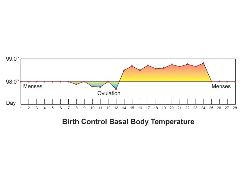
Normal temperature also varies predictably. It is lowest between 0400 and 0600 and highest between 1600 and 1800, swinging 0.5 to 1.0 °C across the day; it rises 0.3 to 0.6 °C after ovulation; it rises with exercise, heavy clothing and a hot environment. If you are comparing a morning temperature to an evening one, part of the difference is the clock. To convert, multiply degrees Celsius by 1.8 and add 32; to reverse, subtract 32 and divide by 1.8.
Choosing a route and performing it correctly
Routes are not interchangeable, and the conversions between them are approximations. Chart the route with the number, every time, and compare like with like across the shift.
| Route | Expected adult range | Technique | Avoid when |
|---|---|---|---|
| Oral | 36.5–37.5 °C (97.7–99.5 °F) | Probe in the posterior sublingual pocket, lips closed, mouth breathing avoided; wait 15–30 minutes after hot or cold intake, smoking or chewing gum | Confusion, seizure risk, inability to follow directions, oral surgery or trauma, children under about 4 years |
| Rectal | 0.5 °C (0.9 °F) higher than oral: 37.0–38.0 °C (98.6–100.4 °F) | Lubricated probe, side-lying, insert 3.5 cm (1.5 in) adult or 1.5–2.5 cm (0.5–1 in) infant, hold in place the entire time | Neutropenia, thrombocytopenia or anticoagulation, rectal or perineal surgery, diarrhea, imperforate anus, spinal cord injury with autonomic dysreflexia risk, cardiac patients (vagal stimulation) |
| Tympanic | Close to rectal; about 0.3–0.6 °C above oral | Pull pinna up and back in adults and children over 3, down and back under 3; aim at the tympanic membrane; use a clean cover | Otitis, ear drainage, impacted cerumen, ear surgery, hearing aid in place |
| Temporal artery | About 0.4 °C above oral | Slide across the forehead to the hairline, then behind the ear lobe if the forehead is diaphoretic | Diaphoresis, forehead scar or dressing, recent cold pack |
| Axillary | 0.5 °C (0.9 °F) lower than oral: 36.0–37.0 °C (96.8–98.6 °F) | Probe deep in a dry axilla, arm held firmly against the chest for the full dwell time | Any time accuracy matters — it is the least reliable route; use only when others are contraindicated |
For infants under three months, rectal temperature is the reference standard and the number that decision rules are built on. For most other patients an oral or temporal reading is enough, and the accuracy you gain from a rectal temperature rarely justifies the invasion. Core temperature from an esophageal, bladder or pulmonary artery probe is used in critical care and during therapeutic hypothermia; if such a probe is in place, it is the number the team will act on.
Fever across the lifespan, and what abnormal temperature means
| Finding | Suggests | Action |
|---|---|---|
| Rectal 38.0 °C (100.4 °F) or higher in an infant under 3 months | Possible serious bacterial infection; the infant cannot localize it | Emergency evaluation — notify the provider immediately, expect a full sepsis workup |
| Single oral temperature 38.3 °C (101 °F), or 38.0 °C sustained one hour, in a neutropenic patient | Neutropenic fever — may be the only sign of overwhelming infection | Medical emergency; cultures and antibiotics within one hour |
| New confusion, falls or hypothermia in an older adult | Infection with a blunted febrile response — sepsis may present without fever | Full vital-sign set, look for other sepsis criteria, notify provider |
| Temperature below 36.0 °C (96.8 °F) with tachycardia and tachypnea | Sepsis, environmental exposure, hypothyroidism, burns | Escalate — hypothermia counts toward sepsis criteria exactly as fever does |
| Above 41.0 °C (105.8 °F), hyperpyrexia | Heat stroke, malignant hyperthermia, CNS lesion | Active cooling now; antipyretics will not fix hyperthermia |
| Temperature spike within 30 minutes of starting a transfusion | Febrile or hemolytic transfusion reaction | Stop the transfusion, keep the line open with normal saline, notify provider and blood bank |
Two lifespan cautions deserve emphasis. Infants have a large surface-area-to-mass ratio, little insulating fat and no shivering thermogenesis at birth; they lose heat fast and become hypothermic rather than febrile when septic. Older adults may run a baseline temperature of 36.2 °C, so a “normal” 37.8 °C may already represent a 1.6-degree rise and a real infection. In both groups the absolute number lies more often than the change from baseline does.
Chart the route with the temperature, treat 38.0 °C (100.4 °F) as fever in adults, and remember that infants and older adults may be seriously infected with a normal or low temperature.
Section 3The Pulse
What you are feeling, and how to feel it
Each ventricular contraction ejects a stroke volume into an already-full aorta, and the pressure wave that travels down the arterial tree is what your fingers detect. That means the pulse carries information about rate, about the regularity of the electrical rhythm, and about the force of the contraction reaching the periphery. Palpate with the pads of your index and middle fingers, never the thumb, which has a pulse of its own that you will happily count as the patient’s.
Press gently. Heavy pressure occludes the vessel and erases the very finding you are grading. Count for 30 seconds and multiply by two if the rhythm is regular; count a full 60 seconds if the rhythm is irregular, if the rate is very fast or very slow, if the patient is an infant, or if you are taking an apical pulse. Doubling an irregular 30-second count is how a real rate of 96 becomes a charted 120.
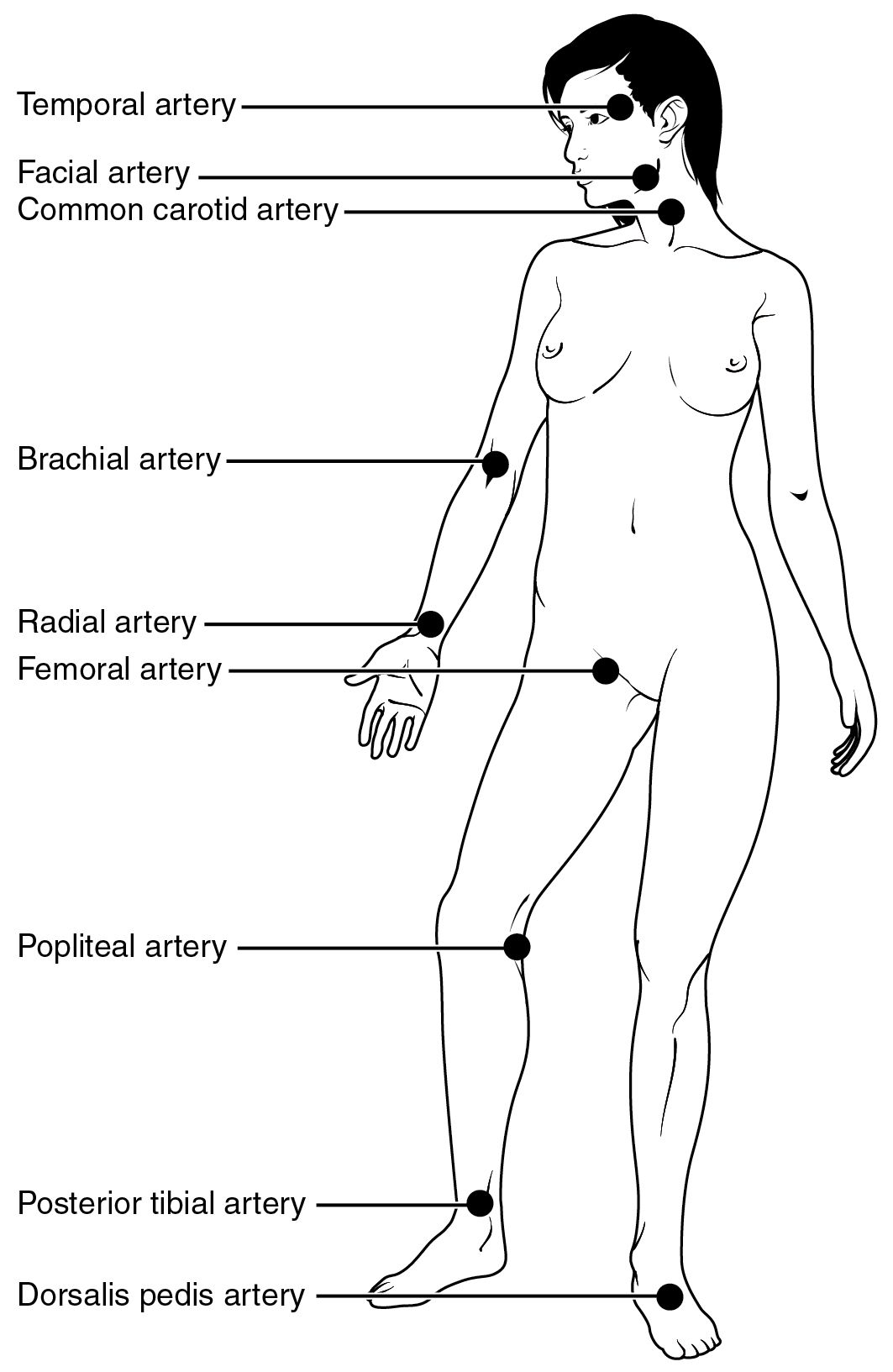
| Site | Where | Used for |
|---|---|---|
| Temporal | Anterior to the ear, over the temporal bone | Children, when the radial is inaccessible |
| Carotid | Medial to the sternocleidomastoid, at the level of the cricoid | Shock and cardiac arrest — the last pulse to disappear. Palpate one side only, gently: bilateral or heavy pressure can trigger a vagal response and bradycardia |
| Brachial | Medial antecubital fossa, groove between biceps and triceps | Blood pressure auscultation; pulse check in infants |
| Radial | Thumb side of the flexor wrist surface | Routine adult pulse |
| Femoral | Below the inguinal ligament, midway between symphysis and anterior superior iliac spine | Shock; assessment after cardiac catheterization |
| Popliteal | Behind the knee, knee slightly flexed | Lower-extremity perfusion; thigh blood pressure |
| Posterior tibial | Behind and below the medial malleolus | Peripheral vascular assessment |
| Dorsalis pedis | Dorsum of the foot, lateral to the extensor hallucis longus tendon | Peripheral perfusion — congenitally absent in up to 10 percent of healthy people, so compare sides and mark the spot you found |
| Apical | Fifth intercostal space, left midclavicular line | Infants and children under 2, irregular rhythms, before digoxin, whenever the radial is unreliable |
Rate, rhythm and amplitude
Report all three. Rate is beats per minute against the expected 60 to 100 in adults, adjusted for age and for the patient’s own baseline — an endurance athlete at 48 and a patient on a beta-blocker at 56 are not necessarily abnormal, while 56 in a patient whose baseline is 88 is a change worth investigating. Rhythm is the spacing between beats: regular, regularly irregular (a repeating pattern, such as every third beat dropped), or irregularly irregular (no pattern at all, the classic finding of atrial fibrillation). Amplitude is the force of the wave, graded on a 0 to 4+ scale; some facilities use a 0 to 3+ scale, so know which one your chart uses before you document a number.
| Grade | What it feels like | Suggests |
|---|---|---|
| 0 | Absent — not palpable by hand; confirm with a Doppler before charting zero, and document whether the signal was present by Doppler only | Arterial occlusion, severe shock, compartment syndrome — a new absent pulse is an emergency |
| 1+ | Weak, thready, easily obliterated | Low stroke volume: hypovolemia, heart failure, peripheral arterial disease |
| 2+ | Normal, expected, not easily obliterated | Expected finding |
| 3+ | Full, increased | Hyperdynamic states: fever, anxiety, exercise, anemia, hyperthyroidism |
| 4+ | Bounding, cannot be obliterated | Aortic regurgitation, patent ductus arteriosus, severe hypertension, fluid overload |
Two special qualities are worth recognizing early. Pulsus paradoxus is an exaggerated fall in systolic pressure — more than 10 mm Hg — during inspiration; the peripheral pulse seems to fade with each breath in. It points toward cardiac tamponade, severe asthma or constrictive pericarditis and warrants immediate escalation. Pulsus alternans alternates strong and weak beats at a regular rhythm and suggests severe left ventricular dysfunction.
The apical pulse and the pulse deficit
The apical pulse is auscultated with the diaphragm at the point of maximal impulse — fifth intercostal space, left midclavicular line in an adult. Each “lub-dub” counts as one beat, and you count for a full minute. Take an apical rather than a radial pulse in any infant or child under two, in any patient with an irregular rhythm, before administering digoxin or another rate-controlling drug, and whenever the peripheral pulse is difficult to feel. In infants the point of maximal impulse sits higher and more lateral — third or fourth intercostal space, at or just lateral to the midclavicular line — because the heart lies more horizontally in the chest.
A pulse deficit is the difference between the apical rate and the radial rate. It exists because some ventricular contractions are too weak, or come too soon after the last one, to fill enough to generate a palpable peripheral wave. You hear them at the apex; the wrist never feels them. The classic causes are atrial fibrillation and frequent premature ventricular contractions, and the size of the deficit is a rough measure of how much of the heart’s work is being wasted.
Measure it properly. The reliable method uses two nurses and one watch: one auscultates the apex, one palpates the radial, both count simultaneously for a full 60 seconds beginning on an agreed signal. Subtract the radial from the apical; the apical is never lower. One nurse taking the two rates sequentially will produce a number, but not a trustworthy one, because the rhythm changes between counts. Document all three values, not just the difference: “Apical 118, irregularly irregular; radial 92; pulse deficit 26. Counted simultaneously for 1 minute with RN Alvarez.”
A pulse has a rate, a rhythm and an amplitude — count irregular rhythms apically for a full minute, and measure a pulse deficit with two nurses counting at the same time.
Section 4Respiration and Oxygen Saturation
Counting a breath without changing it
Breathing is the one vital sign under voluntary control, which means a patient who knows you are counting will breathe differently. Count without announcing it. The standard technique is to finish the radial pulse, keep your fingers on the wrist, and shift your eyes to the chest or abdomen — the patient believes you are still counting the pulse. One full cycle of rise and fall is one respiration. Count for 30 seconds and double if the pattern is regular; count a full 60 seconds if it is irregular, if the rate is abnormal, or if the patient is an infant.
Infants breathe diaphragmatically, so watch the abdomen rather than the chest, and expect an irregular pattern with brief pauses — periodic breathing is normal in newborns, but a pause longer than 20 seconds, or any pause accompanied by bradycardia or cyanosis — judged at the tongue, oral mucosa and conjunctivae rather than by skin color, which is unreliable on brown and black infants — is apnea and is not normal. In older adults, reduced chest wall compliance and lower vital capacity mean a slightly higher resting rate, but a rate above 24 is abnormal at any adult age.
Adult expected values: 12 to 20 breaths per minute, quiet, effortless, with symmetric chest expansion and an inspiration-to-expiration ratio of roughly 1 to 2. Anything that changes that ratio — prolonged expiration in obstructive disease, for example — is a finding even when the rate is normal.
Depth, effort and pattern
Rate alone is half the assessment. Look at depth (shallow, normal or deep), effort (accessory muscle use, nasal flaring, intercostal or subcostal retractions, tripod positioning, pursed-lip breathing, ability to speak in full sentences) and pattern. Retractions and nasal flaring in a child mean respiratory distress regardless of what the number says, and a child who becomes quiet and sleepy after a period of retractions may be tiring toward respiratory failure, not improving.
| Pattern | Description | Suggests |
|---|---|---|
| Eupnea | 12–20/min, regular, unlabored | Expected finding |
| Tachypnea | Over 20/min, often shallow | Fever, pain, anxiety, hypoxemia, pulmonary embolism, early sepsis |
| Bradypnea | Under 12/min, regular | Opioids and sedatives, raised intracranial pressure, severe metabolic alkalosis |
| Kussmaul | Deep, rapid, labored and regular | Metabolic acidosis, classically diabetic ketoacidosis — the lungs blowing off CO2 to compensate |
| Cheyne-Stokes | Crescendo then decrescendo depth, followed by 10–20 seconds of apnea, repeating | Heart failure, raised intracranial pressure, end of life; can be normal in infants and in sleeping older adults |
| Biot (ataxic) | Irregular breaths of varying depth interrupted by irregular apnea | Brainstem injury, meningitis — ominous |
| Apneustic | Prolonged gasping inspiration with a short, insufficient expiration | Pontine lesion |
| Agonal | Slow, irregular gasps | Peri-arrest — treat as no effective breathing and start resuscitation |
Pulse oximetry: what it measures and what it misses
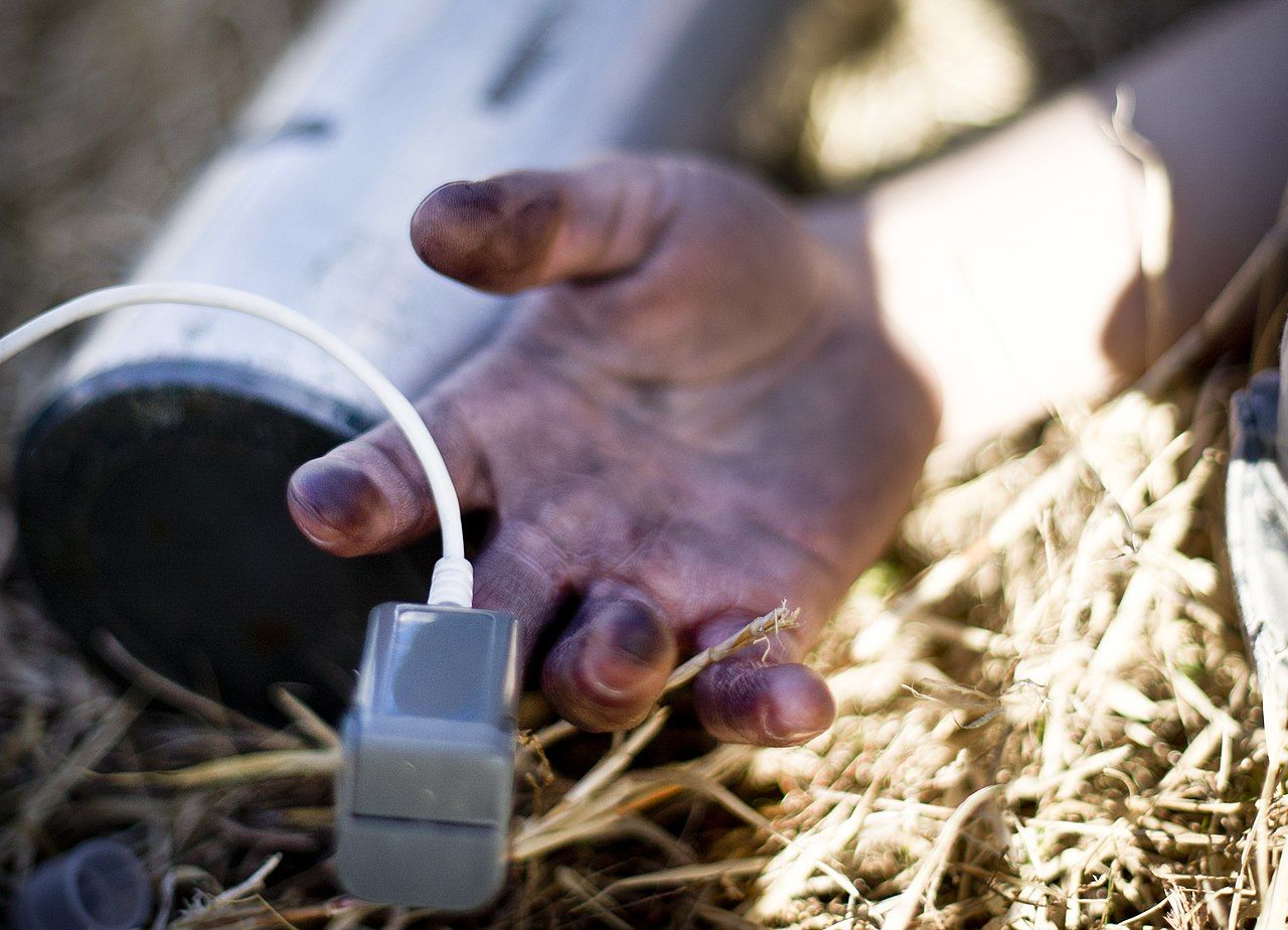
A pulse oximeter shines red and infrared light through a pulsating vascular bed and calculates the percentage of hemoglobin binding sites occupied by oxygen, reported as SpO2. Expected value on room air is 95 to 100 percent. Because of the shape of the oxyhemoglobin dissociation curve, an SpO2 of 90 percent corresponds to a PaO2 of about 60 mm Hg, and below that point the curve falls steeply — small further drops in saturation represent large drops in dissolved oxygen. That is why 90 percent is a threshold rather than a gentle slope. In patients with chronic carbon dioxide retention, a target of 88 to 92 percent is deliberate, not an oversight.
Place the probe on a warm, well-perfused digit with the light source and detector facing each other, and confirm that the displayed pulse rate matches the pulse you palpate and that the waveform or signal-strength indicator looks convincing. A saturation reading attached to a heart rate that does not match the patient is not data.
| Limitation | Effect on the reading | What to do |
|---|---|---|
| Poor perfusion — hypotension, shock, hypothermia, vasopressors, peripheral arterial disease | Low or absent, unreliable signal | Warm the hand, move to earlobe or forehead sensor, correlate with palpated pulse, obtain an arterial blood gas |
| Motion, shivering, tremor | Erratic, artifactually low | Reposition, steady the digit, use a forehead or ear probe |
| Dark nail polish (blue, green, black), artificial nails | Falsely low or no reading | Remove polish, rotate the probe 90 degrees on the digit, or use another site |
| Carbon monoxide poisoning | Falsely normal or high — carboxyhemoglobin is read as oxyhemoglobin | Never trust the SpO2 in a fire or exhaust exposure; obtain co-oximetry and treat clinically |
| Methemoglobinemia (benzocaine, dapsone, nitrates) | Reads near 85 percent regardless of true saturation | Co-oximetry; the classic clue is chocolate-brown blood with a normal PaO2 |
| Severe anemia | Normal percentage, inadequate oxygen content | SpO2 does not measure how much hemoglobin exists — correlate with hemoglobin and clinical status |
| Intravenous dyes (methylene blue, indocyanine green) | Transiently falsely low | Recheck after the dye clears |
| Ambient light, venous congestion, a blood pressure cuff on the same arm | Falsely low or unstable | Shield the probe, use the opposite arm from the cuff |
| Darker skin pigmentation | May overestimate true arterial saturation — occult hypoxemia is measurably more common in patients with darker skin | Do not use a reassuring SpO2 to override work of breathing, mental status or dyspnea; obtain an arterial blood gas when the picture does not fit |
That last row is a patient-safety issue, not a footnote. Occult hypoxemia — a true arterial saturation below 88 percent while the oximeter reads 92 to 96 percent — occurs several times more often in patients with darker skin. Combine that with the fact that cyanosis is harder to appreciate on darkly pigmented skin, and the two tools most likely to catch hypoxemia can fail together in the same patient. Assess the oral mucosa, tongue, conjunctivae, palms and soles, where cyanosis appears as a gray or ashen dullness; count the respiratory rate; listen to whether the patient can finish a sentence; and escalate on the clinical picture even when the number looks acceptable.
Respiratory rate is the earliest sign of deterioration and must be counted, not estimated; pulse oximetry reports saturation only, and it can read falsely reassuring in carbon monoxide exposure, poor perfusion and darker-pigmented skin.
Section 5Blood Pressure, and Knowing When to Call
The technique that makes the number trustworthy
Blood pressure is the force the blood exerts against the arterial walls: the systolic peak during ventricular ejection and the diastolic trough during ventricular relaxation. It is determined by cardiac output and systemic vascular resistance, so it falls with hypovolemia, vasodilation and pump failure, and rises with volume overload, vasoconstriction and increased stroke volume. It is also the vital sign most easily corrupted by technique, and a corrupted blood pressure changes medication decisions.
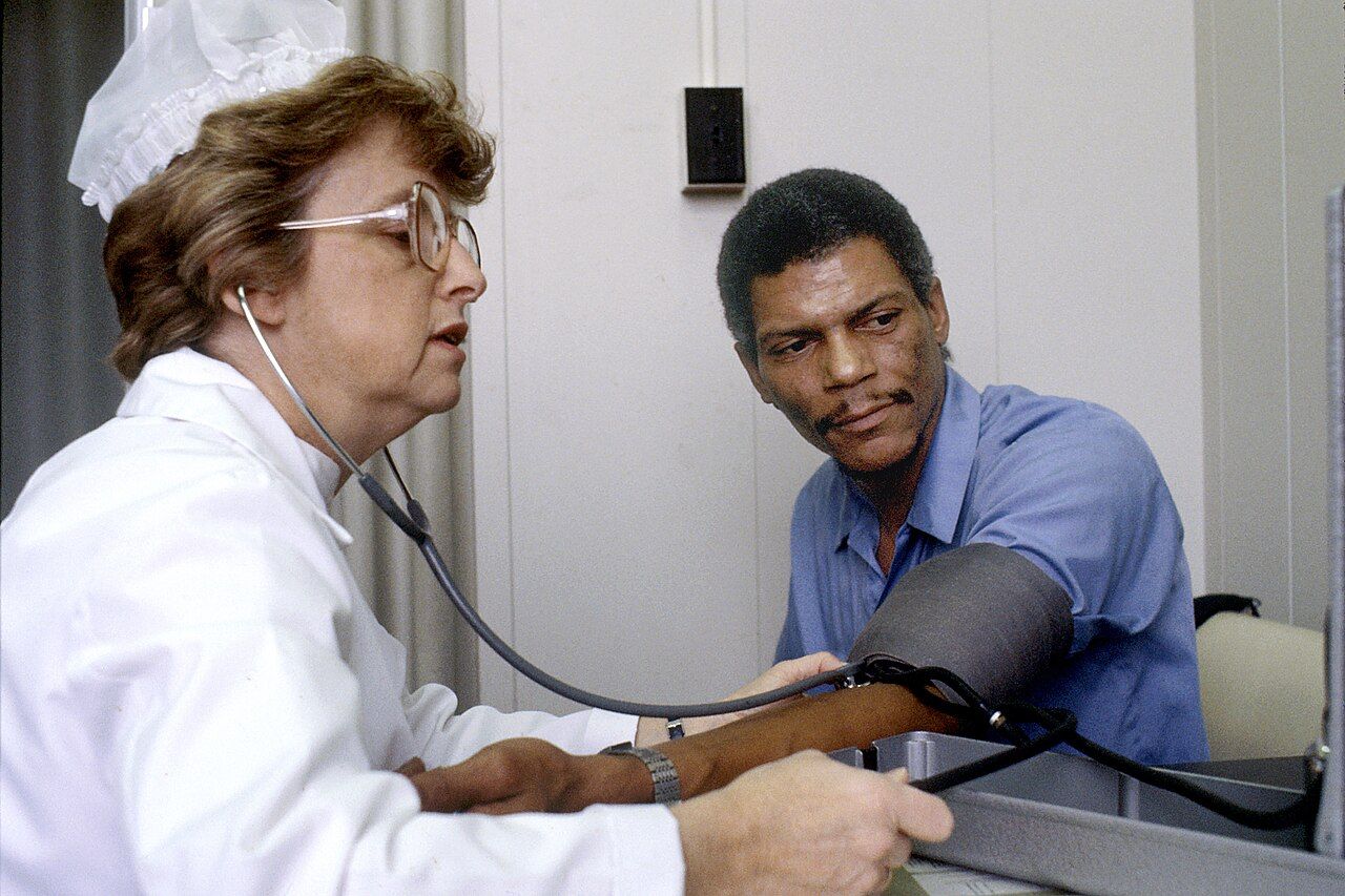
Start with the cuff. The inflatable bladder width should be about 40 percent of the arm circumference at the midpoint of the upper arm, and the bladder length should encircle about 80 percent of the arm. A cuff that is too small requires more pressure to compress the artery and therefore reads falsely high — this is the single most common cause of a spurious hypertension diagnosis, and it happens constantly on patients with large arms. A cuff that is too large reads falsely low. Measure the arm or use the range printed on the cuff; do not eyeball it. If no cuff fits the upper arm, use a correctly sized cuff on the forearm with the radial artery auscultated, and document that you did.
Then prepare the patient. Five minutes of quiet rest, seated, back supported, feet flat on the floor, legs uncrossed, bladder empty, no caffeine or nicotine or exercise in the preceding 30 minutes, and no talking during the measurement — by the patient or by you. Bare the arm; a cuff over a sleeve reads falsely high. Support the arm at heart level, roughly the mid-sternum or fourth intercostal space. An arm hanging below heart level reads falsely high; an arm held above it reads falsely low. Avoid an arm with an arteriovenous fistula or graft, an intravenous infusion or PICC line, lymphedema or axillary node dissection, a cast, injury or paralysis. On first assessment measure both arms and use the higher; a persistent difference greater than 10 to 15 mm Hg between arms is abnormal, and a large acute difference raises the question of aortic dissection.
Now the measurement itself. Palpate the radial pulse and inflate until it disappears, note that number, and deflate fully; wait 30 to 60 seconds. Place the diaphragm (or bell) lightly over the brachial artery in the antecubital fossa, not under the cuff edge. Inflate to 20 to 30 mm Hg above the palpated systolic estimate. Deflate at 2 to 3 mm Hg per second — faster deflation systematically underestimates systolic and overestimates diastolic. The palpation step is not optional: it protects you from the auscultatory gap, a silent interval between the first and second Korotkoff phases seen in some patients with hypertension, which can make you record a systolic pressure 20 to 40 mm Hg too low if you inflated only until sounds vanished.
| Korotkoff phase | Sound | Meaning |
|---|---|---|
| Phase I | First clear tapping | Systolic pressure |
| Phase II | Swishing or blowing murmur | Where the auscultatory gap may appear |
| Phase III | Crisper, more intense tapping | Turbulent flow continues |
| Phase IV | Abrupt muffling | Recorded as diastolic only when sounds continue to zero — document all three numbers, for example 130/72/0 |
| Phase V | Disappearance of sound | Diastolic pressure in adults, in pregnancy and, per current pediatric guidance, in children |
| Error | Effect on the reading |
|---|---|
| Cuff too narrow or too short for the arm | Falsely high |
| Cuff too wide or too long | Falsely low |
| Cuff applied over clothing, or loosely | Falsely high |
| Arm below heart level | Falsely high (about 2 mm Hg per 2.5 cm of drop) |
| Arm above heart level or unsupported | Falsely low or falsely high with isometric effort |
| Legs crossed at the knee | Systolic higher by 2 to 8 mm Hg |
| Back unsupported, feet dangling | Diastolic higher by up to 6 mm Hg |
| Full bladder; talking during measurement; recent caffeine or nicotine | Higher by roughly 10 mm Hg each |
| Deflating faster than 3 mm Hg per second | Systolic falsely low, diastolic falsely high |
| Rechecking immediately without deflating fully and waiting | Venous congestion raises diastolic |
Automated oscillometric devices calculate systolic and diastolic values from the measured mean arterial pressure. They are accurate on stable patients with regular rhythms and unreliable in atrial fibrillation, tremor, shivering, severe hypotension and extremes of arm size. Any automated reading that would change management — especially a very low one — should be confirmed manually before you act on it.
Reading the number: categories, MAP, pulse pressure and orthostatics
| Category (adult, ACC/AHA) | Systolic | Combined by | Diastolic |
|---|---|---|---|
| Normal | Under 120 | and | Under 80 |
| Elevated | 120–129 | and | Under 80 |
| Hypertension stage 1 | 130–139 | or | 80–89 |
| Hypertension stage 2 | 140 or higher | or | 90 or higher |
| Hypertensive crisis | Higher than 180 | and/or | Higher than 120 |
A hypertensive crisis divides on symptoms. Hypertensive urgency is a pressure above 180/120 without evidence of target-organ damage: recheck after five minutes of rest, notify the provider, expect oral therapy and close follow-up. Hypertensive emergency is the same pressure with chest pain, dyspnea, neurologic change, visual change, severe headache, back pain or new oliguria, and it requires immediate intravenous therapy with the mean arterial pressure lowered by no more than about 25 percent in the first hour — dropping it faster can infarct the brain, heart or kidney.
Two derived numbers earn their place at the bedside. Mean arterial pressure approximates the average perfusion pressure across the cardiac cycle and is calculated as the diastolic plus one third of the pulse pressure; normal is 70 to 100 mm Hg, and a MAP below 65 is generally taken as the point at which organ perfusion is threatened. Pulse pressure is systolic minus diastolic, normally 30 to 40 mm Hg. A narrowing pulse pressure — below about 25 mm Hg, or less than 25 percent of the systolic — suggests falling stroke volume from hypovolemia, cardiac tamponade or heart failure, and it often narrows before the systolic pressure falls. A widening pulse pressure suggests arterial stiffening, aortic regurgitation, thyrotoxicosis or, in a head-injured patient, rising intracranial pressure. Cushing triad — rising systolic with a widening pulse pressure, bradycardia and irregular respirations — is a late and emergent sign of brain herniation.
Orthostatic vital signs test whether the patient can maintain pressure against gravity. Perform them when a patient reports dizziness on standing, has fallen, is on antihypertensives or diuretics, is suspected of bleeding or dehydration, or is starting to mobilize after prolonged bed rest. Technique: have the patient lie supine for at least five minutes, then measure blood pressure and heart rate. Assist the patient to sitting, wait one minute, repeat. Assist to standing, and measure at one minute and again at three minutes. Keep a chair behind the patient and a gait belt on; stop immediately and return the patient to bed if symptoms appear. The test is positive when, within three minutes of standing, the systolic falls by 20 mm Hg or more or the diastolic falls by 10 mm Hg or more. A heart-rate rise of 20 beats per minute or more supports volume depletion; a patient who drops pressure without raising the heart rate may have autonomic failure or be beta-blocked. Symptoms — lightheadedness, greying vision, near-syncope — are part of the result and belong in the note.
Charting it so it counts
A vital-sign entry has to let a nurse who has never met the patient reconstruct exactly what you measured, how, and what you did about it. That means values with units, the route or site, the position, the oxygen delivery, the comparison to the prior set, and the action taken. Compare these two entries describing the same patient.
| Vague and indefensible | Precise and defensible |
|---|---|
| “VS stable. Pt appears comfortable. Will continue to monitor.” | “1400 T 38.4 °C (101.1 °F) oral; HR 118 apical, regular; RR 26, unlabored, no accessory muscle use; BP 96/54 right arm, adult regular cuff, seated, arm supported; SpO2 92 percent on room air; pain 3/10 at right flank. Compared with 1000 set (T 37.2, HR 88, RR 18, BP 124/72, SpO2 97 percent RA): febrile, tachycardic, tachypneic, systolic down 28; more confused than at 1000. Meets 3 of 3 qSOFA criteria (RR 26, systolic 96, altered mentation). Provider notified 1408 via SBAR; blood cultures and lactate ordered.” |
| “BP low, held med.” | “0745 BP 88/52 left arm, seated, manual, confirmed after 5 min rest; HR 62. Scheduled metoprolol 25 mg PO held per parameters (hold for SBP under 100). Dr. Nwosu notified 0752, order received to hold and recheck in 1 hour. Patient asymptomatic, denies dizziness, ambulated to bathroom with standby assist.” |
| “Orthostatics positive.” | “Orthostatic VS: supine after 5 min BP 128/78, HR 72; sitting 1 min BP 118/74, HR 84; standing 1 min BP 102/66, HR 96; standing 3 min BP 98/62, HR 104. Patient reported lightheadedness at 3 min standing, no syncope, returned to bed with two-person assist. Positive orthostatic hypotension (SBP drop 30, HR rise 32). Fall precautions in place, provider notified 1120.” |
Chart in the moment, not at the end of the shift; chart what you measured rather than what you concluded (“BP 96/54” rather than “BP soft”); never chart a value you did not personally obtain without naming who did; and never chart in advance. If a value is refused or cannot be obtained, say so and say why: “Rectal temperature declined by patient; temporal 37.9 °C obtained instead.”
Red flags and the SBAR call
Some numbers do not wait for the next scheduled set. Learn these as thresholds that trigger a call, and remember that any acute change from a patient’s own baseline can outrank an absolute value.
| Red flag | Why it matters | Escalation |
|---|---|---|
| Adult respiratory rate under 10 or over 24 — and under 8 or over 30 is critical at any adult age | Earliest marker of deterioration; opioid-induced respiratory depression | Call now; for opioid depression assess sedation, stimulate, oxygen, prepare naloxone |
| SpO2 under 90 percent on room air, or falling despite added oxygen | PaO2 already near 60 mm Hg and on the steep part of the curve | Oxygen, sit upright, reassess airway, call now |
| Systolic under 90, MAP under 65, or a fall of 40 mm Hg from baseline | Perfusion failure — shock of any cause | Rapid response; large-bore access, position supine, prepare fluids |
| Heart rate under 40 or over 130 in an adult | Rate itself now compromises output | Call now; obtain a 12-lead ECG |
| Temperature 38.3 °C or higher, or below 36.0 °C, with tachycardia and tachypnea | Sepsis criteria — hypothermia counts | Sepsis protocol; cultures and lactate before antibiotics |
| Rectal 38.0 °C or higher in an infant under 3 months | Cannot localize infection | Immediate provider notification |
| New confusion, agitation or decreased level of consciousness | Often the first sign of hypoxia or sepsis, and part of qSOFA | Full vital-sign set, glucose, call now |
| Above 180/120 with chest pain, neurologic change, dyspnea or visual change | Hypertensive emergency | Immediate call; expect IV therapy and monitored setting |
| 140/90 or higher after 20 weeks of pregnancy, especially with headache, visual change or epigastric pain | Preeclampsia; 160/110 is severe-range and time-critical | Immediate obstetric notification |
| New absent peripheral pulse with a cold, painful limb that is pale or ashen-grey compared with the other side — compare limbs and check nail beds, palms and soles, since pallor is not reliably visible on darker skin | Acute arterial occlusion or compartment syndrome | Emergency — do not elevate the limb, call immediately |
Escalate in SBAR, and have the chart and the last two sets of vital signs in front of you before you dial. A worked example for the febrile patient above:
S — Situation: “Dr. Nwosu, this is Priya Raman, RN, on 4 West. I’m calling about Mr. Delgado in room 412, a 68-year-old man admitted yesterday with pyelonephritis. His vital signs have changed significantly in the last four hours and I’m concerned he is becoming septic.”
B — Background: “He was admitted at 0900 yesterday, is on IV ceftriaxone, has a history of type 2 diabetes and benign prostatic hyperplasia. At 1000 today he was 37.2 degrees, heart rate 88, respirations 18, blood pressure 124/72, saturation 97 percent on room air.”
A — Assessment: “Right now he is 38.4 oral, heart rate 118 and regular, respirations 26, blood pressure 96/54, saturation 92 percent on room air, and he is more confused than this morning. His mean arterial pressure is 68. He meets all three qSOFA criteria — respiratory rate 26, systolic 96 and altered mentation — and that systolic is down 28 points from this morning. I think he is septic and starting to decompensate.”
R — Recommendation: “I would like you to come and see him now. I am requesting the sepsis order set: blood cultures times two, a lactate, a CBC and chemistry, and a 30 mL/kg crystalloid bolus. I have started oxygen at 2 liters and placed a second large-bore IV. Would you like me to activate the rapid response team while you are on your way? And how often would you like vital signs — I plan on every 15 minutes.”
Then close the loop. Read the orders back, document the time of the call and the response, and if the patient continues to deteriorate or the provider does not respond, move up the chain of command — charge nurse, rapid response team, nursing supervisor. A rapid response team may be activated by any nurse without a provider’s permission, and in most facilities by the patient or family as well.
A blood pressure is only as good as the cuff, the position and the deflation rate — and once you have a trustworthy abnormal number, the job is not charting it but escalating it, in SBAR, now.
The chapter in one breath
Vital signs are physical assessment converted into numbers, and they earn their name because they are the earliest, cheapest and most sensitive warning that a patient is failing. Adults sit at 36.5 to 37.5 °C orally, 60 to 100 beats per minute, 12 to 20 breaths per minute, under 120/80 mm Hg, and 95 to 100 percent saturation; children run faster and lower-pressured, and older adults run cooler with wider pulse pressures. Technique determines whether those numbers mean anything: chart the temperature route, count an irregular pulse apically for a full minute, measure a pulse deficit with two nurses at once, count respirations without telling the patient, and remember that a cuff too small reads falsely high while a hurried deflation drags the systolic down. Pulse oximetry reports saturation only, and it lies in carbon monoxide poisoning, poor perfusion and — importantly — more often in patients with darker skin, so assess mucosa, conjunctivae and work of breathing rather than trusting the display. Interpret every value against the previous set and against that patient’s baseline, delegate measurement on stable patients but never interpretation, document values rather than conclusions, and escalate red flags immediately in SBAR.
Words worth knowing
| Term | What it means |
|---|---|
| Apical pulse | Heart rate auscultated at the point of maximal impulse, fifth intercostal space at the left midclavicular line, counted for a full minute. |
| Auscultatory gap | A silent interval between Korotkoff phases I and II that causes a falsely low systolic reading unless the cuff is first inflated above a palpated estimate. |
| Bradycardia / tachycardia | Adult heart rate under 60 / over 100 beats per minute. |
| Cheyne-Stokes respiration | Cyclical crescendo-decrescendo breathing alternating with apnea; seen in heart failure, raised intracranial pressure and dying. |
| Core temperature | Temperature of the deep tissues, approximated by rectal, tympanic, esophageal, bladder or pulmonary artery measurement. |
| Defervescence | The phase in which the hypothalamic set point falls and the patient diaphoreses and vasodilates as the fever breaks. |
| Eupnea | Normal, quiet, unlabored breathing at 12 to 20 breaths per minute in an adult. |
| Hypertensive emergency | Blood pressure above 180/120 mm Hg with evidence of target-organ damage; requires immediate intravenous treatment. |
| Korotkoff sounds | The five phases of sound heard as a cuff deflates; phase I is systolic, phase V is diastolic. |
| Kussmaul respirations | Deep, rapid, labored breathing compensating for metabolic acidosis, classically diabetic ketoacidosis. |
| Mean arterial pressure (MAP) | Average perfusion pressure, diastolic plus one third of the pulse pressure; normal 70 to 100 mm Hg, with 65 the usual perfusion threshold. |
| Occult hypoxemia | True arterial saturation below 88 percent while pulse oximetry reads 92 percent or higher; more frequent in patients with darker skin pigmentation. |
| Orthostatic hypotension | A fall of 20 mm Hg systolic or 10 mm Hg diastolic within three minutes of standing from supine. |
| Pulse deficit | Apical rate minus radial rate, measured simultaneously by two nurses for one minute; reflects contractions too weak to generate a peripheral wave. |
| Pulse pressure | Systolic minus diastolic pressure; normally 30 to 40 mm Hg, narrowing with falling stroke volume. |
| Pulsus paradoxus | An inspiratory fall in systolic pressure greater than 10 mm Hg; suggests tamponade or severe asthma. |
| Pyrexia | Fever — an adult temperature of 38.0 °C (100.4 °F) or higher, produced by a raised hypothalamic set point. |
| SpO2 | Peripheral oxygen saturation, the percentage of hemoglobin binding sites occupied by oxygen as estimated by pulse oximetry. |
| Thermoregulation | Hypothalamic balancing of heat production against heat loss through radiation, conduction, convection and evaporation. |
| qSOFA | Quick bedside sepsis screen: respiratory rate 22 or higher, systolic 100 mm Hg or lower, altered mentation; two of three indicates high risk. |