Assessing Head, Neck, Eyes, Ears, Nose and Throat
Everything above the clavicles is crowded, and that is exactly why it matters. In a space the size of your two fists sit the airway, four of the great sensory organs, ten lymph node chains, the thyroid, the carotids and eleven of the twelve cranial nerves. A student who learns this region as a list of parts will chart “HEENT within normal limits” and move on. A nurse who learns it as a system will notice that the uvula has shifted to the left, that one pupil is a millimetre larger than it was two hours ago, that the node above the clavicle is hard and will not move, or that a patient who was hoarse this morning is now making noise on inspiration. Those four findings are, in order, a peritonsillar abscess, a herniating brain, a probable malignancy and an airway that is closing. This chapter teaches the technique first — hands, light, sequence — then the findings, then the numbers, then the words you write and the escalation you call.
By the end of this chapter you can
- Perform a systematic head, neck and HEENT examination in the correct order — inspection, palpation, percussion, auscultation — and state the one region where that order changes.
- Palpate the ten cervical lymph node chains and distinguish a reactive node from a node that suggests malignancy, including the supraclavicular (Virchow) node.
- Examine the thyroid by anterior and posterior approach, auscultate for a thyroid bruit, and confirm the trachea is midline.
- Measure distance and near visual acuity, test visual fields by confrontation, assess the six cardinal fields of gaze, and document PERRLA accurately with pupil size in millimetres.
- Perform otoscopy with correct pinna traction for age, identify the normal tympanic membrane landmarks, and interpret the whispered voice, Weber and Rinne tests.
- Inspect the nose, sinuses, oral mucosa, tongue and pharynx, grade tonsils 1+ through 4+, and identify leukoplakia and oral candidiasis.
- State expected vital-sign values for adults and across the lifespan, and describe how HEENT findings differ in infants, children, pregnancy and older adults.
- Assess cyanosis, jaundice, pallor and erythema in patients of every skin tone using oral mucosa, conjunctivae, sclera and palms and soles.
- Recognize HEENT red flags that require escalation now, communicate them using SBAR, and correctly delegate — or refuse to delegate — tasks to unlicensed assistive personnel.
Section 1The Head, Face and Neck
The order of the exam, and the one place it changes
Physical assessment has a fixed order: inspection, palpation, percussion, auscultation. You look before you touch, you touch before you tap, and you tap before you listen. The reason is not tradition. Looking costs nothing and biases nothing; touching a tender area first makes the patient guard, tense and stop cooperating; and percussion sets tissue moving in ways that can change what you hear afterward. In the head and neck you will use inspection and palpation constantly, percussion almost never, and auscultation in exactly two places — over the carotid arteries and over an enlarged thyroid.
There is one region of the body where this order is deliberately broken. The abdomen is the exception: inspect, then AUSCULTATE, then percuss, then palpate. Bowel sounds are produced by peristalsis, and pressing on the abdomen mechanically stimulates or suppresses that peristalsis. If you palpate first, the bowel sounds you then auscultate are sounds you created, not sounds the patient had. Memorize the exception now, because it is the single most commonly missed sequencing item on licensure examinations and the single most commonly botched sequence at the bedside. Everywhere else — head, neck, thorax, extremities — the standard order holds.
Before you touch the patient, set the stage. Perform hand hygiene, introduce yourself, use two identifiers, explain what you are about to do, and position the patient sitting upright with their head at your eye level. Sitting matters: it lets you compare both sides of the face and neck simultaneously, and symmetry is the single most powerful assessment tool you have in this region. Almost every abnormality above the clavicles announces itself first as an asymmetry.
Skull, face and the temporomandibular joint
Inspect the skull for size, shape and symmetry. The expected finding is normocephalic and atraumatic — a rounded, symmetric skull with no depressions, bossing or lesions, and no evidence of injury. Then palpate the entire scalp with the pads of your fingers in small circles, front to back, feeling for tenderness, masses, depressions and step-offs in the bone. Part the hair to look at the scalp itself; you are checking for lesions, scaling, lice and, in an older adult who has fallen, a boggy hematoma hidden under thick hair. Palpate the temporal arteries just anterior to each ear: they should be smooth, non-tender and equally pulsatile.
Inspect the face at rest and in motion. At rest, look at the palpebral fissures, nasolabial folds and mouth corners for symmetry. Then test the muscles of facial expression, which is cranial nerve VII (facial): ask the patient to raise their eyebrows, close their eyes tightly while you try to open them, smile, show their teeth and puff out their cheeks. A drooping mouth corner with an intact forehead points to a central lesion such as a stroke; a droop that includes the forehead on the same side points to a peripheral seventh nerve palsy such as Bell palsy. That distinction — forehead spared or not — changes the entire escalation pathway, and you can make it in eight seconds. While you are there, test cranial nerve V (trigeminal): palpate the masseter and temporalis muscles while the patient clenches, and check light touch sensation on the forehead, cheek and jaw bilaterally.
Palpate the temporomandibular joint by placing your index fingertips just anterior to the tragus of each ear and asking the patient to open and close the mouth slowly. You should feel a smooth, symmetric glide. The mouth should open 3 to 6 cm between the incisors, roughly the width of three of the patient’s own fingers held vertically. Audible clicking is common and by itself not alarming. Crepitus, pain, locking, or an opening of less than 3 cm is abnormal. An inability to open the mouth at all — trismus — in a patient with a sore throat is not a dental problem; it is a deep neck space infection until proven otherwise, and it belongs in Section 4 and in your escalation list.
Lymph nodes: the ten chains and what a bad node feels like
Palpate the cervical nodes using the pads of your index and middle fingers, both hands at once, moving the skin over the underlying tissue in gentle circles. Use light pressure — heavy pressure pushes a small node out of your way and you will miss it. Work in a fixed order every time so you never skip a chain: preauricular (in front of the ear), posterior auricular (over the mastoid), occipital (base of the skull), tonsillar (angle of the mandible), submandibular (halfway between the angle and the tip of the jaw), submental (behind the tip of the chin, one hand), superficial cervical (over the sternocleidomastoid), deep cervical chain (under the sternocleidomastoid — hook your fingers around the muscle and ask the patient to tilt the head toward the side you are examining to relax it), posterior cervical (along the anterior edge of the trapezius) and supraclavicular (in the hollow above the clavicle; ask the patient to shrug and then relax, and have them take a deep breath and hold it to bring a deep node up under your finger).
Most healthy adults have no palpable nodes at all, and many have a few small nodes that are less than 1 cm, soft, mobile, discrete and non-tender. When you find a node, describe six things: location, size in centimetres, shape, consistency, mobility and tenderness. Those six words separate a viral pharyngitis from a lymphoma. The pattern that reassures is tender, warm, soft, mobile and bilateral — that is inflammation, and it usually has an obvious source such as a throat, ear or scalp infection. The pattern that worries is hard, fixed, matted, painless and unilateral — that is how malignancy presents, and painlessness is a feature, not a reassurance.
Two rules override everything else. First, a palpable supraclavicular node is abnormal at any size, in any patient, at any age. The left supraclavicular node — the Virchow node — drains the thoracic duct and can be the first sign of a gastric, pancreatic, esophageal or pulmonary malignancy. Second, size is not the only trigger for referral. Any node that is hard, fixed, matted or painless needs a provider evaluation at any size; so does any node that has been enlarging for more than two to four weeks without an infectious explanation, any node larger than 1 cm that does not settle as the infection settles, and any node larger than 2 cm regardless of how it feels. Do not wait for a suspicious node to reach 2 cm before you report it.
| Finding | Expected | Abnormal | What the abnormal suggests |
|---|---|---|---|
| Skull | Normocephalic, atraumatic, no tenderness | Depression, step-off, boggy area, focal tenderness | Fracture, hematoma; in an older adult, an unwitnessed fall |
| Temporal artery | Smooth, non-tender, equal pulses | Thickened, nodular, tender artery in a patient over 50 | Giant cell arteritis — vision-threatening, urgent |
| Face (CN VII) | Symmetric at rest and with movement | Droop with forehead SPARED | Central lesion — suspect stroke, activate stroke pathway |
| Face (CN VII) | Symmetric at rest and with movement | Droop INCLUDING forehead, one side | Peripheral CN VII palsy (Bell palsy); eye needs protection |
| TMJ | Smooth glide, opens 3–6 cm, painless | Crepitus, pain, locking, opening <3 cm | TMJ disorder; trismus with sore throat suggests abscess |
| Cervical nodes | Non-palpable, or <1 cm, soft, mobile, non-tender | Tender, warm, mobile, bilateral | Reactive — infection somewhere in the drainage field |
| Cervical nodes | Non-palpable, or <1 cm, soft, mobile, non-tender | Hard, fixed, matted, painless, unilateral — at ANY size | Malignancy until proven otherwise |
| Supraclavicular node | Never palpable | Any palpable node | Thoracic or abdominal malignancy; always significant |
| Neck ROM | Flexion 45°, extension 55°, lateral bend 40°, rotation 70° | Nuchal rigidity, pain on passive flexion | Meningeal irritation — with fever and headache, emergency |
Thyroid, trachea and carotids
Confirm the trachea is midline before anything else. Place your index finger in the suprasternal notch and slip it gently to each side of the trachea; the space between the trachea and the sternocleidomastoid should be equal on both sides. A deviated trachea is never a minor finding. Air or fluid under pressure pushes the trachea away from the affected side — that is tension pneumothorax and large pleural effusion. Loss of lung volume pulls the trachea toward the affected side — that is atelectasis, fibrosis and pneumonectomy. A patient who is short of breath with a trachea deviated away from the side with absent breath sounds is a rapid-response call, not a documentation entry.
Now the thyroid. The isthmus lies across the second and third tracheal rings, with a lobe curving back on either side. Inspect first: with the patient’s neck slightly extended, hand them a cup of water and watch them swallow. Thyroid tissue rises with the swallow; lymph nodes and most other neck masses do not. That single manoeuvre — watch it move with the swallow — is the most useful thing you will do at the neck. For the posterior approach, stand behind the seated patient, place the fingers of both hands on the neck with your index fingers just below the cricoid cartilage, ask the patient to flex the neck slightly forward and tilt it toward the side you are palpating (this relaxes the sternocleidomastoid), displace the trachea gently toward that side with the opposite hand, and ask for a sip of water. For the anterior approach, stand in front, use the same displacement and swallow, and palpate with your thumbs. Use whichever you can do reliably; consistency beats elegance.
A normal thyroid is often not palpable at all. When it is, the lobes feel smooth, firm, non-tender and roughly the size of the distal phalanx of the patient’s thumb. Enlargement is a goiter. If you feel any enlargement, place the bell of your stethoscope over each lobe and ask the patient to hold their breath. A soft blowing swishing sound is a thyroid bruit, and it means increased vascularity — classically hyperthyroidism and Graves disease. Palpate gently; vigorous manipulation of a hyperactive gland is avoided. Finally, auscultate each carotid with the bell while the patient holds their breath, listening for a bruit. Never palpate both carotid arteries at the same time and never massage the carotid sinus; you can drop the heart rate and the cerebral perfusion of the patient in front of you.
Assess the head and neck by inspection then palpation, comparing both sides at once, and treat a deviated trachea, a hard fixed node or any palpable supraclavicular node as findings that leave the room with you.
Section 2Assessing the Eyes
Visual acuity and visual fields
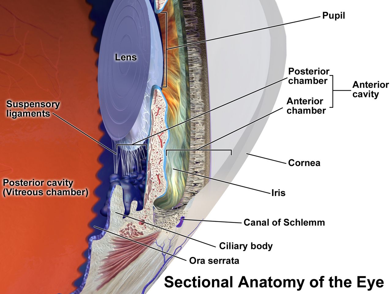
Visual acuity is the vital sign of the eye, and like any vital sign it is measured, not estimated. Use a Snellen chart at 20 feet (6 metres) in good light. Test each eye separately with the other eye covered by an occluder or the patient’s palm — never by their fingers, because patients peek — then test both eyes together. If the patient wears glasses or contacts for distance, leave them on and chart the result as corrected. Record the smallest line the patient reads correctly, plus the number of letters missed on that line: 20/30 − 1 means they read the 20/30 line and missed one letter. The numerator is your testing distance; the denominator is the distance at which a person with normal vision could read that line. So 20/20 is expected adult acuity, and 20/40 means your patient must stand at 20 feet to see what a normal eye sees at 40 feet. Legal blindness in the United States is 20/200 or worse in the better eye with best correction. If a patient cannot read the largest letter, move them closer and record the distance; if they still cannot, test counting fingers, then hand motion, then light perception, in that order.
Test near vision with a handheld Jaeger card held at 14 inches (35 cm); normal is J1 or 14/14. Difficulty with near vision beginning in the forties is presbyopia, a normal age-related stiffening of the lens, and it is the reason your fifty-year-old patient holds the discharge instructions at arm’s length. That is a teaching moment about large-print materials, not a pathology.
Screen peripheral vision by confrontation. Sit facing the patient at eye level about 60 cm (2 feet) away. Cover your right eye and have them cover their left, so you and the patient are looking directly at one another with the opposite eyes. Keep your eyes locked, then bring a wiggling finger in from the far periphery in each of four quadrants, midway between you and the patient, and ask them to say “now” the moment they see it. Because you are the control, you should see the finger at the same instant. Normal fields extend approximately 50° superiorly, 60° nasally, 70° inferiorly and 90° temporally. A quadrant the patient consistently misses is a field cut and needs a provider — and it also needs a safety plan, because a patient with a left field cut will walk into the left side of the doorway and will not see the food on the left half of the tray.
The external eye, the pupils and extraocular movement
Inspect the eyebrows, lids and lashes for symmetry, ptosis, edema, lesions and lid margin inflammation. The upper lid normally covers the top 1 to 2 mm of the iris; the sclera should not be visible above the iris. Inspect the conjunctivae by having the patient look up while you pull the lower lid down: the palpebral conjunctiva should be pink and moist, and the bulbar conjunctiva clear with fine visible vessels. Sclera should be white. In patients with darker skin, small tan or brown melanin deposits are commonly seen in the sclera near the limbus and are a normal variant — do not mistake them for jaundice. When you are screening for jaundice, look at the sclera immediately adjacent to the iris, at the hard palate and under the tongue; the peripheral sclera can carry a yellowish tint from fat and carotene in perfectly healthy people.
Assess the cornea by shining a penlight obliquely across it; it should be smooth, clear and glossy, with no cloudiness or abrasion. A thin greyish-white ring around the outer iris in an adult over 60 is arcus senilis and is a normal aging finding; the same ring in a patient under 40 warrants a lipid evaluation. Test the corneal light reflex by shining a penlight at the bridge of the nose from about 30 cm: the reflection should land on exactly the same spot of each cornea. Asymmetry means the eyes are not aligned.
Pupils are round, equal and, in normal room light, 3 to 5 mm in diameter (the full normal range is roughly 2 to 6 mm depending on light and age). Chart the actual millimetre size using the gauge printed on your penlight or pupil card — “pupils equal” is not data. Darken the room, bring the light in from the side, and check four things: direct constriction of the lit eye, consensual constriction of the other eye, then accommodation and convergence as the patient shifts gaze from a distant object to your finger 10 cm from their nose. That is PERRLA: pupils equal, round, reactive to light and accommodation. About one in five people has a benign difference of up to 1 mm between pupils that is stable over time — physiologic anisocoria. What is never benign is a new difference, especially a unilateral dilated pupil that reacts sluggishly or not at all in a patient with a declining level of consciousness. That is third nerve compression from an expanding intracranial mass, and it is a call to the rapid response team while you are still standing at the bedside.
Assess extraocular movement through the six cardinal fields of gaze, which test cranial nerves III, IV and VI together. Hold your finger about 30 cm away, ask the patient to hold their head still and follow with their eyes only, and move slowly through an H pattern — lateral, up-and-lateral, down-and-lateral on each side. Both eyes should move together, smoothly and fully, with parallel alignment. A few beats of nystagmus at extreme lateral gaze are normal; sustained nystagmus in any other position is not.
| Pupil finding | Description | Most likely cause | Nursing action |
|---|---|---|---|
| Expected | 3–5 mm, equal, brisk direct and consensual response | Normal | Chart size in mm and reactivity |
| Pinpoint (1–2 mm), bilateral | Constricted, may be minimally reactive | Opioid effect; pontine hemorrhage | Assess respirations and sedation; opioid effect needs naloxone consideration and provider notification |
| Unilateral dilated, fixed | One pupil >6 mm, non-reactive, with decreasing level of consciousness | CN III compression — uncal herniation | EMERGENCY. Rapid response now; do not wait for the next round |
| Bilateral fixed and dilated | Both >6 mm, no reaction | Severe hypoxic or anoxic brain injury; anticholinergics; late herniation | EMERGENCY; assess airway, breathing, circulation and call |
| Mid-dilated, fixed, oval; painful red eye | Hard globe, halos around lights, nausea and vomiting | Acute angle-closure glaucoma | EMERGENCY ophthalmology referral; vision is lost in hours |
| Stable unequal pupils, both reactive | Difference up to 1 mm, unchanged over time, patient alert | Physiologic anisocoria | Document the baseline so the next nurse does not panic |
The internal eye, the red reflex and eye emergencies
The funduscopic examination is performed by advanced practice providers in most settings, but you need to understand it because you will assist with it, position for it and read the results. Darken the room. Hold the ophthalmoscope in your right hand, use your right eye, and examine the patient’s right eye; then switch everything for the left. Start about 25 cm away and 15° lateral to the patient’s line of sight, find the orange glow of the red reflex, then follow it in to within 3 to 5 cm. The optic disc is creamy yellow-orange with sharp margins and a cup that occupies no more than about half of the disc diameter. Arterioles are lighter and narrower than venules, in roughly a 2:3 ratio. The macula lies about two disc diameters temporal to the disc. Blurred disc margins — papilledema — indicate raised intracranial pressure.
The red reflex is the finding nurses genuinely own, because it is checked at every newborn and well-child assessment. It should be present, equal and symmetric in both eyes. A white reflex — leukocoria — is an urgent referral: retinoblastoma, congenital cataract and retinopathy of prematurity all present this way, and retinoblastoma is a childhood cancer where time to treatment determines whether the child keeps the eye and the life. If a parent shows you a phone photo in which one of their baby’s eyes glows white while the other glows red, you escalate that photo.
Three ocular situations demand action before assessment. A chemical splash is irrigated immediately — do not stop to test acuity, do not look for the bottle first. Remove contact lenses, hold the lids open and irrigate with normal saline or lactated Ringer solution for at least 15 to 20 minutes, or until the conjunctival pH is 7.0 to 7.4, directing flow from the inner to the outer canthus so you do not wash the chemical into the other eye. Alkali burns penetrate deeper and irrigate longer than acid. Never attempt to neutralize a chemical with another chemical. An impaled foreign body is never removed at the bedside: stabilize it, shield both eyes to stop conjugate movement, keep the patient still and call. And sudden painless loss of vision in one eye — a curtain coming down, a shower of floaters, a shade closing — is a retinal artery occlusion or a retinal detachment and is a same-hour emergency.
Visual acuity is measured with a chart and charted as a fraction, pupils are charted in millimetres, and a new unilateral fixed dilated pupil, a white red reflex, a chemical splash or sudden painless vision loss are all escalations rather than assessments.
Section 3Assessing the Ears and Hearing
The external ear: inspection and the two-second diagnosis
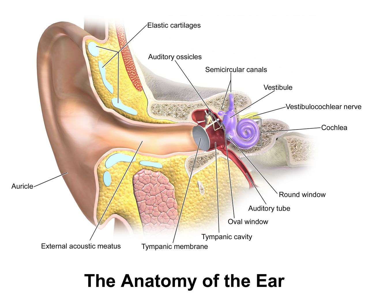
Inspect both auricles for size, shape, symmetry, position and skin condition. The top of the auricle should meet an imaginary line drawn from the outer canthus of the eye to the occiput, and the auricle should sit within 10° of vertical. Low-set or rotated ears in an infant are associated with congenital renal and chromosomal anomalies and are reported, not just noted. Inspect the external canal opening for discharge, redness, swelling and cerumen. Note the colour and character of any discharge: purulent drainage suggests infection, and clear or blood-tinged watery drainage after head trauma is cerebrospinal fluid until proven otherwise.
Then palpate. Move the auricle up and down, press the tragus inward, and press over the mastoid process. This takes two seconds and does real diagnostic work. Pain when you move the pinna or press the tragus points to otitis externa, an infection of the canal. Ear pain with no tenderness on external manipulation points to otitis media, behind the drum. And tenderness, redness or swelling over the mastoid, especially with an auricle that is pushed forward and outward, is mastoiditis — a complication that can extend intracranially and needs urgent evaluation.
Otoscopy: how to hold it, what you should see
Choose the largest speculum that will fit the canal comfortably; a speculum that is too small gives you a keyhole view and is more likely to touch the sensitive canal wall. Hold the otoscope like a pencil, upside down, braced against the patient’s head with the ulnar side of your hand, so that if the patient moves suddenly your hand moves with them and the speculum does not lacerate the canal. Straighten the canal by pulling the auricle: up and back for adults and children 3 years and older, down and back for infants and children under 3, because their canal points upward. Insert slowly, downward and forward, about 1 to 1.5 cm in an adult, watching your path the whole way in.
The normal tympanic membrane is pearly grey, translucent, intact and slightly concave. You should identify the cone of light — the triangular light reflex that points anteroinferiorly, at roughly 5 o’clock in the right ear and 7 o’clock in the left — along with the umbo, the handle of the malleus and the short process. On pneumatic insufflation the membrane moves. Cerumen is normal and protective and does not need removal unless it blocks the view or the hearing; when irrigation is ordered, it is contraindicated if there is any suspicion of perforation, tympanostomy tubes, current infection or a history of ear surgery.
In children, the ear examination and the throat examination are performed last, after everything that requires cooperation, because both are intrusive and both end cooperation. Infants and toddlers have shorter, wider, more horizontally oriented eustachian tubes, which is why otitis media peaks between 6 and 24 months and why a bottle-propped supine feeding raises the risk.
| Tympanic membrane appearance | Interpretation | Typical accompanying findings |
|---|---|---|
| Pearly grey, translucent, cone of light intact, mobile | Expected | Hearing intact, no pain |
| Red, bulging, landmarks obscured, immobile | Acute otitis media | Fever, ear pain, recent upper respiratory infection, irritable infant pulling at ear |
| Amber or yellow, air-fluid level or bubbles visible | Otitis media with effusion (serous) | Muffled hearing, popping, usually afebrile |
| Retracted, landmarks unusually prominent | Eustachian tube dysfunction, negative middle-ear pressure | Fullness, mild hearing loss |
| Dense chalky white patches | Tympanosclerosis — old scarring | Usually asymptomatic; history of repeated infections or tubes |
| Visible dark perforation or purulent drainage in canal | Perforated tympanic membrane | Sudden relief of pain, drainage; NO irrigation, NO drops without an order |
| Blood behind an intact drum (hemotympanum) | Basilar skull fracture until proven otherwise | Head trauma, Battle sign, raccoon eyes — escalate now |
Testing hearing: whisper, Weber and Rinne
Start with the simplest and most sensitive screen: the whispered voice test. Stand about an arm’s length (roughly 60 cm) behind and to the side of the patient so they cannot read your lips. Occlude the non-tested ear by pressing and gently rubbing the tragus. Exhale fully — this ensures you are truly whispering — then whisper a combination of three numbers and letters, such as “4–K–2.” A patient with normal hearing repeats all three correctly. If they miss any, repeat with a different set; failing to repeat at least three of six items is a failed screen. Test each ear separately.
Tuning fork tests separate conductive loss (a problem in the outer or middle ear — cerumen, effusion, perforation, otosclerosis) from sensorineural loss (a problem in the cochlea or eighth nerve — aging, noise exposure, ototoxic drugs). Use a 512 Hz fork. For the Weber test, strike the fork, place the base on the midline of the top of the skull or the mid-forehead, and ask where the sound is heard. Normal is equal in both ears with no lateralization. Sound that lateralizes to the bad ear means conductive loss in that ear (blocking room noise makes bone conduction seem louder there). Sound that lateralizes to the good ear means sensorineural loss in the other ear.
For the Rinne test, place the vibrating fork on the mastoid process, ask the patient to say when the sound disappears, then immediately move the still-vibrating tines about 2 cm in front of the ear canal. Normally, air conduction is better than bone conduction by about a 2:1 time ratio, so the patient hears the fork again in front of the ear — charted as AC > BC, positive Rinne. When bone conduction is equal to or better than air conduction, the Rinne is negative and there is conductive loss on that side. Nurses do not diagnose from these tests; nurses use them to decide how urgently to route a patient and to communicate a precise finding.
Two hearing changes need speed. Sudden sensorineural hearing loss — hearing that disappears in one ear over hours to a day or two, often with the patient describing fullness and tinnitus — is an emergency, not a wait-and-see. Outcomes are best when corticosteroids begin as early as possible — ideally within about 72 hours — and treatment is still offered up to two weeks from onset, so a patient who calls the clinic reporting that one ear stopped working overnight is triaged as urgent, and a patient who reports it five days later is still worked in today rather than dismissed as too late. And any patient on an ototoxic medication — aminoglycosides, high-dose loop diuretics, cisplatin, high-dose salicylates, vancomycin — who reports new tinnitus, hearing loss or vertigo needs the medication reported and levels reviewed before the next dose.
Pull the pinna up and back over age 3 and down and back under age 3, expect a pearly grey drum with a cone of light at 5 and 7 o’clock, and treat sudden one-sided hearing loss, mastoid tenderness and post-traumatic ear drainage as urgent rather than routine.
Section 4Nose, Sinuses, Mouth and Throat
Nose and sinuses
Inspect the external nose for symmetry, deformity and lesions. Test patency one side at a time: occlude one naris, ask the patient to close their mouth and sniff, then repeat on the other side. Then use an otoscope with a wide short speculum, or a nasal speculum with a penlight, tilting the patient’s head back slightly. You should see a midline septum, pink moist mucosa, and the inferior and middle turbinates as smooth reddish-pink ridges on the lateral wall. Do not confuse a turbinate with a polyp; polyps are pale, grey, glistening and mobile, and they do not hurt when touched.
Abnormal nasal findings are unusually informative. Pale, boggy, bluish-grey turbinates with clear watery discharge indicate allergic rhinitis. Bright red swollen mucosa suggests infection. A perforated septum raises questions about intranasal cocaine use, chronic topical decongestant use or trauma. Unilateral foul-smelling purulent discharge in a toddler is a nasal foreign body until someone looks. And clear watery drainage from one nostril after head trauma may be cerebrospinal fluid — do not pack it, do not insert a nasogastric tube, and report it immediately.
Only the frontal and maxillary sinuses are accessible. Palpate the frontal sinuses by pressing up under the bony brow ridges, never directly on the eyes, and the maxillary sinuses by pressing up under the cheekbones. Expected finding: no tenderness. Sinuses may also be transilluminated in a darkened room, but this is a low-yield technique in practice. The ethmoid and sphenoid sinuses cannot be examined at the bedside. Cranial nerve I (olfactory) is tested only when there is a reason — head trauma, suspected neurologic disease, new anosmia — by occluding one nostril and asking the patient to identify a familiar non-irritating odour such as coffee or vanilla with their eyes closed.
For epistaxis, sit the patient upright and leaning forward — never tilted back, which sends blood into the airway and the stomach and causes vomiting. Pinch the soft compressible part of the nose just below the nasal bones firmly and continuously for 10 to 15 minutes without releasing to check. Apply ice to the bridge. Bleeding that continues beyond 20 to 30 minutes of correct pressure, occurs in a patient on anticoagulants, or is accompanied by hemodynamic change requires provider evaluation.
Mouth, tongue and the oral cancer check
Put on gloves. Inspect the lips for colour, moisture, cracking and lesions. Then use a tongue blade and a good light to inspect the buccal mucosa, gums, teeth, hard and soft palate, tongue and floor of the mouth. Expected findings: mucosa pink, smooth and moist; gums pink with tight margins and no bleeding; teeth intact with no obvious caries; palate intact; tongue midline, pink, with a papillated dorsal surface and a smooth ventral surface. The parotid (Stensen) duct opens on the buccal mucosa opposite the second upper molar; the submandibular (Wharton) ducts open on either side of the frenulum under the tongue.
Now do the part students skip. Wrap the tip of the tongue in a gauze square, pull it gently forward and to each side, and inspect the lateral borders and the floor of the mouth, then palpate the floor of the mouth bimanually. Those two areas are where oral cancers most commonly begin, and they are the two areas that a quick “say ahh” never shows you. A thick white patch that does not scrape off is leukoplakia, a precancerous lesion requiring biopsy, and it is strongly associated with tobacco, smokeless tobacco and alcohol. By contrast, white curd-like plaques that do scrape off, leaving a red raw base that may bleed, are oral candidiasis. It is common in infants but it is never a normal finding: report it, expect an antifungal order such as nystatin, and assess feeding, bottle and pacifier hygiene and maternal nipple candidiasis. Distinguish it from milk residue, which wipes away without leaving a raw base, and treat thrush that persists or recurs beyond infancy as a reason to ask about immune status. In an adult it demands a question about inhaled corticosteroids without rinsing, recent antibiotics, diabetes or immunosuppression. Any oral ulcer or lesion present for more than two weeks is referred.
Several normal variants are commonly mistaken for pathology, and most of them are more frequent in patients with darker skin. Leukoedema is a filmy greyish-white translucent appearance of the buccal mucosa that disappears when the mucosa is stretched. Physiologic melanin pigmentation produces brown or blue-grey patches on the gingiva, buccal mucosa or tongue, present for years and asymptomatic. Torus palatinus is a hard bony midline ridge on the hard palate. Fordyce granules are small yellow-white sebaceous spots on the buccal mucosa. None require action; all require that you not chart them as lesions. What does require action is new pigmentation, a pigmented lesion that is changing, or a lesion that bleeds.
Pharynx, tonsils and the airway you are protecting
Depress the middle third of the tongue with a blade — pressing too far back triggers gagging — and ask the patient to say “ahh.” Inspect the tonsils, anterior and posterior pillars, uvula and posterior pharyngeal wall. Expected: mucosa pink and moist, tonsils without exudate, uvula midline, soft palate rising symmetrically and the uvula staying midline as it rises. That rise tests cranial nerves IX and X; a uvula that deviates to one side on phonation indicates weakness on the opposite side. Then ask the patient to stick out their tongue: it should protrude midline, which tests cranial nerve XII. Deviation of the protruded tongue points toward the side of the lesion.
Grade the tonsils on a standard scale, because “big tonsils” means nothing to the next clinician. 1+ tonsils are just visible beyond the pillars. 2+ extend halfway to the uvula. 3+ touch the uvula. 4+ touch each other in the midline — so-called kissing tonsils. Note colour, symmetry and exudate separately from size. In a sore throat, the findings that raise the probability of group A streptococcus are the Centor criteria: fever above 38°C, tonsillar exudate, tender anterior cervical adenopathy and the absence of cough. Cough, hoarseness, coryza and conjunctivitis point toward a virus. Nurses do not prescribe from this — but nurses do decide who gets seen this afternoon and who gets fluids and a follow-up call.
The throat exam has one absolute stop rule. If a patient — particularly a child — presents with sudden high fever, severe sore throat, drooling, muffled “hot potato” voice, refusal to lie down, sitting forward in a tripod position, and any stridor, do not put a tongue blade in that mouth. Do not lie them down. Do not draw blood or start an IV. Do not agitate the child. This picture is epiglottitis or another airway-threatening infection, and instrumenting the pharynx can precipitate complete obstruction. Keep the patient upright and calm, ideally in the parent’s arms, give oxygen in the least disturbing way possible, and get the airway team — anaesthesia and ENT — to the bedside immediately. The same restraint applies to a patient with a rapidly swelling tongue or lips from angioedema, and to a patient with severe unilateral throat pain, trismus and a uvula pushed to the opposite side, which is a peritonsillar abscess.
| Region | Expected finding | Abnormal finding | What it suggests |
|---|---|---|---|
| Nasal mucosa | Pink, moist; septum midline; both nares patent | Pale, boggy, bluish-grey turbinates | Allergic rhinitis |
| Nasal drainage | Scant clear mucus | Unilateral foul purulent drainage in a child | Nasal foreign body |
| Nasal drainage | Scant clear mucus | Clear watery drainage after head trauma | CSF leak — no packing, no NG tube, escalate |
| Lips and oral mucosa | Pink, moist, intact | Dry, sticky mucosa; tongue furrowed | Dehydration (the most reliable site in any skin tone) |
| Buccal mucosa | Pink and moist; leukoedema and melanin pigment are normal variants | White plaques that scrape off, red base beneath | Candidiasis — ask about steroids, antibiotics, immunosuppression |
| Tongue and floor of mouth | Midline, pink, no lesions | White patch that will NOT scrape off | Leukoplakia — precancerous, needs biopsy |
| Gums | Pink, tight margins, no bleeding | Overgrown gingiva covering tooth surfaces | Drug-induced gingival hyperplasia — phenytoin, calcium channel blockers, cyclosporine |
| Uvula and soft palate | Midline; palate rises symmetrically with “ahh” | Uvula deviated, trismus, unilateral swelling | Peritonsillar abscess — urgent |
| Tonsils | 1+ to 2+, pink, no exudate | 3+ to 4+ with exudate, fever, tender anterior nodes, no cough | Probable streptococcal pharyngitis — needs testing |
| Voice and breathing | Clear voice, quiet breathing | Stridor, drooling, tripod posture, muffled voice | AIRWAY EMERGENCY — no tongue blade, call the airway team |
Inspect the lateral tongue and floor of the mouth deliberately, grade tonsils 1+ to 4+, confirm the uvula is midline and rises symmetrically, and never introduce a tongue blade into the mouth of a drooling, stridorous, tripoding patient.
Section 5Numbers, Variation, Charting, Red Flags and Delegation
The numbers, across the lifespan
HEENT complaints rarely stand alone. A sore throat with a respiratory rate of 16 is a clinic visit; the same sore throat with a respiratory rate of 34 and a heart rate of 128 is a different problem entirely. You cannot make that distinction without knowing expected values cold, and expected values change dramatically with age. Adult resting norms are a temperature of 36.5–37.5°C (97.7–99.5°F), heart rate 60–100 beats per minute, respiratory rate 12–20 breaths per minute, blood pressure less than 120/80 mmHg, and oxygen saturation 95–100% on room air. Pain is assessed and documented alongside them.
| Age group | Heart rate (awake, beats/min) | Respiratory rate (breaths/min) | HEENT-relevant note |
|---|---|---|---|
| Newborn (0–1 month) | 100–205 | 30–60 | Obligate nose breather; any rectal temp ≥38.0°C (100.4°F) is an emergency workup |
| Infant (1–12 months) | 100–180 | 30–53 | Anterior fontanel open; otitis media risk peaks 6–24 months |
| Toddler (1–2 years) | 98–140 | 22–37 | Pull pinna DOWN and back until age 3; foreign bodies in nose and ear common |
| Preschool (3–5 years) | 80–120 | 20–28 | Tonsils are proportionally large through childhood, peaking about age 6–10 |
| School age (6–11 years) | 75–118 | 18–25 | Snellen chart usable; vision and hearing screening standard |
| Adolescent (12–15 years) | 60–100 | 12–20 | Screen noise exposure and vaping; adult technique applies |
| Adult | 60–100 | 12–20 | Visual acuity 20/20; presbyopia begins in the forties |
A few structural numbers belong with these. Newborn head circumference is 32–38 cm, measured over the widest point just above the eyebrows and pinnae, and is tracked routinely to about 24–36 months. The anterior fontanel is 2.5–4 cm at its widest and closes between 12 and 18 months; the posterior fontanel is 0.5–1 cm and closes by 1 to 2 months. Assess a fontanel with the infant calm and upright: a bulging, tense fontanel at rest suggests increased intracranial pressure, and a sunken fontanel suggests dehydration. Transient bulging while crying is normal.
Variation across skin tone and life stage
Colour changes are described in most textbooks as if every patient were pale. That is a patient-safety problem, because cyanosis, jaundice, pallor and erythema are all harder to see in richly pigmented skin and are all routinely missed. The solution is to move your assessment to sites where pigmentation is minimal and vascularity is high — and almost all of those sites are in this chapter’s territory.
| Finding | Where to look in ALL skin tones | What you see in lighter skin | What you see in darker skin |
|---|---|---|---|
| Central cyanosis | Lips, tongue, oral mucosa, conjunctivae, nail beds | Blue-grey lips and tongue | Ashen or grey lips and tongue; mucosa loses its normal pink undertone |
| Pallor / anemia | Palpebral conjunctiva, oral mucosa, palms, soles, nail beds | Skin appears white or waxy | Skin looks ashen or yellow-brown; conjunctiva and mucosa look pale rather than pink |
| Jaundice | Sclera adjacent to the iris, hard palate, under the tongue, palms and soles | Yellow skin and sclera | Yellow best seen in hard palate and sclera near the limbus; peripheral scleral yellow and small brown scleral deposits are normal variants |
| Erythema / inflammation | Compare bilaterally; PALPATE for warmth, induration and tautness | Redness readily visible | Redness may be invisible; rely on warmth, swelling, tightness and the patient’s report |
| Dehydration | Oral mucous membranes, tongue furrowing, tear production, fontanel in infants | Dry lips and mucosa | Identical — oral mucosa is skin-tone independent and is the reliable site |
Life stage changes the exam as much as skin tone does. In pregnancy, estrogen-driven mucosal edema causes nasal congestion and epistaxis, and the gums become hyperemic and bleed easily; the thyroid may enlarge modestly. What is never normal in pregnancy is new blurred vision, scotomata or photophobia with headache after 20 weeks — that combination with a blood pressure of 140/90 mmHg or higher is preeclampsia, and 160/110 mmHg or higher is severe-range and requires immediate escalation. In older adults, expect presbyopia, presbycusis, arcus senilis, decreased tear and saliva production (often worsened by anticholinergic medications), thinner drier nasal mucosa, reduced smell and taste, and drooping upper lids. Expect them — but do not dismiss them. Xerostomia causes caries, poor denture fit, altered nutrition and impaired swallowing, and it is very often a medication side effect that can be reduced.
Charting it: vague versus precise
Documentation is not paperwork; it is the instrument by which your assessment reaches the next nurse and the provider at 3 a.m. The test of a good HEENT entry is simple: could a colleague who has never seen this patient reconstruct exactly what you found and detect a change tomorrow? Objective findings, measured values, laterality and negatives that matter — those are what make an entry usable.
| Vague — do not write this | Precise — write this |
|---|---|
| “HEENT within normal limits.” | “Head normocephalic, atraumatic. Face symmetric at rest and with movement. Neck supple, full active ROM, trachea midline. Thyroid non-palpable, no bruit. Cervical nodes non-palpable bilaterally.” |
| “Swollen gland in neck.” | “Right anterior cervical node 1.5 × 1.0 cm, firm, mobile, tender, warm; overlying skin intact. Left anterior cervical chain non-palpable. No supraclavicular nodes.” |
| “Pupils equal and reactive.” | “Pupils 4 mm bilaterally, equal, round, brisk direct and consensual response to light; accommodation intact. EOMs intact through six cardinal fields, no nystagmus.” |
| “Vision blurry.” | “Pt reports blurred vision OS × 2 hours, painless, no floaters. Corrected acuity 20/25 OD, 20/70 OS, 20/30 OU by Snellen at 20 ft. Visual fields full to confrontation OD; misses lower temporal quadrant OS.” |
| “Sore throat, tonsils big.” | “Oral mucosa pink and moist. Tonsils 3+ bilaterally with white exudate; uvula midline, soft palate rises symmetrically. No trismus, no drooling, voice clear. Temp 38.6°C oral; tender anterior cervical nodes bilaterally; no cough reported.” |
| “Ear looks infected.” | “Right TM erythematous and bulging, landmarks and cone of light obscured; canal without drainage. Left TM pearly grey, cone of light at 7 o’clock, intact. Tenderness with right tragal pressure absent; no mastoid tenderness. Whispered voice 3/3 left, 1/3 right.” |
Red flags, SBAR and who may do what
Some findings are charted. Some findings are called. Learning which is which is the difference between a nurse who documents an event and a nurse who prevents one. The following list is not exhaustive, but every item on it means you stop what you are doing and escalate.
| Red flag | Why it is urgent | Immediate nursing action |
|---|---|---|
| Stridor, drooling, tripod posture, muffled voice | Impending airway obstruction | No tongue blade, no supine position, keep calm and upright, oxygen, call airway team NOW |
| New unilateral dilated non-reactive pupil with declining LOC | Uncal herniation compressing CN III | Rapid response, elevate head of bed 30°, prepare for imaging, neuro checks |
| Tracheal deviation with dyspnea and absent breath sounds | Tension pneumothorax | Rapid response, high-flow oxygen, prepare for needle decompression |
| Sudden painless vision loss, curtain, shower of floaters | Retinal artery occlusion or detachment | Same-hour ophthalmology; keep patient still, nothing by mouth pending decision |
| Severe eye pain, halos, hard globe, mid-dilated fixed pupil, vomiting | Acute angle-closure glaucoma | Emergency ophthalmology; do not instill dilating drops |
| Chemical splash to the eye | Ongoing corneal destruction, worst with alkali | Irrigate immediately 15–20 min BEFORE acuity testing; remove contacts |
| Headache with fever, photophobia and nuchal rigidity | Meningitis | Isolation precautions, provider now, prepare for lumbar puncture and antibiotics |
| New temporal artery tenderness, jaw claudication, vision change, age over 50 | Giant cell arteritis — irreversible blindness within days | Urgent provider notification, ESR/CRP, steroids are time-critical |
| Expanding neck swelling or hoarseness after thyroid or carotid surgery | Hematoma compressing the airway | Rapid response, sit upright, oxygen, stay with patient, emergency airway equipment at bedside |
| Sudden one-sided hearing loss over hours | Sudden sensorineural hearing loss; steroids work best started early (ideally <72 h) and are still offered up to 2 weeks | Urgent ENT/audiology referral, do not schedule routinely |
| Clear rhinorrhea or otorrhea after head trauma, with Battle sign or raccoon eyes | Basilar skull fracture with CSF leak | No nasal packing, no NG tube (orogastric only), head of bed elevated, provider now |
| Rectal temperature ≥38.0°C (100.4°F) in an infant under 3 months | Serious bacterial infection risk | Immediate provider notification and full sepsis evaluation |
Escalate with SBAR, because a structured report gets a decision and an unstructured one gets questions. Here is a HEENT escalation written out in full, the way you would actually say it into the phone:
| SBAR | What you say |
|---|---|
| S — Situation | “This is Dana Reyes, RN, on 4 West. I’m calling about Mr. Alvarez in 412. He has new swelling of the anterior neck and his voice has changed in the last 30 minutes. I’m concerned about his airway.” |
| B — Background | “He is a 62-year-old man, post-operative day zero from a total thyroidectomy at 0900. He was stable through recovery. He is on enoxaparin prophylaxis.” |
| A — Assessment | “At 1615 his anterior neck is visibly fuller than at 1545 and the dressing is taut. He is hoarse and reports throat tightness. Respirations 26 and slightly noisy on inspiration, heart rate 112, blood pressure 148/86, SpO2 93% on room air, temperature 37.1°C. He is anxious and sitting forward. Trachea appears midline. This looks like an expanding neck hematoma.” |
| R — Recommendation | “I need you to come to the bedside now. I have him upright, oxygen at 4 L by nasal cannula, an emergency airway tray and a suture removal set at the bedside, and I’ve called anaesthesia. Do you want the wound opened at the bedside if he deteriorates, and may I make him nothing by mouth?” |
Finally, delegation. The rule is a rule of scope, not of workload. The nursing assessment, the interpretation of findings, patient teaching, evaluation of outcomes, and the care of any unstable or unpredictable patient stay with the registered nurse and cannot be delegated. Unlicensed assistive personnel may perform tasks that are routine, standardized and have a predictable outcome on a stable patient, and they may — and must — report what they observe. Delegating well means being specific: not “keep an eye on him,” but “tell me right away if his voice changes, if he starts drooling, or if he says his throat feels tight.”
| May be delegated to UAP | RN only (never delegated) |
|---|---|
| Positioning the patient upright, adjusting lighting, gathering the otoscope, penlight and Snellen chart | Performing and interpreting the HEENT assessment, including acuity, pupils, nodes and otoscopy findings |
| Cleaning and disinfecting reusable equipment; disposing of specula | Deciding what a finding means and whether it requires escalation |
| Measuring and recording vital signs on a STABLE patient | Assessing vital signs on an unstable patient, or interpreting any trend |
| Assisting with oral care, denture care and hygiene | Assessing oral mucosa, swallow safety and aspiration risk |
| Applying a warm compress or an ice pack per an existing order | Eye irrigation for a chemical splash; instilling any medication |
| Escorting, transporting and assisting a patient with a visual field deficit | Teaching the patient about the deficit and creating the safety plan |
| Reporting observations: “his eye is red,” “she said her ear hurts,” “his voice sounds different” | Acting on those observations and documenting the assessment |
Know the expected numbers for every age, assess colour changes at the mucous membranes and conjunctivae rather than the skin, chart measured values with laterality, escalate red flags using SBAR, and keep assessment, interpretation, teaching and unstable patients in your own hands.
The chapter in one breath
The head and neck are examined by inspection then palpation, comparing both sides simultaneously, with percussion essentially unused and auscultation reserved for the carotids and an enlarged thyroid — and with the standing reminder that the abdomen is the one region where the order becomes inspect, auscultate, percuss, palpate. Ten cervical node chains are palpated in a fixed order; tender, mobile, bilateral nodes suggest infection while hard, fixed, painless, unilateral nodes suggest malignancy, and any supraclavicular node is abnormal. The thyroid rises with a swallow, and a goiter is auscultated with the bell for a bruit. The trachea is midline. Eyes are measured, not guessed: Snellen at 20 feet, Jaeger at 14 inches, confrontation fields, pupils in millimetres, six cardinal fields of gaze, and the red reflex in every infant. Ears require correct pinna traction — up and back over 3, down and back under 3 — a pearly grey drum with the cone of light at 5 and 7 o’clock, and whisper, Weber and Rinne to separate conductive from sensorineural loss. The mouth demands inspection of the lateral tongue and floor, tonsil grading 1+ to 4+, and a midline uvula that rises symmetrically. Findings are charted with numbers and laterality, red flags are escalated with SBAR, and assessment, teaching, evaluation and unstable patients never leave the RN.
Words worth knowing
| Term | What it means |
|---|---|
| Normocephalic, atraumatic | Standard charting phrase for a skull of normal size and shape with no evidence of injury. |
| Lymphadenopathy | Enlarged lymph nodes; described by location, size in centimetres, shape, consistency, mobility and tenderness. |
| Virchow node | An enlarged left supraclavicular node draining the thoracic duct; a classic sign of abdominal or thoracic malignancy. |
| Goiter | Any visible or palpable enlargement of the thyroid gland. |
| Thyroid bruit | A soft blowing sound heard with the bell over an enlarged, hypervascular thyroid; suggests hyperthyroidism. |
| Trismus | Inability to open the mouth fully because of muscle spasm; with a sore throat it suggests a deep space or peritonsillar abscess. |
| Snellen chart | Distance visual acuity chart read at 20 feet (6 m); results charted as a fraction such as 20/40. |
| Presbyopia | Age-related loss of near focusing ability beginning in the forties as the lens stiffens. |
| PERRLA | Pupils Equal, Round, Reactive to Light and Accommodation — always charted with actual pupil size in millimetres. |
| Consensual response | Constriction of the opposite pupil when light is shone into one eye. |
| Anisocoria | Unequal pupil size; up to about 1 mm and stable is physiologic, but a new difference is an emergency. |
| Red reflex | The orange-red glow seen through the ophthalmoscope; absence or a white reflex (leukocoria) in an infant is an urgent referral. |
| Papilledema | Swelling of the optic disc with blurred margins, indicating raised intracranial pressure. |
| Arcus senilis | Greyish-white ring at the corneal periphery; normal after 60, needs lipid evaluation under 40. |
| Cone of light | Triangular light reflex on the tympanic membrane at 5 o’clock on the right and 7 o’clock on the left. |
| Weber test | Tuning fork on the midline skull; lateralizes to the bad ear in conductive loss and to the good ear in sensorineural loss. |
| Rinne test | Compares air to bone conduction; normal is AC greater than BC in about a 2:1 ratio. |
| Presbycusis | Age-related high-frequency sensorineural hearing loss; speak lower and slower rather than louder. |
| Leukoplakia | A thick white oral patch that does NOT scrape off; precancerous and requiring biopsy. |
| Leukoedema | Filmy greyish-white buccal mucosa that disappears on stretching; a normal variant, more common in darker-skinned patients. |