CONCEPTION, FETAL DEVELOPMENT AND GENETICS
Every patient you will ever meet began as a single cell that had to divide correctly, travel correctly, implant correctly and then build an entire human body in thirty-eight weeks. Most of the time this happens without anyone watching. When it does not, the consequences arrive in your care — as a newborn who cannot be roused, a toddler with an unexplained developmental delay, a family holding a genetic report they cannot read. This chapter follows the biology in the order the body performs it: gametes and fertilisation, implantation, the embryonic window when organs form and teratogens do their worst, the placenta and the fetal circulation that sustain the pregnancy, and the inheritance patterns that explain why a condition appears in one sibling and not another. Then it turns practical — what each prenatal test can and cannot say, what you do first in a cord prolapse, and how to speak with a family about genetics without ever implying that anyone is at fault.
By the end of this chapter you can
- Trace the sequence from gametogenesis through fertilisation, cleavage, blastocyst formation and implantation, and state the day range for each step.
- Distinguish gestational age from fertilisation age and calculate an estimated date of birth using Naegele rule.
- Identify the embryonic period as the window of maximum teratogenic vulnerability and match named teratogens to the structures they damage.
- Explain placental transfer, the placental hormones, amniotic fluid function and the three fetal shunts, including how each closes after birth.
- Sequence the immediate nursing actions for umbilical cord prolapse, magnesium sulfate toxicity and early postpartum haemorrhage, naming the first action in each.
- Predict recurrence risk for autosomal dominant, autosomal recessive and X-linked recessive conditions, and describe nondisjunction, translocation, imprinting and trinucleotide repeat expansion.
- Compare prenatal screening with diagnostic testing, including timing, accuracy and the meaning of a positive cell-free DNA result.
- Demonstrate non-directive, family-centred communication about genetic risk, and state which nursing actions may not be delegated.
Section 1From Gametes to Implantation
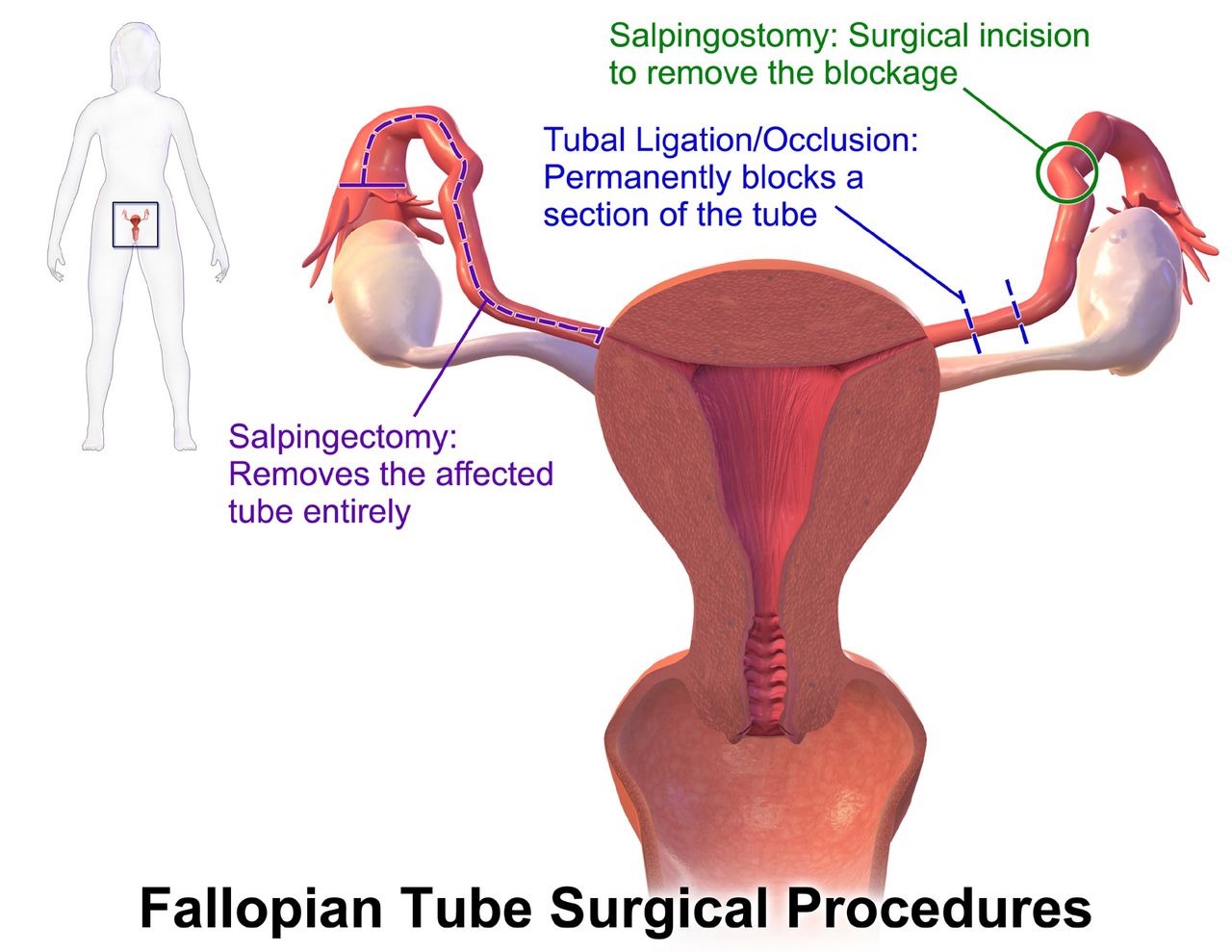
Gametogenesis and the chromosomal arithmetic of a new person
Human somatic cells carry forty-six chromosomes arranged in twenty-three pairs: twenty-two pairs of autosomes and one pair of sex chromosomes. Gametes cannot carry forty-six, or every generation would double the count, so germ cells undergo meiosis — two divisions that halve the chromosome number to twenty-three and, along the way, shuffle the parental chromosomes through crossing over. Fertilisation restores the diploid number. That single sentence contains most of what goes wrong in clinical genetics: if meiosis distributes the chromosomes unevenly, the resulting gamete carries twenty-four or twenty-two, and the conceptus is trisomic or monosomic from the first cell onward.
Oogenesis and spermatogenesis are not symmetrical, and the difference matters at the bedside. Oogenesis begins before birth: a female fetus contains six to seven million primary oocytes at around twenty weeks of gestation, roughly one to two million at birth, and about three hundred thousand to four hundred thousand at puberty, of which only four hundred or so will ever be ovulated. Those primary oocytes sit arrested in prophase of the first meiotic division for decades, and the spindle apparatus that must eventually separate the chromosomes ages with the person. This is the accepted explanation for the steep rise in nondisjunction with maternal age — the risk of a liveborn infant with trisomy 21 is approximately 1 in 1250 at age twenty-five, 1 in 350 at thirty-five, 1 in 100 at forty and 1 in 30 at forty-five. Note how the language should be framed for families: the ovum did not fail, and the person carrying it did nothing wrong.
Spermatogenesis is continuous from puberty, with each cycle taking roughly sixty-four to seventy-four days — which is why a change in health, temperature, illness or medication shows up in semen parameters about three months later, and why preconception counselling for the sperm-producing partner needs at least that much lead time. Continuous division carries its own cost: advanced paternal age is associated with new (de novo) autosomal dominant mutations such as achondroplasia, Marfan syndrome and some craniosynostosis syndromes. The sperm also determines chromosomal sex, since the ovum always contributes an X, and the SRY gene on the short arm of the Y chromosome initiates testicular differentiation from about week seven.
Fertilisation, cleavage and implantation: the first ten days
Ovulation releases a secondary oocyte that remains capable of fertilisation for roughly twelve to twenty-four hours. Sperm, once in the female reproductive tract, survive up to five days, so the fertile window in a cycle is about six days — the five days before ovulation plus the day of ovulation itself. Fertilisation almost always occurs in the ampulla, the outer third of the fallopian tube. Sperm must first undergo capacitation, several hours of enzymatic change in the female tract that makes the acrosome reactive; the acrosomal enzymes then digest a path through the corona radiata and zona pellucida. The instant one sperm nucleus enters, the zona reaction hardens the outer coat and blocks polyspermy. The fused nuclei form the zygote, a complete forty-six chromosome cell with a unique genome.
Cleavage begins within about thirty hours: two cells, then four, then a solid sixteen-cell ball called the morula that enters the uterine cavity around day three to four. By day five, fluid has collected inside and the structure is a blastocyst with two distinct populations — the inner cell mass (embryoblast), which becomes the embryo and amnion, and the outer trophoblast, which becomes the chorion and placenta. The blastocyst hatches from the zona pellucida on day five or six and burrows into the secretory endometrium, usually in the upper posterior uterine wall, between days six and ten. Roughly a quarter of patients notice light implantation bleeding around this time and may mistake it for a scant period, which is one reason self-reported dating is unreliable.
The trophoblast differentiates into cytotrophoblast and syncytiotrophoblast, and the syncytiotrophoblast begins secreting human chorionic gonadotropin (hCG) almost immediately. hCG rescues the corpus luteum so that it keeps producing progesterone and the endometrium is not shed; it is detectable in maternal serum about eight to eleven days after conception and in urine a few days later. Levels roughly double every forty-eight to seventy-two hours in early normal pregnancy and peak at eight to ten weeks. By weeks eight to ten the placenta assumes progesterone production and the corpus luteum regresses — the so-called luteal-placental shift. When implantation occurs anywhere other than the uterine cavity, most often in a fallopian tube, the pregnancy is ectopic; this is covered in the high-risk chapter, but recognise here that the biology of implantation is what makes it possible.
Twinning: the timing of division decides the risk
Dizygotic (fraternal) twins arise from two ova fertilised by two sperm. They are genetically ordinary siblings who happen to share a uterus, they may be of different sexes, and they always have two chorions and two amnions. Incidence rises with maternal age, with a family history on the maternal side, and markedly with ovulation induction and assisted reproduction. Monozygotic (identical) twins arise from a single zygote that splits, occur at a fairly constant rate worldwide of about 1 in 250 births, and are genetically identical apart from later somatic mutations and differences in gene expression.
For monozygotic twins the clinically decisive variable is when the split occurred, because that determines how many membranes and how many placentas the twins share. Shared circulation is the danger. Monochorionic twins are at risk of twin-to-twin transfusion syndrome, in which vascular anastomoses in the shared placenta shunt blood from one fetus to the other, leaving a plethoric, polyhydramniotic recipient and an anaemic, oligohydramniotic donor. Monoamniotic twins add the risk of cord entanglement, and are typically monitored intensively and delivered by caesarean before term.
| Day of division (post-fertilisation) | Result | Placentas / sacs | Clinical significance |
|---|---|---|---|
| Days 1–3 (before morula) | Dichorionic diamniotic | 2 placentas, 2 sacs | Lowest risk of the monozygotic group |
| Days 4–8 (blastocyst) | Monochorionic diamniotic | 1 placenta, 2 sacs | Most common; risk of twin-to-twin transfusion |
| Days 8–13 (after amnion forms) | Monochorionic monoamniotic | 1 placenta, 1 sac | Cord entanglement; high fetal loss; planned caesarean |
| After day 13 | Incomplete division | 1 placenta, 1 sac | Conjoined twins |
Fertilisation occurs in the ampulla within twenty-four hours of ovulation, the blastocyst implants on days six to ten while secreting hCG to sustain the corpus luteum, and for monozygotic twins the day of division determines chorionicity and therefore risk.
Section 2The Embryo, the Fetus and the Windows of Vulnerability
The embryonic period: weeks three to eight, when everything is built
Development divides into three stages with very different vulnerabilities. The pre-embryonic period covers the first two weeks after fertilisation, when the conceptus is a dividing ball of undifferentiated cells. Injury here tends to follow an all-or-none pattern: either enough cells are destroyed that the pregnancy is lost, often before it is recognised, or the remaining totipotent cells replace the losses and development proceeds normally. Structural malformation from a first-fortnight exposure is uncommon, which is a genuinely reassuring thing to be able to tell a patient who drank alcohol before she knew she was pregnant — paired, always, with counselling to stop now.
The embryonic period runs from week three through week eight post-conception, and it is the period of organogenesis. In these six weeks the flat embryonic disc folds into a body, the neural tube closes, the heart begins to beat, limbs bud and rotate, the face and palate fuse, and every major organ system is at least sketched. This is why the embryonic period is the window of maximum teratogenic sensitivity: a structure can only be malformed while it is being formed. The neural tube closes by about day twenty-six to twenty-eight post-conception, that is, roughly six weeks gestational age — frequently before a person has missed a second period or booked a first prenatal visit. The heart begins contracting around day twenty-one to twenty-two and cardiac septation is complete by about day forty. The palate fuses between roughly days forty-two and fifty-five, which is why cleft palate can result from an exposure that occurs weeks after the heart and neural tube are already safe.
Each structure therefore has its own critical period, and the same drug taken at two different gestational ages produces two different outcomes. Angiotensin-converting enzyme inhibitors, for example, are relatively less damaging in the first trimester but cause fetal renal failure, oligohydramnios and skull hypoplasia when taken in the second and third trimesters, because the injury is to a functioning kidney rather than to a forming one. When a patient asks whether an exposure has harmed the pregnancy, the two questions that determine the answer are what and exactly when — and a teratogen information service, not an internet search, should supply the risk estimate.
The fetal period: growth, maturation and the edge of viability
From week nine post-conception until birth, the fetus grows and its organs mature functionally. Nothing new is built, but a great deal becomes capable of working. Kidneys produce urine from about week nine to twelve, and fetal urine becomes the dominant source of amniotic fluid after roughly sixteen weeks — which is why renal agenesis or urinary obstruction produces severe oligohydramnios. External genitalia are distinguishable on ultrasound by around twelve to fourteen weeks. Fetal movement is usually first felt (quickening) at sixteen to eighteen weeks by someone who has been pregnant before, and eighteen to twenty weeks in a first pregnancy, because experience is what makes the sensation recognisable.
Lung maturation dominates the third trimester and dominates neonatal outcome. Type II pneumocytes begin producing surfactant at around twenty-four weeks, but production is scanty and unstable until roughly thirty-two to thirty-four weeks and functionally mature at about thirty-five weeks, when the lecithin-to-sphingomyelin ratio reaches 2:1. Periviability begins around twenty-two to twenty-five weeks and survival improves week by week within that band; antenatal corticosteroids (betamethasone 12 mg intramuscularly, two doses twenty-four hours apart, or dexamethasone 6 mg every twelve hours for four doses) accelerate surfactant production and reduce respiratory distress syndrome, intraventricular haemorrhage and necrotising enterocolitis. Brown fat, the tissue the newborn will later draw on for non-shivering thermogenesis, is deposited mostly in these last weeks, which is another reason the preterm infant cannot hold a temperature.
| Gestational age | Approximate size | Landmark developments |
|---|---|---|
| 6 weeks (4 post-conception) | 4–5 mm | Neural tube closing; heart tube begins to beat; limb buds appear |
| 10 weeks (8 post-conception) | 3 cm, 2 g | All organ systems present; recognisably human; embryonic period ends |
| 12 weeks | 8 cm, 45 g | Fetal heart audible by Doppler; kidneys produce urine; sex distinguishable |
| 16 weeks | 14 cm, 200 g | Lanugo appears; meconium forms; quickening may be felt (multipara) |
| 20 weeks | 25 cm, 300–450 g | Vernix caseosa; fetal heart audible by fetoscope; anatomy survey performed |
| 24 weeks | 30 cm, 600–800 g | Surfactant production begins; alveolar ducts form; edge of viability |
| 28 weeks | 35–38 cm, 1100–1300 g | Eyes open; strong respiratory effort possible with support; testes descending |
| 32 weeks | 40 cm, 1800–2000 g | Bone fully developed; more subcutaneous fat; better temperature control |
| 36 weeks | 45 cm, 2500–2750 g | Lanugo disappearing; sole creases forming; lecithin/sphingomyelin approaching 2:1 |
| 40 weeks | 48–53 cm, 3000–3600 g | Full term at 39 0/7 to 40 6/7; vernix in creases only; ready for extrauterine life |
Teratogens, TORCH infections and the conversation that prevents harm
A teratogen is any agent — drug, infection, chemical, radiation, maternal metabolic state — that disturbs development and produces a permanent structural or functional abnormality. Teratogenic effect depends on the agent, the dose, the timing relative to organogenesis and the genetic susceptibility of both fetus and pregnant person. The old letter categories (A, B, C, D, X) were retired in favour of the Pregnancy and Lactation Labeling Rule in 2015, which replaced a falsely precise letter with a narrative risk summary; the letters still circulate in older references and in student memory, so recognise them but do not rely on them.
Alcohol deserves separate emphasis because it is the most common preventable cause of intellectual disability in the developed world, and because fetal alcohol spectrum disorder includes central nervous system effects that occur across all three trimesters, not only during organogenesis. No safe amount, no safe trimester and no safe type of alcohol has ever been established. The counselling stance is non-judgemental and forward-facing: what is drunk today is what matters now, screening with a validated tool is routine for everyone rather than targeted at people who look like they might drink, and referral is offered rather than imposed.
The TORCH infections — toxoplasmosis, other (syphilis, varicella, parvovirus B19, Zika, hepatitis, HIV), rubella, cytomegalovirus and herpes simplex — cross the placenta and cause a shared pattern of microcephaly, intracranial calcification, hearing loss, chorioretinitis, hepatosplenomegaly and growth restriction. Cytomegalovirus is the most common congenital infection and the leading infectious cause of congenital sensorineural hearing loss; it is spread mainly by the saliva and urine of young children, so hand hygiene teaching for pregnant people who work in childcare or already have toddlers is genuinely preventive nursing. Rubella vaccine is a live vaccine and cannot be given during pregnancy; a non-immune patient is vaccinated postpartum, before discharge, and advised to avoid pregnancy for four weeks.
| Agent | Characteristic effect | Highest-risk window | Nursing action |
|---|---|---|---|
| Alcohol | Fetal alcohol spectrum disorder: growth restriction, smooth philtrum, thin upper lip, short palpebral fissures, lifelong cognitive and behavioural effects | All trimesters | Universal screening; non-blaming counselling; stopping at any point helps |
| Isotretinoin | Craniofacial, cardiac, thymic and CNS malformations; high loss rate | Weeks 3–8 | Pregnancy prevention programme; two contraceptive methods; negative tests before, during and after |
| Warfarin | Nasal hypoplasia, stippled epiphyses, CNS anomalies, fetal bleeding | Weeks 6–12 and third trimester | Transition to low-molecular-weight heparin, which does not cross the placenta |
| ACE inhibitors and ARBs | Renal failure, oligohydramnios, skull hypoplasia, limb contracture | Second and third trimesters | Discontinue and substitute a pregnancy-compatible antihypertensive |
| Valproate / carbamazepine | Neural tube defects (1–2 percent with valproate), craniofacial anomalies, reduced IQ | Weeks 3–4 for the neural tube | Preconception neurology review; high-dose folic acid; never stop an antiepileptic abruptly |
| Lithium | Ebstein anomaly of the tricuspid valve | First trimester | Risk-benefit review with psychiatry; fetal echocardiography |
| Tetracyclines | Permanent tooth discoloration, enamel hypoplasia | After 18 weeks | Substitute a safe antibiotic class |
| Methotrexate | Craniofacial and limb anomalies; pregnancy loss | Weeks 6–8 | Contraception counselling before prescribing |
| Uncontrolled maternal diabetes | Caudal regression, cardiac and neural tube defects; macrosomia later | Weeks 3–8 | Preconception glycaemic control; target HbA1c below 6.5 percent where achievable |
| Cytomegalovirus | Microcephaly, sensorineural hearing loss, chorioretinitis | Any, worst in first half | Hand hygiene after nappy changes and toddler saliva contact |
| Toxoplasma gondii | Hydrocephalus, intracranial calcification, chorioretinitis | Any; severity worst early | Avoid cat litter, undercooked meat, unwashed produce |
| Parvovirus B19 | Severe fetal anaemia and hydrops fetalis | First half of pregnancy | Serology after exposure; serial Doppler for anaemia |
Organs are built in weeks three to eight post-conception, so that is when structural teratogens do their damage; the fetal period grows and matures what already exists, and the timing of an exposure predicts the injury more reliably than the agent alone.
Section 3The Placenta, Membranes, Cord and Fetal Circulation
The placenta as endocrine organ and imperfect filter
The placenta forms where the chorion frondosum meets the decidua basalis; it is structurally established by the end of week twelve and continues to enlarge until term, when it weighs roughly 500 grams — about one-sixth of the fetal weight — and contains fifteen to twenty cotyledons on the dull, red maternal surface. The fetal surface is smooth and glistening with radiating vessels under the amnion. At term the placenta receives roughly 500 to 750 mL of maternal blood per minute. Maternal and fetal blood do not mix; they are separated by the placental membrane, across which exchange occurs by four mechanisms. Simple diffusion moves oxygen, carbon dioxide, water, electrolytes and most drugs; facilitated diffusion moves glucose; active transport moves amino acids, calcium, iron and water-soluble vitamins against a gradient; and pinocytosis moves large molecules including immunoglobulin G.
That last mechanism has a clinical corollary worth memorising. Maternal IgG crosses the placenta, mainly in the third trimester, and gives the newborn passive immunity for the first months of life — which is why the preterm infant, who missed that transfer, is more susceptible to infection, and why maternal pertussis and influenza vaccination protects the newborn. Maternal IgM does not cross. Therefore elevated IgM in a newborn was made by the newborn and indicates intrauterine infection. The placenta is often described to patients as a barrier; it is more honestly described as a selective filter that alcohol, nicotine, most medications and many viruses cross freely.
The placenta is also the largest endocrine organ the body ever produces. hCG maintains the corpus luteum and is the basis of pregnancy testing. Progesterone maintains the endometrium and keeps the myometrium quiescent. Oestrogen drives uterine growth, breast ductal development and increased uteroplacental blood flow. Human placental lactogen (hPL) is an insulin antagonist that deliberately raises maternal blood glucose so that more glucose is available to the fetus — the physiological basis of gestational diabetes, and the reason screening is timed at twenty-four to twenty-eight weeks when hPL is rising steeply. Relaxin softens the cervix and pelvic ligaments, and also explains the altered balance and joint discomfort patients report in later pregnancy.
Amniotic fluid, membranes and the cord
Amniotic fluid is derived early from maternal plasma and later, after about sixteen weeks, predominantly from fetal urine; it is cleared chiefly by fetal swallowing and by absorption across the membranes. Volume rises from about 30 mL at ten weeks to 800 to 1000 mL at thirty-six to thirty-eight weeks, then declines. It cushions the fetus from trauma, maintains a stable temperature, prevents adhesion and permits symmetrical growth, allows the free movement that develops the musculoskeletal system, prevents cord compression and — critically — is required for lung development, because the fetus breathes fluid in and out to expand the developing airways.
Volume is measured either as the amniotic fluid index (the sum of the deepest vertical pocket in four quadrants, normal 5 to 24 cm) or as the single deepest pocket (normal 2 to 8 cm). Oligohydramnios suggests reduced fetal urine output or fluid loss — renal agenesis, posterior urethral valves, ruptured membranes, placental insufficiency with growth restriction, post-term pregnancy, or exposure to an ACE inhibitor — and predisposes to cord compression, pulmonary hypoplasia and limb deformity. Polyhydramnios suggests the fetus cannot swallow or is producing excess urine: oesophageal atresia, tracheo-oesophageal fistula, anencephaly, poorly controlled maternal diabetes, multiple gestation or fetal anaemia. Green or greenish-brown fluid is meconium-stained and must be reported, since it signals a fetus that has passed stool in utero and raises the possibility of meconium aspiration.
The umbilical cord contains two arteries and one vein wrapped in Wharton jelly, which resists compression and kinking. Remember the direction, because it is counterintuitive: the single vein carries oxygenated blood to the fetus and the two arteries carry deoxygenated blood away to the placenta. Average cord length is about 55 cm, with a normal range of roughly 30 to 90 cm. A two-vessel cord (single umbilical artery) occurs in about one percent of births and is associated with renal and cardiac anomalies, so its identification obliges a careful newborn assessment and often a renal ultrasound. Nuchal cord — the cord looped around the neck — is found in about a quarter of births and is usually managed simply by slipping it over the head at delivery.
Fetal circulation and the switch that happens at the first breath
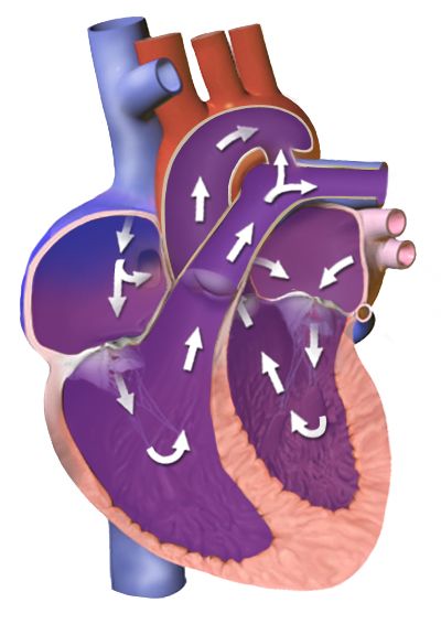
In utero the lungs are fluid-filled and pulmonary vascular resistance is high, while the placenta provides a low-resistance circuit; three shunts exploit this arrangement. The ductus venosus carries the well-oxygenated umbilical vein blood past the liver directly into the inferior vena cava. The foramen ovale lets that blood cross from right atrium to left atrium, so the best-oxygenated blood goes preferentially to the coronary arteries and brain. The ductus arteriosus shunts blood from the pulmonary artery into the descending aorta, bypassing the lungs. Fetal oxygen saturation is highest in the umbilical vein at roughly eighty percent, and the fetus compensates for the modest saturation with fetal haemoglobin, which has a higher affinity for oxygen, and with a haemoglobin concentration around 15 to 20 g/dL.
At birth the sequence reverses in seconds. The first breaths expand the alveoli and drop pulmonary vascular resistance; clamping the cord removes the low-resistance placenta and raises systemic vascular resistance. Left atrial pressure now exceeds right atrial pressure and the foramen ovale closes functionally, becoming the fossa ovalis over the following months. Rising arterial oxygen tension and falling prostaglandin levels constrict the ductus arteriosus, usually within twelve to twenty-four hours and almost always by seventy-two hours, leaving the ligamentum arteriosum. The ductus venosus closes within days to become the ligamentum venosum. This physiology is directly actionable: an infant with a ductal-dependent cardiac lesion deteriorates as the duct closes, and prostaglandin E1 is infused at 0.05 to 0.1 micrograms per kilogram per minute to keep it open — for a 3 kg newborn that is 3 multiplied by 0.05, or 0.15 micrograms per minute at the starting rate. Apnoea is a common and expected adverse effect, so airway equipment stays at the bedside.
| Structure | Function before birth | Closure | Becomes |
|---|---|---|---|
| Ductus venosus | Umbilical vein blood bypasses the liver | Days after cord clamping | Ligamentum venosum |
| Foramen ovale | Right atrium to left atrium, bypassing the lungs | Functional at first breaths; anatomic over months | Fossa ovalis |
| Ductus arteriosus | Pulmonary artery to aorta, bypassing the lungs | Functional 12–24 hours, usually complete by 72 hours | Ligamentum arteriosum |
| Umbilical vein | Carries oxygenated blood to the fetus | Obliterates after birth | Ligamentum teres (round ligament of the liver) |
| Umbilical arteries | Carry deoxygenated blood to the placenta | Obliterate after birth | Medial umbilical ligaments |
When the cord or placenta fails: sequences that save the life
Umbilical cord prolapse occurs when the cord slips below the presenting part, most often after spontaneous or artificial rupture of membranes with an unengaged presenting part, in malpresentation, prematurity, polyhydramnios or multiple gestation. The cord is then compressed between the presenting part and the pelvis, and fetal oxygenation stops. The presentation is a sudden prolonged deceleration or bradycardia immediately after membranes rupture, sometimes with a palpable or visible cord. The instinctive action — to push the cord back in — is exactly wrong, because handling the cord causes vasospasm and worsens the very hypoxia you are trying to relieve.
Vasa previa is the related catastrophe: unprotected fetal vessels crossing the internal os rupture when membranes rupture, producing painless vaginal bleeding with immediate fetal bradycardia or a sinusoidal fetal heart rate pattern. The blood lost is fetal blood, and a fetus has only about 250 mL in total, so exsanguination is measured in minutes. This is an immediate caesarean birth. Uterine rupture presents with a sudden tearing abdominal pain, loss of the fetal station on examination, an abnormal fetal heart rate pattern, cessation of contractions and signs of maternal hypovolaemia; it demands immediate surgical delivery and volume resuscitation.
When the placenta separates and the uterus does not contract down onto the open spiral arteries, the result is early postpartum haemorrhage — defined as cumulative blood loss of 1000 mL or more, or any loss accompanied by signs of hypovolaemia, within twenty-four hours of birth. Uterine atony causes seventy to eighty percent of cases, so the first action is to massage the fundus, not to start a drug and not to call the provider. Massage while a second nurse establishes access and empties the bladder, since a full bladder displaces the uterus and prevents contraction. Only then do the uterotonics follow: oxytocin 10 to 40 units in 500 to 1000 mL of crystalloid intravenously or 10 units intramuscularly; methylergonovine 0.2 mg intramuscularly, contraindicated in hypertension; carboprost 250 micrograms intramuscularly, avoided in asthma; misoprostol 600 to 1000 micrograms rectally or sublingually; and tranexamic acid 1 g intravenously, which works best given within three hours of birth. Quantify the loss by weighing, do not estimate it by eye.
The placenta is a selective filter and the largest endocrine organ of pregnancy, three shunts close within hours to days of the first breath, and in cord prolapse, magnesium toxicity and postpartum haemorrhage the first action — lift the presenting part, stop the infusion, massage the fundus — is what changes the outcome.
Section 4Patterns of Inheritance and Chromosomal Variation

Mendelian patterns: dominant, recessive and X-linked
In autosomal dominant inheritance a single altered copy of a gene is enough to produce the condition. An affected person has a fifty percent chance of passing the altered allele to each child, independent of sex, and the condition typically appears in every generation of a pedigree. Two features complicate the picture. Penetrance describes what proportion of people carrying the allele show any signs at all, and expressivity describes how severely they show them — which is why one family member with neurofibromatosis type 1 has a few café-au-lait macules and a relative has plexiform neurofibromas. Familiar autosomal dominant conditions include Huntington disease, Marfan syndrome, achondroplasia, neurofibromatosis type 1, familial hypercholesterolaemia, autosomal dominant polycystic kidney disease and hereditary breast and ovarian cancer syndrome caused by pathogenic BRCA1 or BRCA2 variants.
In autosomal recessive inheritance two altered copies are required, one from each parent. Carriers have one copy and are clinically unaffected, so the condition appears to skip generations and often surprises a family with no known history. When both parents are carriers, each pregnancy carries a twenty-five percent chance of an affected child, a fifty percent chance of an unaffected carrier and a twenty-five percent chance of a child with two typical copies — so two-thirds of the unaffected children are carriers. Consanguinity raises the risk because relatives share alleles. Carrier frequencies differ by ancestry: cystic fibrosis carriers are roughly 1 in 25 among people of Northern European descent, sickle cell trait affects roughly 1 in 12 Black Americans, and Tay-Sachs carriers are about 1 in 30 in Ashkenazi Jewish and certain French-Canadian and Cajun populations. Ancestry-based figures guide screening offers but must never be used to assume a person’s ancestry or to decline screening a family requests.
X-linked recessive inheritance is the pattern students most often get backwards, so work it from the chromosomes. A carrier mother has one altered X and one typical X. Each son receives one of her X chromosomes, so fifty percent of sons are affected; each daughter receives one of her X chromosomes plus a typical X from her father, so fifty percent of daughters are carriers and essentially none are affected. An affected father passes his X to every daughter, so all his daughters are carriers, and passes his Y to every son, so no sons are affected — there is no father-to-son transmission of an X-linked condition, and finding it in a pedigree rules the pattern out. Haemophilia A and B, Duchenne muscular dystrophy, glucose-6-phosphate dehydrogenase deficiency and red-green colour vision deficiency follow this pattern.
| Pattern | Risk per pregnancy | Pedigree clue | Examples |
|---|---|---|---|
| Autosomal dominant | 50 percent of children affected if one parent affected | Present in every generation; male and female equally | Huntington disease, Marfan syndrome, achondroplasia, BRCA1/2 |
| Autosomal recessive | 25 percent affected, 50 percent carrier when both parents carry | Skips generations; consanguinity increases risk | Cystic fibrosis, sickle cell disease, phenylketonuria, Tay-Sachs |
| X-linked recessive | Carrier mother: 50 percent of sons affected, 50 percent of daughters carriers | Males affected; no father-to-son transmission | Haemophilia A, Duchenne muscular dystrophy, G6PD deficiency |
| X-linked dominant | Affected father: all daughters affected, no sons | Rare; may be lethal in males | Rett syndrome, X-linked hypophosphataemic rickets |
| Multifactorial | 2–5 percent recurrence after one affected child | Clusters in families without a clean pattern | Neural tube defects, cleft lip and palate, pyloric stenosis, clubfoot |
| Mitochondrial | Affected mother: all children at risk; affected father: none | Transmitted only through the maternal line | MELAS, Leber hereditary optic neuropathy |
Numerical and structural chromosome changes
Nondisjunction — failure of a chromosome pair to separate during meiosis — produces a gamete with an extra or a missing chromosome, and therefore a trisomic or monosomic conceptus. Most autosomal trisomies are lethal early; three are compatible with live birth. Trisomy 21 (Down syndrome) occurs in roughly 1 in 700 live births and results from nondisjunction in about ninety-five percent of cases, from an unbalanced Robertsonian translocation in three to four percent and from mosaicism in one to two percent. The clinical picture includes generalised hypotonia, upslanting palpebral fissures with epicanthal folds, a flat nasal bridge and small low-set ears, a single transverse palmar crease, brachycephaly, and a wide sandal gap between the first and second toes. Nursing priorities follow the associated conditions rather than the appearance: forty to fifty percent have a congenital heart defect, most often an atrioventricular septal defect, so every infant needs an echocardiogram; duodenal atresia presents with bilious vomiting in the first day; congenital hypothyroidism and later thyroid disease are common; there is an increased leukaemia risk; and atlantoaxial instability requires evaluation before contact sports and careful neck positioning during intubation or transfer.
Trisomy 18 (Edwards syndrome), roughly 1 in 6000 to 8000 births, presents with severe growth restriction, a prominent occiput, micrognathia, clenched hands with overlapping index and fifth fingers, rocker-bottom feet and major cardiac defects. Trisomy 13 (Patau syndrome), roughly 1 in 10,000 to 16,000, produces midline defects — holoprosencephaly, cleft lip and palate, microphthalmia — along with polydactyly and cardiac anomalies. Both carry high early mortality, and care is a shared decision with the family that may reasonably range from full intervention to comfort-focused care; the nurse’s role is to make sure the family understands the options, that the infant is treated as a person with a name, and that memory-making and palliative support are offered without being imposed.
Sex chromosome variations behave differently because a missing or extra sex chromosome is better tolerated. Turner syndrome (45,X), about 1 in 2500 female births, presents with short stature, a webbed neck, a low posterior hairline, lymphoedema of the hands and feet in the newborn, coarctation of the aorta or a bicuspid aortic valve, streak ovaries with primary amenorrhoea and infertility, and typically normal intelligence with a specific weakness in spatial and mathematical reasoning; growth hormone and later oestrogen replacement are standard. Klinefelter syndrome (47,XXY), about 1 in 500 to 1000 male births, is often not recognised until adolescence or an infertility evaluation: tall stature with long limbs, small firm testes, gynaecomastia, sparse body hair and language-based learning difficulties, treated with testosterone from puberty. Structural rearrangements complete the picture — deletions such as cri-du-chat (5p minus) and the 22q11.2 deletion (conotruncal cardiac defect, abnormal facies, thymic hypoplasia with immunodeficiency, cleft palate and hypocalcaemia), duplications, inversions, and balanced translocations in which the carrier is clinically well but has recurrent pregnancy loss.
Beyond Mendel: multifactorial, mitochondrial, imprinting and repeat expansion
Multifactorial conditions arise from several genes interacting with the environment and account for most common congenital anomalies: neural tube defects, cleft lip and palate, congenital heart disease, pyloric stenosis, clubfoot and developmental dysplasia of the hip. They cluster in families without following a clean pattern, and empiric recurrence risk after one affected child is generally two to five percent, rising with each additional affected relative and with the severity of the defect. This is exactly the situation in which folic acid supplementation, glycaemic control and avoidance of specific teratogens genuinely lower risk.
Mitochondrial inheritance follows the ovum. Mitochondria carry their own small genome and the sperm contributes essentially none, so an affected mother may pass the condition to all of her children while an affected father passes it to none — a pattern that instantly identifies mitochondrial disease in a pedigree. Because cells carry many mitochondria with a variable proportion of altered copies (heteroplasmy), severity varies widely even within one family, and tissues with the highest energy demand — brain, heart, skeletal muscle — are affected first.
Genomic imprinting means some genes are expressed only from the maternally inherited copy and others only from the paternal copy, so the parent of origin changes the phenotype. The classic pair involves the same region of chromosome 15: loss of the paternal contribution produces Prader-Willi syndrome, which presents in the newborn as profound hypotonia and poor feeding requiring gavage, then converts in early childhood to insatiable hyperphagia with obesity; loss of the maternal contribution produces Angelman syndrome, with severe intellectual disability, minimal speech, ataxic movements and a happy, excitable demeanour. Finally, trinucleotide repeat expansion explains conditions that worsen down the generations (anticipation): fragile X syndrome from CGG expansion in FMR1 is the most common inherited cause of intellectual disability, with normal alleles below 45 repeats, premutation at 55 to 200 and full mutation above 200; Huntington disease from CAG expansion; and myotonic dystrophy from CTG expansion. Premutation carriers are clinically important in their own right, being at risk of primary ovarian insufficiency and of a late-onset tremor-ataxia syndrome.
Dominant conditions carry a fifty percent risk per pregnancy, recessive conditions twenty-five percent when both parents carry, X-linked recessive conditions affect half of a carrier’s sons with no father-to-son transmission, and trisomies arise from nondisjunction — except when a parental translocation raises recurrence risk enough to change counselling.
Section 5Screening, Diagnosis and Family-Centred Genetic Nursing
Screening tests: what they can and cannot say
The single most consequential teaching point in prenatal genetics is the difference between a screen and a diagnosis. A screening test estimates probability in a healthy population and can never be positive or negative for a condition — it returns a risk. A diagnostic test examines fetal cells and answers the question. Patients routinely hear a high-risk screen as a diagnosis, make irreversible decisions on it, and are then devastated or falsely reassured. Every conversation about a screening result should include the sentence: this is a screen, and it must be confirmed by a diagnostic test before anything is decided.
The first-trimester combined screen is performed at 11 0/7 to 13 6/7 weeks and pairs an ultrasound measurement of nuchal translucency with maternal serum PAPP-A and free beta-hCG, detecting roughly eighty-five percent of trisomy 21. Cell-free DNA screening (non-invasive prenatal testing) analyses placental DNA fragments circulating in maternal blood from ten weeks onward; for trisomy 21 it detects over ninety-nine percent with a false-positive rate under one in a thousand, and it also reports fetal sex and fetal RhD status. Even so, its positive predictive value depends on how common the condition is in that population, so a positive cell-free DNA result in a young patient screening for a rare trisomy may still be more likely to be a false positive than a true one. Discordant results also arise from confined placental mosaicism, a vanished twin or a maternal chromosomal finding. Cell-free DNA does not screen for neural tube defects.
Second-trimester serum screening is performed at 15 to 20 weeks. Maternal serum alpha-fetoprotein is elevated in open neural tube defects, abdominal wall defects, multiple gestation and simple underestimation of gestational age — and the first step after a high result is always to confirm dates and fetal number by ultrasound, because wrong dating is the commonest explanation. The quadruple screen adds hCG, unconjugated oestriol and inhibin A; the trisomy 21 pattern is low AFP, low oestriol, high hCG and high inhibin A. The detailed anatomy ultrasound at 18 to 22 weeks then evaluates structure directly. Preconceptionally and in early pregnancy, expanded carrier screening panels test the pregnant person first and the partner if a variant is found; a three-generation family history remains the cheapest and most informative genetic tool available and takes about ten minutes to take.
Diagnostic testing and the nursing care around it
Chorionic villus sampling is performed at 10 0/7 to 13 6/7 weeks, transcervically or transabdominally under ultrasound guidance, and samples placental tissue for karyotype, microarray or targeted gene testing. Its advantage is early results; its limitations are that it cannot detect neural tube defects because there is no amniotic fluid to assay, that it carries a procedure-related loss risk of roughly one in five hundred in experienced hands, and that it must not be performed before ten weeks because of an association with limb reduction defects.
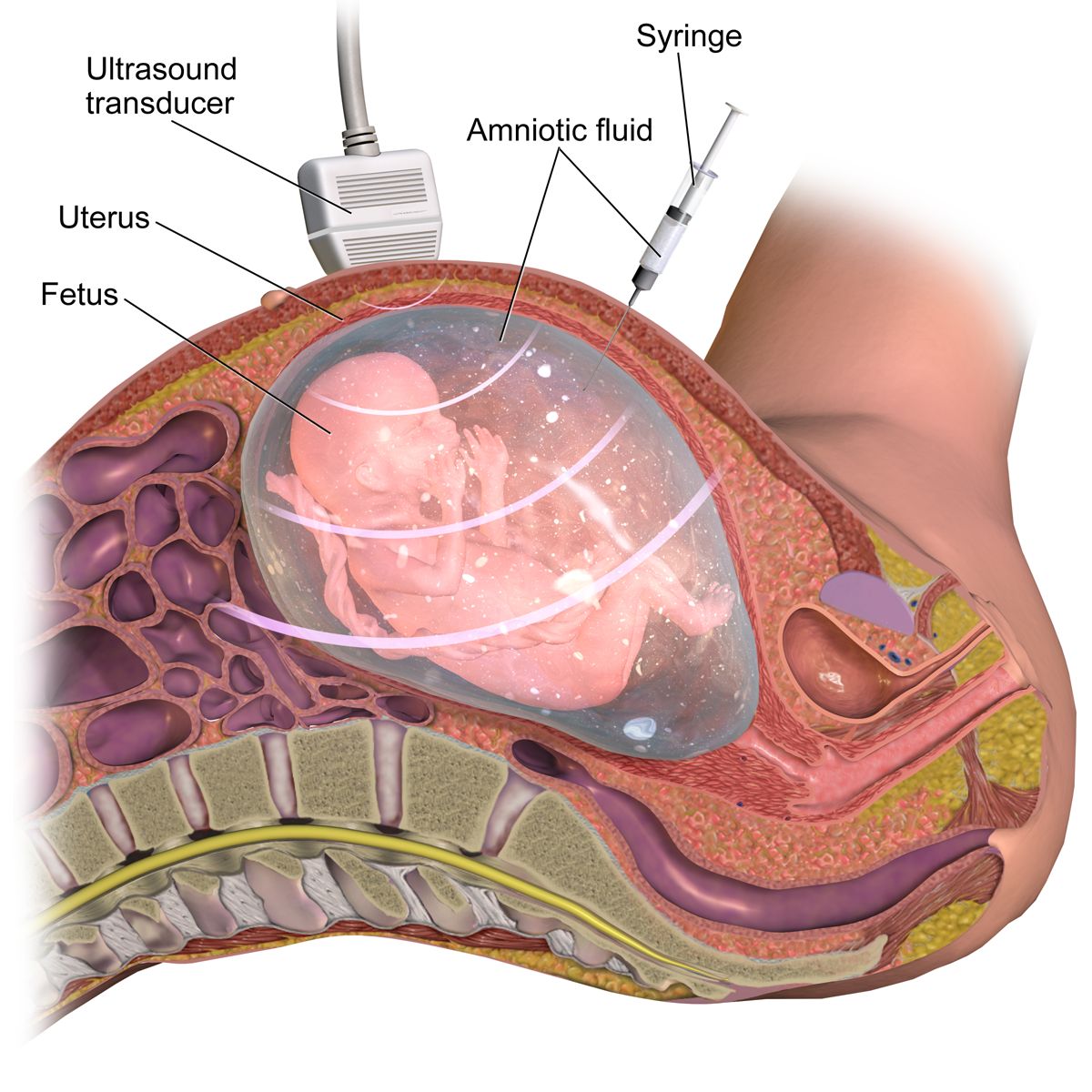
Amniocentesis for genetic indications is performed from 15 to 20 weeks, when there is enough fluid to sample safely, with a procedure-related loss risk of about one in five hundred to one in a thousand. Later in pregnancy the same procedure assesses fetal lung maturity or bilirubin in alloimmunisation, or decompresses polyhydramnios. Nursing care: confirm informed consent has been obtained by the provider and that the patient can explain in her own words what is being tested and what she will do with the result; obtain baseline vital signs and fetal heart rate; ask the patient to empty her bladder before a second- or third-trimester procedure so the bladder is not punctured; support positioning and, more importantly, support the patient, because this is frightening. Afterwards, monitor fetal heart rate and contractions for thirty to sixty minutes, and teach the patient to report leaking of fluid, vaginal bleeding, contractions, fever or decreased fetal movement. For any Rh-negative unsensitised patient, Rh immune globulin is indicated after an invasive procedure and after any potential sensitising event: 300 micrograms intramuscularly covers 30 mL of fetal whole blood and is given within seventy-two hours; a 50 microgram microdose is used for events before thirteen weeks.
| Test | Timing | Screen or diagnostic | What it detects / limits |
|---|---|---|---|
| Cell-free DNA (NIPT) | From 10 weeks | Screen | Trisomies 21, 18, 13, sex chromosomes, RhD; does not detect neural tube defects; must be confirmed |
| Nuchal translucency plus PAPP-A and free beta-hCG | 11 0/7 – 13 6/7 weeks | Screen | Approximately 85 percent detection for trisomy 21 |
| Chorionic villus sampling | 10 0/7 – 13 6/7 weeks | Diagnostic | Karyotype and microarray; no neural tube information; loss risk about 1 in 500 |
| Maternal serum AFP / quad screen | 15 – 20 weeks | Screen | High AFP: neural tube or abdominal wall defect, twins, wrong dates. Low AFP with high hCG and inhibin A: trisomy 21 |
| Amniocentesis | 15 – 20 weeks (genetic) | Diagnostic | Karyotype, microarray, AFP, infection, later lung maturity; loss risk about 1 in 500 to 1 in 1000 |
| Anatomy ultrasound | 18 – 22 weeks | Screen (structural) | Direct visualisation of structure, placenta, fluid volume and growth |
| Newborn blood spot screen | 24 – 48 hours of age | Screen | Metabolic, endocrine, haemoglobin and CF disorders; repeat if drawn before 24 hours |
The first hours after birth: newborn screening
Newborn screening is the largest and most successful public-health genetics programme in existence, and the nurse performs it. The heel-stick blood spot is collected after at least twenty-four hours of feeding, ideally at twenty-four to forty-eight hours of age, because several analytes require protein intake to become abnormal; a specimen drawn before twenty-four hours — increasingly common with early discharge — must be repeated. Technique matters: warm the heel, puncture the lateral or medial aspect of the plantar surface to avoid the calcaneus, allow a large drop to form, fill each printed circle completely from one side of the card only, and never layer successive drops or touch the paper, because layering invalidates the assay and a rejected card delays a diagnosis by days. Panels vary by jurisdiction but generally include phenylketonuria (treated with a phenylalanine-restricted formula, which prevents intellectual disability entirely if started in the first weeks), congenital hypothyroidism (levothyroxine begun within the first two weeks preserves cognition), cystic fibrosis, sickle cell disease, galactosaemia, medium-chain acyl-CoA dehydrogenase deficiency, congenital adrenal hyperplasia and biotinidase deficiency.
Two more screens are completed before discharge. Hearing is screened by otoacoustic emissions or automated auditory brainstem response, because early identification and amplification preserve language development. Critical congenital heart disease is screened by pulse oximetry at twenty-four hours of age or later, measured in the right hand (pre-ductal) and either foot (post-ductal): the infant passes if the saturation is ninety-five percent or higher in the hand or the foot and the difference between the two is three percent or less. A saturation below ninety percent in either limb is an immediate fail requiring urgent evaluation with no repeat; a reading of ninety to ninety-four percent in both limbs, or a difference greater than three percent, is repeated in one hour and becomes a fail if it persists on the third measurement. A fail leads to echocardiography, not to discharge.
Non-directive counselling, privacy, delegation and the family
Genetic counselling in nursing is non-directive. Your task is to make sure the family understands the information, not to steer them toward a decision, and this is harder than it sounds because families frequently and directly ask what you would do. A workable reply acknowledges the question rather than deflecting it: “I can tell you what the test can and cannot show, and what families in your situation usually think about. What matters is what fits your family.” Language carries judgement whether or not it is intended — prefer “the chance is one in four” to “the risk is twenty-five percent”, “a condition” or “a difference” to “an abnormality”, and person-first phrasing such as “an infant with Down syndrome” rather than “a Down baby”. Never describe a family as non-compliant; describe what was recommended, what happened and what barrier the family identified.
Genetic information is family information, which creates the privacy problem unique to this field: a result belongs to the patient, yet it may be medically important to relatives who have not consented to know. The patient decides what is shared. Patients also have a right not to know — a person at risk for Huntington disease may reasonably decline predictive testing, and that decision is respected without repeated offers. In the United States the Genetic Information Nondiscrimination Act of 2008 prohibits the use of genetic information in health insurance and employment decisions, but it does not extend to life, disability or long-term care insurance, and patients considering predictive testing deserve to be told that gap plainly before the blood is drawn.
Finally, delegation. The registered nurse retains assessment, teaching, evaluation and the care of any unstable patient, and in this chapter that means the RN takes the family history, explains what a screening result means, obtains and evaluates post-procedure fetal monitoring, and performs and interprets the newborn assessment. Assistive personnel may weigh and measure the infant, position and comfort the patient, gather supplies, transport specimens and take vital signs on a stable patient and report them — but interpreting a pulse oximetry screening result, teaching a family about recurrence risk, or evaluating a patient who is leaking fluid after an amniocentesis are never delegated. Licensed practical or vocational nurse scope varies by jurisdiction; know yours before you assign.
A screen returns a probability and a diagnostic test returns an answer, newborn screening only works if the blood spot is collected correctly after twenty-four hours of feeding, and genetic counselling stays non-directive with assessment, teaching and evaluation firmly held by the registered nurse.
The chapter in one breath
Meiosis halves forty-six chromosomes to twenty-three so that fertilisation in the ampulla can restore the diploid number, and from that moment the timetable governs everything. The blastocyst implants on days six to ten while secreting hCG to sustain the corpus luteum; weeks three to eight post-conception build every organ, which is why teratogens do structural damage then and why folic acid at 400 micrograms — or 4 mg after a previously affected pregnancy — must already be on board before the neural tube closes at day twenty-eight. The placenta then feeds, filters imperfectly and manufactures hCG, progesterone, oestrogen and human placental lactogen, while the ductus venosus, foramen ovale and ductus arteriosus divert blood past the liver and lungs until the first breath reverses the pressures and closes them. Inheritance explains recurrence: fifty percent for dominant, twenty-five percent for recessive with two carriers, half of a carrier’s sons for X-linked recessive, and a translocation karyotype that changes the counselling entirely. Screens estimate; only chorionic villus sampling and amniocentesis diagnose. And in the emergencies, sequence is survival — lift the presenting part off a prolapsed cord, stop the magnesium before reaching for calcium gluconate, and massage the fundus before any uterotonic.
Words worth knowing
| Term | What it means |
|---|---|
| Blastocyst | Fluid-filled stage reached about day five, with an inner cell mass that becomes the embryo and a trophoblast that becomes the placenta; the stage that implants. |
| Capacitation | Enzymatic conditioning of sperm within the female reproductive tract that must occur before the acrosome can release its enzymes and fertilisation can happen. |
| Cell-free DNA screening | Analysis of placental DNA fragments in maternal blood from ten weeks; highly sensitive for common trisomies but a screen, never a diagnosis. |
| Chorionicity | The number of chorions (and therefore placentas) in a multiple pregnancy; determined by when a monozygotic zygote divides and the strongest predictor of twin risk. |
| Ductus arteriosus | Fetal shunt from pulmonary artery to aorta that bypasses the lungs; closes functionally within 12 to 24 hours of birth to become the ligamentum arteriosum. |
| Foramen ovale | Fetal opening between the right and left atria; closes functionally at the first breaths when left atrial pressure exceeds right. |
| Gestational age | Age counted from the first day of the last menstrual period, approximately two weeks more than fertilisation age; the basis of all obstetric documentation. |
| Human placental lactogen | Placental hormone that antagonises insulin to divert glucose to the fetus; the physiological driver of gestational diabetes. |
| Imprinting | Parent-of-origin-specific gene expression; loss of the paternal chromosome 15 region causes Prader-Willi syndrome and loss of the maternal causes Angelman syndrome. |
| Nondisjunction | Failure of chromosomes to separate during meiosis, producing a gamete with an extra or missing chromosome; the mechanism of most trisomies. |
| Oligohydramnios | Amniotic fluid index below 5 cm or deepest pocket below 2 cm; suggests reduced fetal urine output or ruptured membranes and risks cord compression and pulmonary hypoplasia. |
| Organogenesis | Formation of organ systems during weeks three to eight post-conception; the window of maximum teratogenic vulnerability. |
| Penetrance | The proportion of people carrying a genetic variant who show any clinical sign of it; distinct from expressivity, which describes severity. |
| Polyhydramnios | Amniotic fluid index above 24 cm or deepest pocket above 8 cm; associated with impaired fetal swallowing, anencephaly, maternal diabetes and fetal anaemia. |
| Robertsonian translocation | Fusion of two acrocentric chromosomes; a balanced carrier parent is healthy but recurrence risk for translocation Down syndrome is far higher than the age-related risk. |
| Surfactant | Phospholipid produced by type II pneumocytes from about 24 weeks that lowers alveolar surface tension; functionally mature near 35 weeks at a lecithin/sphingomyelin ratio of 2:1. |
| Teratogen | Any agent that disturbs development and causes permanent structural or functional abnormality; effect depends on agent, dose, timing and genetic susceptibility. |
| Trinucleotide repeat expansion | Growth of a repeated three-base sequence across generations, causing anticipation; the mechanism of fragile X syndrome, Huntington disease and myotonic dystrophy. |
| Wharton jelly | Gelatinous connective tissue surrounding the umbilical vessels that resists compression and kinking of the cord. |