The Antepartum Period: Adaptation and Prenatal Care
Pregnancy is not a disease, and the antepartum period is not a waiting room. Across roughly forty weeks a healthy body remodels nearly every organ system: blood volume climbs by almost half, the kidneys filter faster than they ever will again, the diaphragm rises about four centimetres, and insulin resistance is deliberately manufactured so that glucose crosses the placenta to the fetus. Prenatal care exists because those adaptations look, on paper, exactly like pathology — the physiologic anaemia, the glycosuria, the dependent oedema, the soft systolic murmur. The nurse’s work is to know which changes are expected, which are early warnings, and which demand escalation within minutes. This chapter builds that discrimination: dating and history first, then adaptation system by system, then the schedule of visits and screens that turns adaptation into surveillance, then the teaching that keeps a family safe between visits, and finally the danger signs whose sequence of response saves a life.
By the end of this chapter you can
- Classify the presumptive, probable and positive signs of pregnancy and explain why only the positive signs confirm a fetus.
- Calculate an estimated date of birth using Naegele’s rule and record an obstetric history accurately in GTPAL format.
- Explain the mechanism behind at least eight maternal physiological adaptations and distinguish each expected finding from its pathological look-alike.
- Describe the standard prenatal visit schedule and state what is measured at every visit, including fundal height and fetal heart rate.
- Match each routine antepartum screen — cell-free DNA, the anatomy ultrasound, the glucose challenge, the group B streptococcus culture, Rh immune globulin — to its correct gestational window.
- Apply weight-gain, energy and micronutrient recommendations by pre-pregnancy body mass index, including the ten-fold difference between routine and high-risk folic acid dosing.
- Teach the antepartum danger signs and escalate an abnormal finding using SBAR, naming the correct first action for cord prolapse, magnesium toxicity, postpartum haemorrhage, shoulder dystocia and uterine rupture.
- Apply delegation limits, keeping assessment, teaching, evaluation and the unstable patient with the registered nurse.
Section 1Confirming, Dating and Beginning: The First Prenatal Visit
Presumptive, probable and positive signs
Pregnancy is confirmed in tiers, and the tiers exist because each lower tier has impostors. Presumptive signs are what the pregnant person notices: amenorrhoea, nausea and vomiting, breast tenderness and darkening of the areolae, urinary frequency, fatigue, and quickening — the first perceived fetal movement, typically at sixteen to twenty weeks in a first pregnancy and as early as fourteen to sixteen weeks in a person who has been pregnant before. Every one of these has another explanation. Amenorrhoea follows stress, thyroid disease, extreme exercise and perimenopause; nausea follows gastroenteritis; quickening can be intestinal gas. Presumptive signs are subjective, and they belong in the history, not in the diagnosis.
Probable signs are what the examiner detects. Goodell sign is softening of the cervix at about six weeks. Chadwick sign is the blue-violet discolouration of the cervix and vaginal wall from venous congestion, at six to eight weeks. Hegar sign is softening of the lower uterine segment, palpable at six to twelve weeks. Ballottement, a positive pregnancy test on urine or serum, uterine enlargement and Braxton Hicks contractions also sit here. A serum human chorionic gonadotrophin level is still only probable: hydatidiform mole and some germ-cell tumours secrete the same hormone. Early in a normal intrauterine pregnancy, human chorionic gonadotrophin roughly doubles every forty-eight to seventy-two hours, peaks around eight to ten weeks, then falls and plateaus — a doubling pattern that flattens is a reason to look for ectopic implantation or early pregnancy loss.
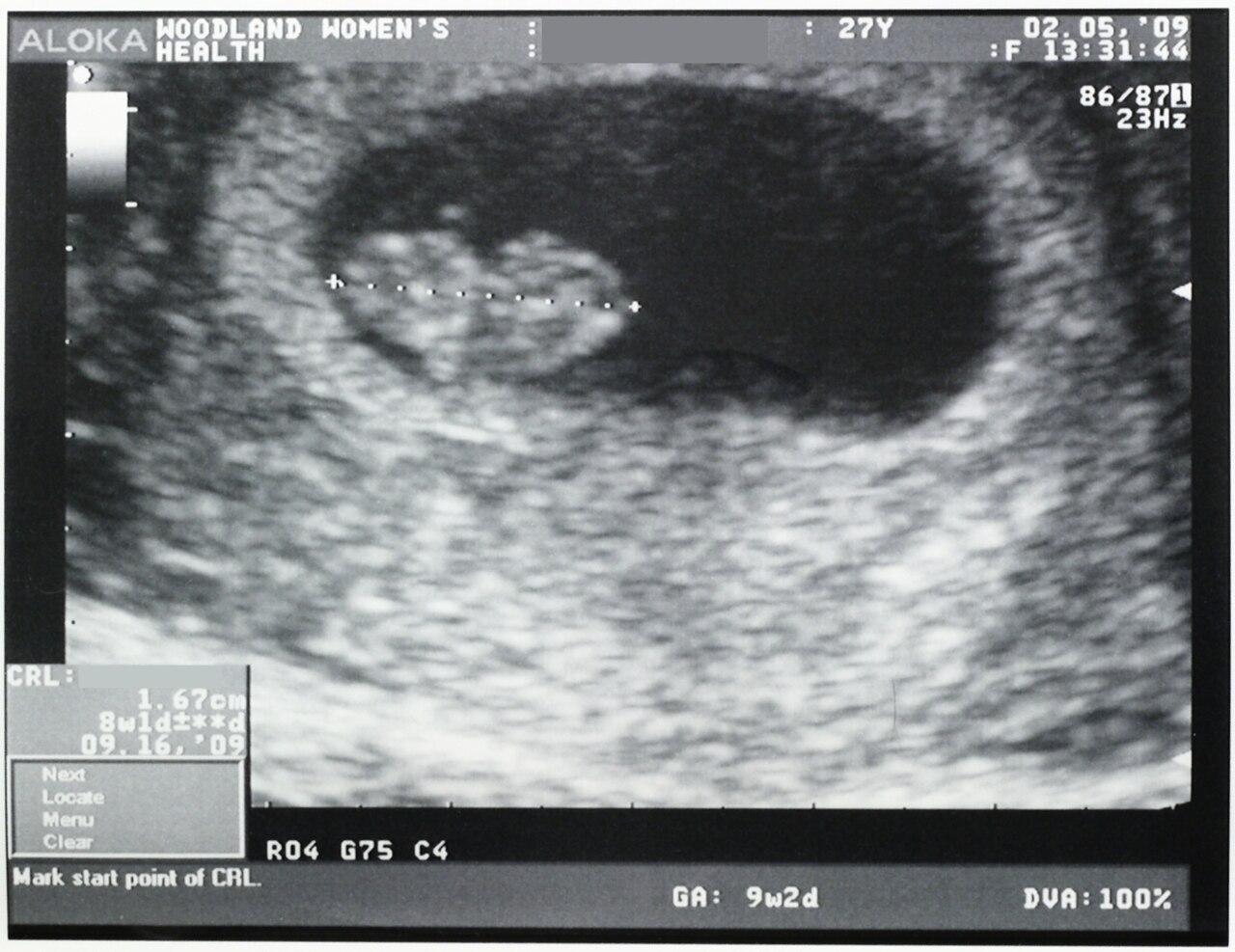
Only three findings are positive signs, and all three come directly from the fetus: visualisation of the fetus by ultrasound (a gestational sac from about five weeks by transvaginal scan, cardiac activity from about six weeks), auscultation of the fetal heart (Doppler at ten to twelve weeks, fetoscope at eighteen to twenty weeks), and fetal movement palpated by a trained examiner. Fetal heart rate baseline is 110 to 160 beats per minute. If what you hear matches the pregnant person’s radial pulse, you are listening to the uterine souffle, not the fetus — palpate the radial pulse while you auscultate, every time.
Dating, gravidity and parity
Dating drives everything downstream: when to screen, when to worry about growth, when a birth is preterm, when an induction is safe. Naegele’s rule estimates the date of birth from the first day of the last menstrual period: subtract three months, add seven days, add one year. A last menstrual period beginning 10 September 2025 gives 10 June 2025, then 17 June 2025, then an estimated date of birth of 17 June 2026 — the seven days are the step most often dropped. The rule assumes a regular twenty-eight-day cycle with ovulation on day fourteen, so it drifts in anyone with irregular or long cycles. A first-trimester crown-rump length by ultrasound is accurate to within five to seven days and supersedes menstrual dating when the two disagree by more than that margin; accuracy degrades to one to two weeks in the second trimester and two to three weeks in the third, which is why an early scan is worth more than a late one.
Obstetric history is recorded as GTPAL. Gravida is the total number of pregnancies including the current one, counting a multiple gestation once. Term births are those at thirty-seven weeks zero days and beyond; Preterm births are from twenty weeks zero days through thirty-six weeks six days; Abortions covers all losses, spontaneous or induced, before twenty weeks; Living is the number of children currently alive. A person pregnant now, with one birth at thirty-nine weeks, one set of twins born at thirty-four weeks, and one miscarriage at nine weeks, with all three children alive, is G4 T1 P1 A1 L3. Note that twins counted once in gravidity and once in parity still contribute two living children — parity counts births, not babies.
What the first visit actually contains
The first prenatal visit is the longest of the pregnancy and is ideally completed before ten weeks. It gathers a menstrual, obstetric, medical, surgical, medication, substance, genetic and psychosocial history; establishes dating; performs a full physical including a pelvic examination; and draws a baseline laboratory panel. That panel routinely includes blood type and Rh with antibody screen, complete blood count, rubella immunity, varicella immunity, hepatitis B surface antigen, syphilis serology, human immunodeficiency virus (opt-out testing), urinalysis with culture, and chlamydia and gonorrhoea screening. Hepatitis C screening is now recommended in every pregnancy. Tuberculosis, haemoglobinopathy and thyroid testing are added by risk.
Two of those results carry immediate action. Asymptomatic bacteriuria — a positive urine culture with no symptoms — is treated in pregnancy even though it would be left alone outside pregnancy, because the dilated, progesterone-relaxed ureters let bacteria ascend, and untreated bacteriuria progresses to pyelonephritis in a substantial minority and is associated with preterm birth and low birth weight. A negative rubella titre means the person is susceptible; the vaccine is a live virus and is contraindicated during pregnancy, so the intervention is to document susceptibility, counsel on avoiding exposure, and give the vaccine in the immediate postpartum period, ideally before hospital discharge.
Only ultrasound visualisation, auscultated fetal heart tones and examiner-palpated fetal movement confirm a pregnancy; everything else — including a positive hormone test — is presumptive or probable and must be dated by first-trimester ultrasound to be clinically useful.
Section 2Maternal Physiological Adaptation, System by System
Cardiovascular and haematological adaptation
Total blood volume rises forty to fifty per cent by about thirty-two weeks. The rise is disproportionate: plasma volume increases roughly fifty per cent while red cell mass increases only twenty to thirty per cent. Dilution follows, and the haemoglobin and haematocrit fall — the physiologic anaemia of pregnancy. This is a laboratory appearance, not a disease; oxygen-carrying capacity has actually increased in absolute terms. True anaemia is diagnosed at a haemoglobin below 11 g/dL in the first and third trimesters and below 10.5 g/dL in the second. The extra volume is not waste. It perfuses the placental bed, it dissipates the heat of fetal metabolism, and it is the reserve that lets a person lose 500 mL at a vaginal birth without becoming unstable.
Cardiac output rises thirty to fifty per cent, peaking around twenty-five to thirty weeks, driven early by stroke volume and later by heart rate, which climbs ten to twenty beats per minute above baseline. Systemic vascular resistance falls under the influence of progesterone, relaxin, nitric oxide and prostacyclin, so blood pressure — particularly diastolic — drops five to fifteen millimetres of mercury, reaching its nadir around twenty-four to twenty-eight weeks before returning toward baseline at term. A blood pressure that rises in the second trimester is therefore moving against the physiology and deserves attention. The heart is displaced upward and to the left, the point of maximal impulse moves laterally, and a soft systolic ejection murmur appears in most pregnancies; a diastolic murmur never becomes normal in pregnancy.
From about twenty weeks, lying supine allows the uterus to compress the inferior vena cava, cutting venous return and cardiac output. Supine hypotensive syndrome presents as dizziness, pallor, clamminess, nausea and hypotension, and it compromises uteroplacental perfusion before the pregnant person feels much at all. The intervention is immediate and mechanical: turn the patient to the left lateral position, or wedge the right hip so the uterus falls off the vessels. Pregnancy is also a hypercoagulable state — fibrinogen rises by about fifty per cent, clotting factors VII, VIII, IX and X rise, and fibrinolysis is suppressed — which protects against haemorrhage at birth and raises venous thromboembolism risk roughly four to five-fold. Unilateral calf pain and swelling in a pregnant patient is a thrombosis until proven otherwise, not a muscle strain.
Respiratory, renal and gastrointestinal adaptation
Progesterone acts directly on the respiratory centre, so tidal volume rises thirty to fifty per cent while respiratory rate barely changes. Minute ventilation rises with it and produces a compensated respiratory alkalosis: arterial carbon dioxide falls to about twenty-seven to thirty-two millimetres of mercury, and the kidney excretes bicarbonate to hold pH near 7.40 to 7.45. That gradient drives carbon dioxide from fetus to parent. Meanwhile the growing uterus elevates the diaphragm about four centimetres, the subcostal angle widens, functional residual capacity falls about twenty per cent, and oxygen consumption rises twenty to thirty per cent. The practical consequence is dangerous: a pregnant patient desaturates far faster during apnoea than a non-pregnant one. Physiologic dyspnoea of pregnancy is mild, gradual and does not disturb sleep or accompany chest pain; dyspnoea that is sudden, positional at night, or paired with chest pain, cough or tachycardia is not physiologic.
Renal plasma flow and glomerular filtration rate rise forty to fifty per cent by the end of the first trimester. Serum creatinine and blood urea nitrogen therefore fall: a normal pregnant creatinine is about 0.4 to 0.8 mg/dL, so a creatinine of 1.0 mg/dL — unremarkable in a non-pregnant adult — signals renal impairment in pregnancy. Progesterone-mediated smooth muscle relaxation and mechanical compression by the dextrorotated uterus dilate the collecting system, right side more than left, producing the physiologic hydronephrosis that makes ascending infection easy. Glycosuria is common because filtered glucose exceeds tubular reabsorption; trace glycosuria is expected, but persistent or heavy glycosuria still prompts glucose evaluation. Proteinuria is not in the same category: a dipstick of 1+ or more after twenty weeks must be measured properly and interpreted alongside blood pressure.
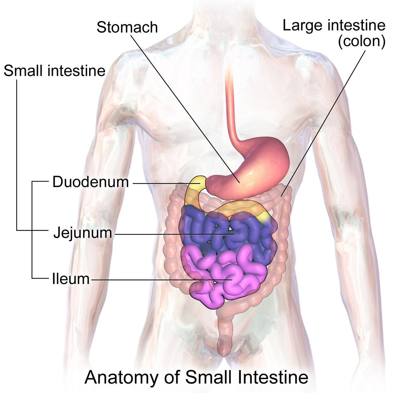
In the gastrointestinal tract, progesterone relaxes the lower oesophageal sphincter and slows transit. Reflux, constipation, bloating and haemorrhoids follow, and gallbladder stasis raises the risk of cholelithiasis. Nausea and vomiting affect up to seventy per cent of pregnancies and usually resolve by sixteen to twenty weeks; hyperemesis gravidarum is different in kind, defined by persistent vomiting with weight loss of more than five per cent of pre-pregnancy weight, dehydration, ketonuria and electrolyte disturbance, and it requires intravenous rehydration rather than dietary advice. Gum hypertrophy and bleeding (epulis gravidarum) are oestrogen effects; dental care is safe and should be encouraged, not deferred.
Endocrine, musculoskeletal and integumentary adaptation
The placenta is an endocrine organ. Human placental lactogen, along with placental growth hormone, progesterone and cortisol, is deliberately diabetogenic: it antagonises insulin so that maternal glucose stays available for placental transfer. Insulin requirements consequently fall in early pregnancy — a person with pre-existing type 1 diabetes is at real risk of hypoglycaemia in the first trimester — and then rise steeply in the second half. Thyroid-binding globulin rises under oestrogen so total thyroxine rises, while free thyroxine stays near normal; the thyroid enlarges modestly but a frankly nodular or markedly enlarged gland is not a normal finding. Human chorionic gonadotrophin has weak thyroid-stimulating activity, which is one reason severe early vomiting sometimes travels with transient biochemical hyperthyroidism.
Relaxin and progesterone loosen ligaments, including the sacroiliac joints and pubic symphysis. Combined with the forward shift of the centre of gravity, this produces the lordosis, waddling gait, low back pain and increased fall risk of late pregnancy, and it explains diastasis recti, the midline separation of the rectus abdominis. Fluid retention in confined spaces produces carpal tunnel symptoms and nasal stuffiness with epistaxis. Skin changes are melanocyte-stimulating: linea nigra down the midline, melasma (the “mask of pregnancy”) over the face, darkened areolae, and striae gravidarum. Palmar erythema and spider angiomas reflect the oestrogen-driven rise in cutaneous blood flow. Most of these fade postpartum, though striae persist as silvery lines and melasma improves with sun protection.
| System change | Expected finding | Not normal — investigate |
|---|---|---|
| Plasma volume up ~50 per cent, red cell mass up ~25 per cent | Haemoglobin 11–12 g/dL; mild dilutional fall | Haemoglobin below 11 g/dL (first or third trimester) or below 10.5 g/dL (second) |
| Systemic vascular resistance falls | Diastolic pressure drops 5–15 mm Hg, nadir 24–28 weeks | Blood pressure 140/90 mm Hg or higher on two readings four hours apart after 20 weeks |
| Cardiac output up 30–50 per cent; heart displaced left | Soft systolic ejection murmur; heart rate up 10–20 beats/min | Any diastolic murmur; resting heart rate above 110; chest pain |
| Tidal volume up 30–50 per cent; diaphragm rises 4 cm | Mild gradual dyspnoea; carbon dioxide 27–32 mm Hg | Sudden dyspnoea, orthopnoea, haemoptysis, oxygen saturation below 95 per cent |
| Glomerular filtration rate up 40–50 per cent | Creatinine 0.4–0.8 mg/dL; trace glycosuria | Creatinine 1.0 mg/dL or higher; proteinuria 1+ or more after 20 weeks |
| Ureteral dilation, urinary stasis | Urinary frequency without dysuria | Positive urine culture (treat even without symptoms); flank pain with fever |
| Hypercoagulability; fibrinogen up ~50 per cent | White cell count 5,000–15,000/mm³; dependent oedema of feet and ankles | Unilateral calf pain or swelling; sudden facial or hand oedema |
| Insulin antagonism by human placental lactogen | Rising insulin needs after 20 weeks | Fasting glucose 92 mg/dL or higher; abnormal glucose challenge at 24–28 weeks |
Nearly every antepartum adaptation has a pathological look-alike, so the assessment question is never “is this abnormal?” but “is this the expected magnitude, in the expected direction, at the expected gestational age?”
Section 3The Rhythm of Prenatal Care: Visits, Screening and Fetal Surveillance
The visit schedule and the four measurements taken every time
For an uncomplicated pregnancy the traditional schedule is every four weeks until twenty-eight weeks, every two weeks from twenty-eight to thirty-six weeks, and weekly from thirty-six weeks until birth — about twelve to fourteen visits. The spacing is not arbitrary: it tightens exactly when hypertensive disease, growth restriction and preterm labour become most likely. Reduced-visit and group prenatal care models are safe in low-risk pregnancies and often improve attendance and satisfaction; what matters is not the number of visits but that the surveillance items happen in their windows.
Four things are checked at essentially every return visit: blood pressure, weight, urine dipstick for protein and glucose, and fetal heart rate. From about eighteen or twenty weeks a fifth is added, fundal height: measured in centimetres from the upper border of the symphysis pubis over the curve of the abdomen to the top of the fundus, with the patient supine and the bladder empty. Landmarks worth memorising are the fundus at the symphysis at about twelve weeks and at the umbilicus at about twenty weeks. Between eighteen and thirty-six weeks the measurement in centimetres approximates the gestational age in weeks within about two centimetres. A discrepancy greater than two to three centimetres prompts an ultrasound: too small suggests growth restriction, oligohydramnios or a dating error; too large suggests multiple gestation, polyhydramnios, macrosomia, fibroids or, again, a dating error.
Leopold manoeuvres — four systematic palpations to determine fetal lie, presentation, position and engagement — are performed from about the third trimester, and they tell you where to place the Doppler. Fetal heart tones are best heard through the fetal back: in a cephalic presentation, below the umbilicus on the side where the back lies; in a breech presentation, at or above the umbilicus. Always count the pregnant person’s pulse simultaneously so that a maternal tachycardia is not mistaken for a fetal bradycardia.
Screening, trimester by trimester
Genetic screening is offered, not imposed, and the distinction between screening and diagnosis must be explicit in the teaching. Screening tests estimate risk; diagnostic tests give a karyotype. Cell-free DNA (non-invasive prenatal screening) analyses placental DNA fragments in maternal blood from ten weeks and has very high sensitivity for trisomy 21, but a “positive” result is still a probability and must be confirmed by chorionic villus sampling at ten to thirteen weeks or amniocentesis at fifteen to twenty weeks. A patient who hears “positive” and believes it is a diagnosis has been failed by the counselling, not by the test.
Maternal serum alpha-fetoprotein, drawn as part of the quad screen at fifteen to twenty-two weeks, is a useful mental model for how these markers behave. A high value suggests an open neural tube defect, abdominal wall defect, multiple gestation or, most commonly, inaccurate dating. A low value is associated with trisomy 21 and trisomy 18. The single most frequent cause of an abnormal alpha-fetoprotein is a wrong gestational age, which is one more argument for early dating.
| Window | Screen or intervention | What the nurse teaches or watches |
|---|---|---|
| First visit, ideally before 10 weeks | Blood type, Rh, antibody screen, complete blood count, rubella and varicella immunity, hepatitis B and C, syphilis, human immunodeficiency virus, urine culture, chlamydia and gonorrhoea | Treat asymptomatic bacteriuria; defer live vaccines to postpartum; opt-out human immunodeficiency virus testing |
| From 10 weeks | Cell-free DNA screening (optional) | Screening, not diagnosis; a positive result requires confirmatory testing |
| 10–13 weeks | Chorionic villus sampling (diagnostic, optional) | Rh-negative patients need Rh immune globulin after the procedure |
| 11–13 weeks 6 days | First-trimester combined screen: nuchal translucency plus serum markers | Requires precise dating; results are risk ratios |
| 12–28 weeks (start before 16 if possible) | Low-dose aspirin 81 mg daily for those at elevated pre-eclampsia risk | Continue daily until birth; explain it is prophylaxis, not treatment |
| 15–22 weeks | Quad screen; amniocentesis from 15 weeks if diagnostic testing is chosen | Post-amniocentesis: report leaking fluid, bleeding, fever, contractions |
| 18–22 weeks | Anatomy ultrasound | Full bladder may be requested; fetal sex disclosure is the family’s choice |
| 24–28 weeks | Gestational diabetes screening; repeat complete blood count and antibody screen | See the glucose criteria below |
| 28 weeks | Rh immune globulin 300 micrograms intramuscularly if Rh-negative and antibody-screen negative | Repeat within 72 hours of birth if the newborn is Rh-positive; also give after bleeding, trauma, amniocentesis, ectopic or loss |
| 27–36 weeks | Tdap in every pregnancy; maternal respiratory syncytial virus vaccine at 32–36 weeks in season | Tdap is repeated each pregnancy to transfer pertussis antibody to the newborn |
| 36 weeks 0 days – 37 weeks 6 days | Rectovaginal group B streptococcus culture | A positive culture means intrapartum intravenous antibiotics, not treatment now |
| Any trimester, seasonally | Inactivated influenza vaccine; COVID-19 vaccine per current guidance | Inactivated vaccines are safe in pregnancy; live attenuated influenza vaccine is not |
Gestational diabetes screening at twenty-four to twenty-eight weeks uses one of two approaches. In the two-step method, a non-fasting 50 g glucose challenge is followed by a plasma glucose at one hour; a value at or above 130 to 140 mg/dL (institutional threshold) leads to a fasting 100 g three-hour tolerance test, and gestational diabetes is diagnosed when two or more values are abnormal: fasting 95 mg/dL or higher, one hour 180 or higher, two hours 155 or higher, three hours 140 or higher. In the one-step method, a fasting 75 g two-hour test is diagnostic when any one value is abnormal: fasting 92 or higher, one hour 180 or higher, two hours 153 or higher. Patients with risk factors — prior gestational diabetes, obesity, a previous macrosomic newborn, polycystic ovary syndrome, strong family history — are screened at the first visit as well as at twenty-four to twenty-eight weeks.
Fetal surveillance in the third trimester
The simplest and cheapest surveillance tool belongs to the pregnant person. Fetal movement counting is taught from about twenty-eight weeks: lie on the left side after a meal at roughly the same time each day and count distinct movements; ten movements within two hours is reassuring. Teach the pattern, not just the number — a fetus has sleep cycles of twenty to forty minutes, so a quiet half-hour is not an alarm, but a definite decrease from that fetus’s own established pattern is a reason to be assessed the same day. Never advise a patient to “wait until tomorrow and see”.
The non-stress test records fetal heart rate and movement for twenty minutes. It is reactive at thirty-two weeks or more when there are two accelerations of at least fifteen beats per minute above baseline lasting at least fifteen seconds within twenty minutes; before thirty-two weeks the criterion is ten beats for ten seconds, because an immature autonomic nervous system produces smaller accelerations. A non-reactive test is most often a sleeping fetus, so the test is extended to forty minutes and vibroacoustic stimulation may be used before escalating. The biophysical profile adds ultrasound assessment of fetal breathing movements, gross body movement, tone and amniotic fluid volume to the non-stress test; each of the five components scores 0 or 2. A score of 8 to 10 is reassuring, 6 is equivocal and warrants repeat or delivery depending on gestation, and 4 or less indicates significant risk. Amniotic fluid volume is the component that reflects chronic placental function, because a hypoxic fetus shunts blood from the kidneys and makes less urine.
Every antepartum screen has a gestational window, and the window is the point: cell-free DNA from ten weeks, anatomy scan at eighteen to twenty-two, glucose at twenty-four to twenty-eight, Rh immune globulin at twenty-eight, Tdap at twenty-seven to thirty-six, and group B streptococcus culture at thirty-six weeks zero days to thirty-seven weeks six days.
Section 4Nutrition, Weight, Discomforts and Medication Safety
Weight gain and energy needs
Recommended gestational weight gain is set by pre-pregnancy body mass index, not by a single number, because the risks at either extreme differ. Too little gain is associated with growth restriction, preterm birth and low birth weight; too much is associated with macrosomia, caesarean birth, gestational diabetes, hypertensive disease and postpartum weight retention. Discuss weight in terms of fetal growth and health, never as a moral matter, and be aware that weight conversations are a common reason patients disengage from prenatal care.
| Pre-pregnancy body mass index | Total gain, singleton | Rate in 2nd and 3rd trimesters | Total gain, twins |
|---|---|---|---|
| Underweight, below 18.5 | 12.5–18 kg (28–40 lb) | ~0.5 kg (1 lb) per week | Individualised |
| Normal, 18.5–24.9 | 11.5–16 kg (25–35 lb) | ~0.4–0.5 kg (1 lb) per week | 17–25 kg (37–54 lb) |
| Overweight, 25.0–29.9 | 7–11.5 kg (15–25 lb) | ~0.3 kg (0.6 lb) per week | 14–23 kg (31–50 lb) |
| Obese, 30.0 or higher | 5–9 kg (11–20 lb) | ~0.2 kg (0.5 lb) per week | 11–19 kg (25–42 lb) |
Energy needs do not rise meaningfully in the first trimester. They rise by about 340 kcal per day in the second trimester and about 452 kcal per day in the third, above the pre-pregnancy requirement — roughly a sandwich and a glass of milk, not a second dinner. Protein requirement rises to about 71 g per day. Fluid intake of about 2.3 to 3 litres daily supports the expanded plasma volume and reduces constipation. “Eating for two” is a phrase to retire in teaching; the fetus is small and the surplus required is modest.
The micronutrients that change outcomes
Folic acid is the single clearest example of prevention in obstetrics, and it carries a dosing trap. Routine supplementation is 400 to 800 micrograms daily, started at least one month before conception and continued through the first trimester, because the neural tube closes by day twenty-eight — often before a pregnancy is recognised. A person with a previous pregnancy affected by a neural tube defect is prescribed 4 mg (4000 micrograms) daily, a ten-fold higher dose, beginning one to three months before conception and continuing through the first trimester. Certain anticonvulsants and other folate antagonists also raise the requirement. The trap is the unit: 4 mg is not 400 micrograms, and the high dose is never achieved by taking more prenatal vitamins, because that would deliver a toxic amount of preformed vitamin A.
Iron requirement rises to 27 mg elemental iron daily, and therapeutic dosing for established iron-deficiency anaemia is higher, typically 60 to 200 mg of elemental iron per day in divided doses. Teach absorption: take on an empty stomach or with a vitamin C source such as orange juice; do not take with milk, calcium supplements, antacids, tea or coffee; expect dark stools, and manage constipation with fluid, fibre and a stool softener rather than stopping the iron. Calcium is 1000 mg daily (1300 mg if the pregnant person is eighteen or younger, whose own skeleton is still mineralising). Iodine is 220 micrograms daily and matters for fetal neurodevelopment. Vitamin D deficiency is common and is screened by risk. Preformed vitamin A (retinol) above about 10,000 international units daily is teratogenic; beta-carotene from food is not.
Food safety teaching is concrete. Avoid unpasteurised dairy and juices, soft cheeses made from unpasteurised milk, deli meats and refrigerated pâtés unless heated until steaming, raw sprouts, and undercooked meat, eggs and fish — the target is Listeria monocytogenes, which crosses the placenta and causes loss, preterm birth and neonatal sepsis. Avoid high-mercury fish (shark, swordfish, king mackerel, tilefish, bigeye tuna) but do eat eight to twelve ounces weekly of low-mercury fish, because omega-3 fatty acids support fetal brain and retinal development. Limit caffeine to under 200 mg daily — about one 12-ounce brewed coffee. Avoid changing cat litter and wear gloves for gardening because of Toxoplasma gondii. There is no known safe amount or safe timing of alcohol.
Discomforts and self-care
Most antepartum complaints are managed with positioning, timing and food before they are managed with drugs. Nausea responds to small frequent dry carbohydrate meals, avoiding an empty stomach, ginger, vitamin B6 (pyridoxine) 10 to 25 mg three or four times daily, and doxylamine added if needed. Heartburn responds to small meals, remaining upright for an hour after eating, and avoiding late meals, fatty and spicy food; sodium bicarbonate is avoided because of the sodium load. Constipation responds to fluid, fibre and walking; docusate is preferred to stimulant laxatives, and mineral oil is avoided because it blocks fat-soluble vitamin absorption. Backache responds to pelvic tilt exercises, supportive low-heeled shoes, side-lying with a pillow between the knees and squatting rather than bending. Varicosities and dependent oedema respond to elevation, support hose applied before rising, and avoiding crossed legs and constrictive clothing. Leg cramps respond to dorsiflexing the foot with the knee extended. Supine hypotension responds to the left lateral position, always.
Medications, substances and immunisation
The old letter categories (A, B, C, D, X) have been replaced by narrative Pregnancy and Lactation Labeling Rule summaries describing risk, clinical considerations and data. The teaching point survives the change: no medication, prescription or over-the-counter or herbal, is taken in pregnancy without checking. Known teratogens include isotretinoin, warfarin, angiotensin-converting enzyme inhibitors and angiotensin receptor blockers, methotrexate, valproic acid, thalidomide, lithium (Ebstein anomaly), tetracyclines, live vaccines and high-dose retinol. Non-steroidal anti-inflammatory drugs are avoided from twenty weeks because of oligohydramnios and premature ductus arteriosus constriction; paracetamol/acetaminophen is the usual analgesic of choice at the lowest effective dose. Antidepressants are frequently continued because untreated perinatal depression carries its own substantial risk — that decision belongs to a shared conversation, not to a blanket rule.
Substance use is screened with a validated tool at the first visit and at least once each trimester, using the same non-judgemental, universal script for every patient. Opioid use disorder in pregnancy is treated with buprenorphine or methadone; medically supervised withdrawal is not recommended because relapse risk and fetal risk both rise. Smoking cessation support at any gestation improves birth weight. Tobacco, alcohol and stimulant use are health conditions, and the language used about them determines whether a patient tells you the truth at the next visit.
Routine folic acid is 400 to 800 micrograms daily, but a prior neural-tube-defect-affected pregnancy requires 4 mg daily — a ten-fold difference that is never met by taking extra prenatal vitamins, because that would deliver a teratogenic amount of preformed vitamin A.
Section 5Danger Signs, Escalation and the Sequences That Save Lives

Teaching the danger signs and escalating what you find
Between visits, the family is the monitoring system, so danger-sign teaching is a clinical intervention, not a courtesy. Teach these as “call us today, do not wait for your appointment”: any vaginal bleeding; a gush or steady trickle of fluid; a severe or persistent headache that does not respond to paracetamol; visual changes such as blurring, spots or flashes; epigastric or right upper quadrant pain; sudden swelling of the face or hands; persistent vomiting with inability to keep fluids down; fever of 38 °C (100.4 °F) or higher; painful urination; regular contractions before thirty-seven weeks; a definite decrease in fetal movement; and any abdominal trauma, including a fall or a motor vehicle crash even at low speed. Give the teaching in writing and in the family’s language, and confirm understanding by asking what they would do rather than asking whether they understood.
Three of these carry particular weight. Headache, visual change, epigastric pain and sudden facial oedema cluster as the warning signature of pre-eclampsia with severe features, which is diagnosed by blood pressure at or above 140/90 mm Hg on two occasions four hours apart after twenty weeks with proteinuria (300 mg per 24 hours, a protein-to-creatinine ratio of 0.3 or more, or dipstick 2+), and which becomes severe at 160/110 mm Hg or with platelets below 100,000/mm³, creatinine above 1.1 mg/dL, transaminases at twice normal, pulmonary oedema, or new neurological symptoms. Any abdominal trauma in an Rh-negative patient requires Rh immune globulin and a minimum of four hours of electronic fetal monitoring, because placental abruption may declare itself hours after the impact. And a decrease in fetal movement is assessed today, not tomorrow.
Escalate with SBAR, and put the ask in the recommendation. Situation: “Ms Oyelaran, 34 weeks, in triage, blood pressure 168 over 112 on two readings fifteen minutes apart.” Background: “Gravida 2 para 1, no chronic hypertension, on aspirin 81 mg since 13 weeks; she has a frontal headache and sees spots.” Assessment: “This looks like pre-eclampsia with severe features; fetal heart rate is 145 with moderate variability, reflexes 3+, urine dipstick 3+ protein.” Recommendation: “I have her in left lateral with a large-bore line and I need an order now for magnesium sulfate seizure prophylaxis and an antihypertensive, plus severe-range preeclampsia labs. Can you come to the bedside?” Say the number, name the concern, request the specific order, and state a timeframe.
The emergencies where the sequence itself is the treatment
In obstetrics the correct first action is frequently not the intuitive one, and doing the intuitive thing first costs time that a fetus does not have. These sequences are introduced here and developed in the intrapartum and postpartum chapters; learn the first action now.
| Emergency | Recognition | What you do FIRST | Then, and never |
|---|---|---|---|
| Postpartum haemorrhage | Blood loss 1000 mL or more, or bleeding with signs of hypovolaemia; soft “boggy” fundus, often displaced upward and to the right by a full bladder | Massage the fundus — one hand supporting the lower uterine segment, the other massaging the fundus. Uterine atony causes the great majority of cases and massage treats it in seconds | Then empty the bladder, call for help, start oxytocin (10–40 units in 500–1000 mL crystalloid), add second-line uterotonics and tranexamic acid within 3 hours. Never give methylergonovine in hypertension, never carboprost in asthma, never start the intravenous line before you have massaged |
| Magnesium sulfate toxicity | The therapeutic serum magnesium range for seizure prophylaxis is 4–7 mEq/L (about 5–8 mg/dL). Above it the signs appear in order: flushing, warmth, lethargy and slurred speech; then loss of deep tendon reflexes (about 9–12 mg/dL); then respiratory rate below 12 (about 12–18 mg/dL); then cardiac arrest (about 24–30 mg/dL). Reflexes disappear before breathing slows, which is why the patellar reflex is the earliest bedside warning. Urine output below 30 mL/hour is not a stage of toxicity but a cause of it — magnesium is cleared renally, so oliguria lets the level climb | Stop the infusion immediately, then assess airway and support ventilation | Then give the antidote, calcium gluconate 1 g (10 mL of a 10 per cent solution) intravenously over at least 3 minutes, draw a magnesium level and notify the provider. Never continue the infusion “until the level comes back”. Hold the infusion if reflexes are absent, respirations are under 12, or urine output is under 30 mL/hour |
| Umbilical cord prolapse | Sudden prolonged fetal deceleration or bradycardia, usually right after membranes rupture; a pulsating cord felt or seen at the introitus | Relieve the pressure of the presenting part on the cord: insert a gloved hand and lift the presenting part off the cord, and keep it there until birth | Then place the patient in knee-chest or steep Trendelenburg or modified Sims, give oxygen at 10 L/min by non-rebreather, call for immediate caesarean, and cover an exposed cord with sterile saline-moistened gauze. Never push the cord back in and never leave the bedside to call — use the call light or send someone |
| Shoulder dystocia | The head delivers and retracts against the perineum (“turtle sign”); the shoulders do not follow with the next contraction | McRoberts manoeuvre — two people hyperflex the thighs sharply onto the abdomen, which rotates the symphysis and flattens the sacral promontory | Then add suprapubic pressure, then internal rotational manoeuvres and delivery of the posterior arm; call for help and assign someone to announce elapsed time. Never apply fundal pressure — it drives the shoulder harder into the symphysis and risks uterine rupture |
| Uterine rupture | Abnormal fetal heart rate — usually a prolonged deceleration or bradycardia — is often the earliest sign; then sudden sharp “tearing” pain, loss of fetal station, palpable fetal parts through the abdominal wall, cessation of contractions, and signs of hypovolaemic shock | Prepare for immediate surgical birth: call for help, stop oxytocin, give oxygen, position for uteroplacental perfusion, and start volume resuscitation through large-bore access | Then notify the surgical and neonatal teams and initiate the massive transfusion protocol. Never continue oxytocin, and never treat a prolonged deceleration after a previous caesarean as merely positional |
Psychosocial adaptation, safety and delegation
Psychological work runs alongside the physiological. Classically the first trimester is ambivalence — even a deeply wanted pregnancy provokes mixed feelings, and normalising that is genuinely therapeutic; the second trimester brings acceptance and the fantasy of the baby as quickening makes the fetus real; the third brings preparation, nesting, and the anticipatory fear of labour. The developmental tasks are to seek safe passage for self and child, to secure acceptance of the child by significant others, to bind in to the unknown child, and to learn to give of oneself. Partners adapt too, and some experience couvade syndrome — nausea, weight gain and backache mirroring the pregnancy. Family-centred care means the pregnant person defines who the family is, and that definition is honoured on the chart and at the bedside.
Screen for intimate partner violence at the first visit and at least once per trimester, always with the partner out of the room and never using a family member as interpreter. Pregnancy is a period of elevated risk, and abuse in pregnancy is associated with abruption, low birth weight, preterm birth and homicide. Screen for perinatal depression and anxiety with a validated instrument such as the Edinburgh Postnatal Depression Scale at least once antenatally and again postpartum, and act on any positive response to the self-harm item immediately. Also assess social determinants directly: food security, housing, transport to appointments, interpersonal racism in care, and immigration-related fear of accessing services all shape outcomes as powerfully as any laboratory result.
Delegation in the antepartum setting follows the rule that runs through this whole book. Assessment, teaching, evaluation, clinical judgement and the unstable patient stay with the registered nurse. Unlicensed assistive personnel may take vital signs on stable patients, obtain weights and clean-catch urine specimens, assist with positioning and ambulation, record intake and output, and provide comfort measures — and they must be told exactly what to report and by when (“tell me immediately if the systolic is 140 or higher”). A licensed practical or vocational nurse may reinforce teaching the registered nurse has already given and administer many medications within scope. Titrating a magnesium sulfate infusion, performing the admission assessment, interpreting a fetal heart tracing, teaching danger signs for the first time and caring for the patient with severe-range blood pressures are not delegated.
In every obstetric emergency the first action is the mechanical one at the bedside — massage the fundus, lift the presenting part off the cord, hyperflex the thighs, stop the magnesium — and the registered nurse never delegates assessment, first teaching, evaluation or the unstable patient.
The chapter in one breath
The antepartum period is a physiological renovation that the nurse must be able to read. Pregnancy is confirmed only by ultrasound visualisation, auscultated fetal heart tones or examiner-palpated movement, dated most accurately by first-trimester crown-rump length, and recorded in GTPAL. Then almost every system changes in a way that mimics disease: plasma volume outruns red cell mass to produce physiologic anaemia; systemic vascular resistance falls so diastolic pressure dips to a nadir at twenty-four to twenty-eight weeks; tidal volume and glomerular filtration rate rise while carbon dioxide and creatinine fall; progesterone slows the gut and dilates the ureters; the placenta manufactures insulin resistance. Prenatal care converts those adaptations into surveillance on a schedule — blood pressure, weight, urine, fetal heart rate and fundal height at every visit, with cell-free DNA from ten weeks, the anatomy scan at eighteen to twenty-two, glucose at twenty-four to twenty-eight, Rh immune globulin at twenty-eight, Tdap at twenty-seven to thirty-six and group B streptococcus culture at thirty-six to thirty-seven weeks. Teaching closes the gap between visits: gain by body mass index, 340 then 452 extra kilocalories, folic acid 400 to 800 micrograms — or 4 mg after a prior neural tube defect — and a written list of danger signs. And when the emergency comes, the hands move first: massage, lift, hyperflex, stop the pump.
Words worth knowing
| Term | What it means |
|---|---|
| Antepartum | The period from conception to the onset of labour; also called the prenatal period. |
| Naegele’s rule | Estimated date of birth from the first day of the last menstrual period: minus three months, plus seven days, plus one year. |
| GTPAL | Gravida, Term, Preterm, Abortions, Living — the standard shorthand for obstetric history; parity counts births, not babies. |
| Quickening | The pregnant person’s first perception of fetal movement, usually 16–20 weeks in a first pregnancy, 14–16 weeks thereafter; a presumptive sign. |
| Goodell, Chadwick and Hegar signs | Probable signs: cervical softening, blue-violet vaginal and cervical discolouration, and softening of the lower uterine segment. |
| Physiologic anaemia of pregnancy | Apparent fall in haemoglobin because plasma volume rises ~50 per cent while red cell mass rises only ~25 per cent; true anaemia is below 11 g/dL (below 10.5 in the second trimester). |
| Supine hypotensive syndrome | Vena caval compression by the uterus when supine after ~20 weeks, reducing cardiac output and uteroplacental perfusion; treated by left lateral position or right hip wedge. |
| Human placental lactogen | Placental hormone that antagonises insulin so glucose crosses to the fetus; the basis of gestational insulin resistance. |
| Fundal height | Symphysis-to-fundus measurement in centimetres, approximating gestational age in weeks from about 18–36 weeks within 2 cm. |
| Leopold manoeuvres | Four systematic abdominal palpations determining fetal lie, presentation, position and engagement, and guiding Doppler placement. |
| Cell-free DNA screening | Analysis of placental DNA fragments in maternal blood from 10 weeks; a screening test that estimates risk and always requires diagnostic confirmation. |
| Non-stress test | Twenty-minute fetal heart tracing; reactive at 32 weeks or more with two accelerations of 15 beats per minute for 15 seconds (10 by 10 before 32 weeks). |
| Biophysical profile | Non-stress test plus ultrasound assessment of fetal breathing, movement, tone and amniotic fluid; each scores 0 or 2, with 8–10 reassuring and 4 or less concerning. |
| Rh immune globulin | 300 micrograms intramuscularly at 28 weeks and within 72 hours of birth of an Rh-positive newborn, plus after bleeding, trauma or invasive procedures, to prevent alloimmunisation. |
| Asymptomatic bacteriuria | A positive urine culture without symptoms; always treated in pregnancy because of the risk of pyelonephritis and preterm birth. |
| Hyperemesis gravidarum | Persistent pregnancy vomiting with more than five per cent weight loss, dehydration, ketonuria and electrolyte disturbance; treated with intravenous rehydration rather than dietary advice. |
| McRoberts manoeuvre | Sharp hyperflexion of the thighs onto the abdomen, the first action in shoulder dystocia; suprapubic — never fundal — pressure is added. |
| Couvade syndrome | Physical symptoms experienced by a partner that mirror the pregnancy, such as nausea, weight gain and backache. |