Cardiovascular
The cardiovascular system is a pump and a plumbing network that never rests, and small failures ripple everywhere. This chapter runs from the pressure in the pipes to the muscle of the pump itself: hypertension, coronary disease and heart attack, heart failure, valve problems, the rhythms on the EKG, and the diseases of the peripheral vessels.
By the end of this chapter you can
- Explain what sets blood pressure and how hypertension damages organs.
- Distinguish stable angina from a myocardial infarction and name its markers.
- Contrast left- and right-sided, systolic and diastolic heart failure.
- Tell valve stenosis from regurgitation.
- Read the basic EKG waves and recognize dangerous arrhythmias and peripheral vascular disease.
Section 1Blood pressure & hypertension
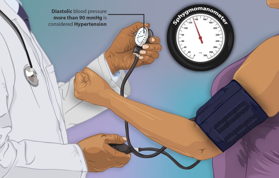
Every time your heart beats, it pushes blood against the walls of your arteries. Blood pressure is simply the force of that push. We write it as two numbers, like 120/80 mmHg: the top (systolic) is the pressure when the heart squeezes, and the bottom (diastolic) is the pressure when the heart relaxes between beats. Healthy pressure keeps blood moving to every organ without hammering the vessels so hard they get damaged.
What actually sets the pressure
Two things multiplied together decide your blood pressure: how much blood the heart pumps per minute and how tight the pipes are. In textbook terms, BP = cardiac output (CO) times systemic vascular resistance (SVR). Cardiac output is heart rate times stroke volume (how much blood leaves the heart with each beat). Systemic vascular resistance is how narrow or constricted the arterioles are. Think of a garden hose: turn up the water flow (more cardiac output) or squeeze the nozzle tighter (more resistance), and pressure rises. This is why a racing heart, extra fluid volume, or clamped-down vessels all push the number up.
How the body keeps it steady
Your body corrects blood pressure minute-to-minute and day-to-day using two main systems.
Baroreceptors are stretch sensors in the carotid arteries and aortic arch. When pressure drops, they sense less stretch and tell the brainstem to speed the heart and constrict vessels; when pressure spikes, they do the opposite. This is the fast, second-by-second reflex, and it is why you may feel dizzy standing up too quickly before the reflex catches up.
The RAAS (renin-angiotensin-aldosterone system) is the slower, longer-term controller. When the kidneys sense low pressure or low blood flow, they release renin. Renin kicks off a chain that makes angiotensin II, a powerful vasoconstrictor that tightens vessels (raising SVR). Angiotensin II also triggers the adrenal glands to release aldosterone, which tells the kidneys to hold onto sodium and water, raising blood volume and therefore cardiac output. Both actions raise pressure. Many blood-pressure medications (ACE inhibitors, ARBs) work by interrupting this exact pathway.
When pressure stays too high: hypertension
Hypertension means blood pressure that stays elevated over time, usually defined as 130/80 mmHg or higher on repeated readings. It shows up in two flavors.
| Feature | Primary (essential) | Secondary |
|---|---|---|
| How common | About 90-95% of cases | Roughly 5-10% of cases |
| Cause | No single identifiable cause; genetics, aging, salt, weight, stress | An identifiable underlying condition |
| Typical triggers | Family history, obesity, high-sodium diet, inactivity | Kidney disease, renal artery narrowing, pheochromocytoma, sleep apnea, certain drugs |
| Onset clue | Gradual, often midlife | Sudden, very high, or resistant to treatment; sometimes young patients |
| Treatment focus | Lifestyle plus medication | Fix the underlying cause |
Primary hypertension has no single fixable cause; it emerges from a mix of genetics, aging vessels, high sodium intake, and often extra weight. Secondary hypertension is driven by a specific, identifiable problem, so treating that problem can actually cure the high pressure.
The silent killer and its targets
Hypertension earns the nickname the silent killer because it usually causes no symptoms for years. There is no headache, no warning; the person feels fine while the elevated pressure quietly grinds away at organs. By the time damage shows, it can be advanced.
That damage is called target-organ damage, and it strikes vessel-rich organs:
- Heart: the left ventricle must pump against higher resistance, so it thickens (left ventricular hypertrophy), and over time can fail; coronary arteries also stiffen, raising heart-attack risk.
- Brain: weakened, pressure-battered vessels can rupture or clot, causing stroke; chronic damage contributes to vascular dementia.
- Kidneys: the tiny filtering vessels scar and narrow, leading to chronic kidney disease and eventually failure.
- Eyes: the retinal vessels are visible windows onto this damage; hypertensive retinopathy can blur or steal vision.
- Blood pressure equals cardiac output times systemic vascular resistance, so more fluid, a faster heart, or tighter vessels all raise it.
- Baroreceptors give fast, moment-to-moment correction; the RAAS gives slower control by constricting vessels (angiotensin II) and retaining salt and water (aldosterone).
- Primary (essential) hypertension is 90-95% of cases with no single cause; secondary hypertension has an identifiable, often treatable cause.
- Hypertension is the silent killer because it causes no symptoms while quietly harming the heart, brain, kidneys, and eyes (target-organ damage).
- Because patients feel fine, adherence teaching is critical; drugs like ACE inhibitors and ARBs work by blocking the RAAS pathway.
Section 2Coronary artery disease, angina & MI
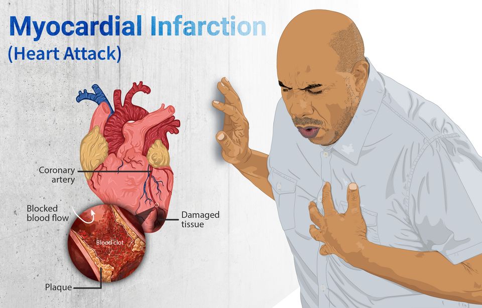
Your heart is a muscle, and like any muscle it needs a steady supply of oxygen-rich blood to keep working. But here is the twist: even though gallons of blood rush through its chambers every hour, the heart muscle itself cannot use that blood directly. Instead, it feeds itself through its own dedicated plumbing, the coronary arteries, which branch off the aorta and wrap around the outside of the heart. When those vessels are open and healthy, supply easily keeps up with demand, and the muscle never notices. Trouble begins when the pipes get narrow.
How the coronary arteries get clogged
Over years, cholesterol, inflammatory cells, and cellular debris build up inside the artery wall, forming a fatty deposit called a plaque. This slow, silent process is atherosclerosis. As the plaque grows, it stiffens the vessel and squeezes the channel (the lumen) smaller, so less blood can get through. This is coronary artery disease (CAD). For a long time the narrowing causes no symptoms because a resting heart does not demand much. The problem shows up when the heart is asked to work harder.
Angina: the heart's cry for oxygen
Angina is chest pain or pressure caused by myocardial ischemia - the muscle is not getting enough oxygen for the work it is doing. It is a demand-versus-supply problem. In stable angina, a fixed plaque leaves just enough room for resting blood flow. But when someone climbs stairs, shovels snow, or gets upset, the heart rate and force climb, oxygen demand outstrips the narrowed supply, and the muscle aches. The classic tip-off is that it is predictable: it comes on with exertion and fades within minutes of rest or a nitroglycerin tablet, which dilates vessels and eases the heart's workload. Importantly, in stable angina the muscle is starved but not yet dying.
Unstable angina is a different, more dangerous animal. Here a plaque has become fragile and partly disrupted, and pain occurs at rest, lasts longer, or is suddenly worse than the patient's usual pattern. It is one of the acute coronary syndromes (ACS) and signals that a heart attack may be brewing.
| Feature | Stable angina | Unstable angina |
|---|---|---|
| Trigger | Predictable exertion or stress | Often at rest, no clear trigger |
| Plaque | Fixed, stable narrowing | Disrupted, unstable plaque |
| Relief | Rest or nitroglycerin, minutes | Poorly relieved, may persist |
| Muscle status | Ischemic, not dying | At high risk, emergency |
When the pipe closes: myocardial infarction
A myocardial infarction (MI) - a heart attack - happens when an unstable plaque ruptures. The body treats the exposed plaque like a wound and rushes platelets and clotting factors to it, forming a blood clot, or thrombus. If that clot completely blocks the artery, blood flow stops cold. The muscle downstream is suddenly cut off from oxygen, and the starved heart muscle begins to die within about 20 to 30 minutes, and the damage spreads over the next few hours. This is infarction - irreversible tissue death (necrosis), not just the reversible starvation of angina. This is why nurses live by the phrase time is muscle: every minute the artery stays blocked, more heart is lost for good.
Recognizing an MI
The hallmark is chest pain - often crushing, heavy, or squeezing pressure, sometimes radiating to the left arm, jaw, or back, and frequently paired with sweating (diaphoresis), nausea, and shortness of breath. But nurses must stay alert for atypical presentations: women, older adults, and people with diabetes may have only fatigue, indigestion, or breathlessness with no dramatic chest pain, because neuropathy and different symptom patterns blunt the classic warning. Three tools confirm the diagnosis:
- ECG: a STEMI (ST-elevation MI) shows a raised ST segment, meaning the full thickness of muscle is being injured by a completely blocked artery - a true emergency needing immediate reopening of the vessel.
- Troponin: when heart cells die, they leak the protein troponin into the blood. A rising troponin level over serial draws is the most specific sign that muscle has actually been damaged.
- The story: the pattern and timing of the pain ties it together.
- Atherosclerosis narrows the coronary arteries over years, reducing blood flow to the heart muscle (coronary artery disease).
- Stable angina is reversible ischemia from a demand-supply mismatch, relieved by rest or nitroglycerin; unstable angina occurs at rest and warns of an impending MI.
- A myocardial infarction results from plaque rupture and a thrombus that blocks the artery, causing irreversible muscle death.
- Recognize an MI by chest pain (watch for atypical presentations), ST-elevation on ECG, and rising troponin - and remember that time is muscle.
Section 3Heart failure
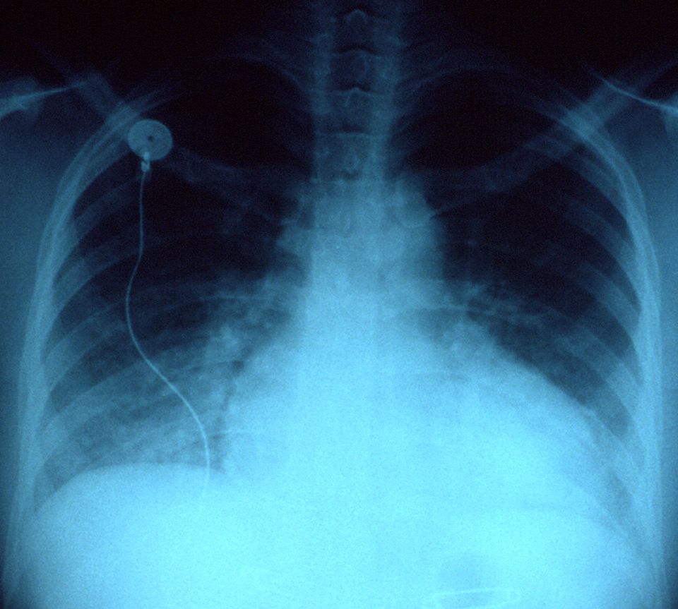
Picture the heart as a two-sided pump working every second of your life. The right side collects oxygen-poor blood returning from the body and pushes it to the lungs; the left side receives freshly oxygenated blood from the lungs and drives it out to every organ. When the heart is healthy, the amount of blood coming in matches the amount going out, and tissues get exactly the oxygen they need. Heart failure is what happens when the pump can no longer keep up with the body's demand. It does not mean the heart has stopped. It means the heart is failing to move blood forward efficiently, so blood backs up behind the weak side and organs downstream go under-supplied.
Left-sided failure: the backup floods the lungs
The left ventricle is the strongest chamber because it has to push blood to the whole body. When it weakens, blood cannot move forward fast enough, so it dams up behind the left side, back into the lungs' capillaries. Rising pressure forces fluid out of those capillaries and into the air sacs, a state called pulmonary congestion or pulmonary edema. This is why left-sided failure is a lung story. Patients feel short of breath (dyspnea), especially on exertion. Lying flat lets fluid redistribute toward the lungs, so they wake up gasping or need to prop up on pillows, which is orthopnea and paroxysmal nocturnal dyspnea. On auscultation you hear crackles (rales), the sound of air bubbling through fluid-filled alveoli. A frothy or pink-tinged cough can appear as fluid leaks into airways.
Right-sided failure: the backup floods the body
When the right ventricle fails, blood dams up behind it, into the systemic veins that were trying to return blood to the heart. Now the congestion shows up in the body, not the lungs. You see jugular venous distention (JVD) as neck veins bulge under the backed-up pressure, peripheral edema in the ankles and legs where gravity pulls the leaked fluid, and hepatomegaly as blood engorges the liver. The single most common cause of right-sided failure is left-sided failure itself: the lung congestion eventually strains the right ventricle, which is why untreated left failure often becomes biventricular.
| Feature | Left-sided failure | Right-sided failure |
|---|---|---|
| Blood backs up into | Lungs (pulmonary circulation) | Body (systemic veins) |
| Hallmark signs | Dyspnea, orthopnea, crackles | JVD, peripheral edema, hepatomegaly |
| Classic complaint | Cannot breathe lying flat | Swollen legs, weight gain |
| Common cause | Hypertension, prior MI | Left-sided failure, lung disease |
Systolic vs diastolic: a squeeze problem or a filling problem
Failure can come from two different mechanical faults. In systolic failure, the ventricle is weak and cannot contract forcefully, so it ejects a smaller fraction of the blood it holds. This is measured as a reduced ejection fraction (EF), often under 40% (normal is usually 55 to 70%), and is called HFrEF (heart failure with reduced ejection fraction). In diastolic failure, the ventricle contracts fine but is stiff and cannot relax and fill properly, so less blood is available to pump even though the squeeze percentage looks normal. This is HFpEF (preserved ejection fraction). The key teaching point: a normal EF does not rule out heart failure.
Compensation that turns against you
When cardiac output drops, the body reacts as if it were bleeding. The sympathetic nervous system fires, raising heart rate and squeezing vessels to prop up blood pressure. The kidneys, sensing low flow, activate the renin-angiotensin-aldosterone system (RAAS), which retains sodium and water and constricts arteries. In the short term these buy time. Over weeks, though, they backfire: the extra fluid worsens the congestion, the higher pressure makes the failing heart work harder, and chronic overdrive drives ventricular remodeling, where the heart enlarges and stiffens. This vicious cycle is why the mainstay drugs (ACE inhibitors, ARBs, beta-blockers) work by blocking these compensations.
The stretched, overworked ventricles release B-type natriuretic peptide (BNP), a hormone that tries to promote fluid loss. Because BNP rises with wall stretch, a high level is a useful marker that shortness of breath is coming from heart failure rather than the lungs. Explore the left- versus right-sided comparison below.
- Heart failure is a pump that cannot meet the body's demand; blood backs up behind the failing side.
- Left-sided failure floods the lungs (dyspnea, orthopnea, crackles); right-sided failure floods the body (JVD, peripheral edema, hepatomegaly).
- Systolic failure (HFrEF) is a weak squeeze with low EF; diastolic failure (HFpEF) is a stiff, poorly filling ventricle with normal EF.
- RAAS and sympathetic activation compensate at first but eventually worsen congestion and drive remodeling.
- BNP rises with ventricular stretch and helps confirm that dyspnea is cardiac in origin.
Section 4Valve disorders
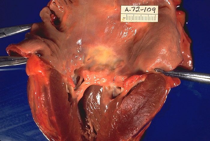
Picture your heart as a house with four rooms and four one-way doors between them. Those doors are your valves, and their whole job is simple: let blood move forward and never let it slip backward. When the heart squeezes, the doors ahead swing open easily; when it relaxes, they snap shut tight so nothing leaks back. Two of these doors do the heavy lifting on the left side of the heart, the side that pushes blood out to the whole body: the mitral valve (between the left atrium and left ventricle) and the aortic valve (between the left ventricle and the aorta). When a valve stops opening well or stops sealing well, the heart has to work harder for every single beat, and over time that extra work reshapes the chamber behind it.
The two ways a valve fails: stenosis vs. regurgitation
There are really only two things that can go wrong with a door. Either it is hard to push open, or it does not shut all the way. That is the entire concept.
Stenosis means the valve has become stiff, thick, or narrowed, so the opening is too small. The chamber behind it must generate much higher pressure to force blood through the tight gap, like trying to breathe through a pinched straw. Regurgitation (also called insufficiency or a leaky/incompetent valve) means the valve does not seal, so some blood sloshes backward with each beat. Now the chamber has to pump the same blood twice, handling both the normal load and the leaked-back volume. Stenosis is mainly a pressure problem; regurgitation is mainly a volume problem, and that distinction predicts how the heart changes.
| Feature | Stenosis | Regurgitation |
|---|---|---|
| Basic defect | Valve too narrow, hard to open | Valve leaks, does not close |
| Main burden | Pressure overload | Volume overload |
| Chamber response | Concentric hypertrophy (thick walls) | Eccentric hypertrophy/dilation (stretched) |
| Classic murmur timing | Systolic (aortic) or diastolic (mitral) | Depends on valve; e.g., holosystolic in MR |
The two lesions you will see most
Aortic stenosis (AS) is the classic pressure problem. The aortic door stiffens, often from age-related calcification, so the left ventricle must slam against a tight opening to eject blood. To make that pressure, the ventricle wall thickens (concentric hypertrophy). The classic triad you learn for symptomatic AS is angina, syncope, and exertional dyspnea, often signaling a narrowed valve area. Patients frequently have a crescendo-decrescendo systolic murmur.
Mitral regurgitation (MR) is the classic volume problem. The mitral door does not seal, so with each squeeze, blood jets backward into the left atrium instead of only moving forward into the aorta. The left atrium and left ventricle stretch and enlarge (eccentric hypertrophy) to hold the extra volume. A holosystolic (pansystolic) murmur best heard at the apex is typical.
Rheumatic heart disease: a delayed autoimmune injury
One cause deserves its own spotlight because it is so testable and so preventable. Rheumatic heart disease follows acute rheumatic fever, which itself follows an untreated group A streptococcal throat infection (think untreated strep throat). The immune system makes antibodies against the bacteria, but through molecular mimicry those antibodies cross-react with the body's own tissues, including heart valves. Years of smoldering inflammation scar and fuse the valve leaflets. The mitral valve is the most commonly damaged, classically producing mitral stenosis. This is why treating strep throat with antibiotics is genuinely heart-protective.
Why chronic valve disease drifts toward heart failure
Here is the through-line that ties it all together. Whether the chamber is straining against high pressure (stenosis) or drowning in extra volume (regurgitation), it copes for a while by remodeling, thickening or stretching to keep output up. This compensation can last years, and patients often feel fine. But muscle that is chronically overworked eventually cannot keep up. Thick, stiff walls relax poorly and fill poorly; stretched, dilated walls squeeze weakly. Either way, the ventricle's ability to move blood forward drops, pressure backs up into the lungs, and you get the picture of heart failure: dyspnea, fatigue, and fluid congestion. Understanding which chamber is overloaded and how tells you exactly where the backup and symptoms will appear. Explore the figure below to see stenosis and regurgitation side by side.
- Two failure modes: stenosis is a stiff, narrowed valve (pressure overload) while regurgitation is a leaky valve that allows backflow (volume overload).
- Signature lesions: aortic stenosis drives left-ventricular concentric hypertrophy; mitral regurgitation dilates the left atrium and ventricle (eccentric hypertrophy).
- Rheumatic heart disease stems from untreated group A strep via molecular mimicry and classically scars the mitral valve, so treating strep throat protects the heart.
- The common endpoint: chronic pressure or volume overload compensates for years, then the overworked chamber fails, backing blood up into the lungs as heart failure.
- Watch symptom onset: a compensated patient can feel well for years; new symptoms (especially in aortic stenosis) signal decompensation and need prompt evaluation.
Section 5The EKG & arrhythmias
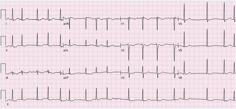
Your heart has its own built-in electrical wiring, and every heartbeat starts as a tiny spark. Before we talk about what goes wrong, let us walk the normal path that spark travels, because every arrhythmia is really just a detour, a delay, or a short-circuit somewhere along that road.
The normal conduction system: the heart's spark and wire
The signal begins in the sinoatrial (SA) node, a patch of cells high in the right atrium. The SA node is the heart's natural pacemaker because it fires fastest, usually 60 to 100 times a minute in an adult at rest. Its spark spreads across both atria and squeezes them, then funnels down to the atrioventricular (AV) node. The AV node is the gatekeeper: it deliberately delays the signal a fraction of a second so the atria can finish emptying blood into the ventricles before the ventricles contract. From there the current races down the bundle of His, splits into the right and left bundle branches, and fans out through the Purkinje fibers, which fire both ventricles almost at once for a strong, coordinated pump.
Reading the P-QRS-T
An EKG (ECG) is simply a voltage recording of that electrical trip. Each normal beat draws three shapes:
- P wave = atrial depolarization (the atria firing and contracting).
- QRS complex = ventricular depolarization (the ventricles firing). It is tall and narrow because the Purkinje system spreads current so fast.
- T wave = ventricular repolarization (the ventricles resetting for the next beat).
To read a strip, check rate and rhythm. For rate, count the QRS complexes in a 6-second strip and multiply by 10, or use the interval between beats. For rhythm, ask three questions: Is there a P wave before every QRS? Is every QRS narrow? Are the intervals regular? A normal sinus rhythm answers yes to all three. When one of those answers turns to no, you have an arrhythmia (dysrhythmia).
Atrial fibrillation and its stroke risk
In atrial fibrillation (AFib) the atria stop firing from the SA node and instead quiver chaotically from many misfiring spots. On the strip the P waves disappear, replaced by a wavy baseline, and the QRS complexes march by irregularly irregular (no pattern at all). The danger is not the fast pulse itself but what happens to the blood: because the atria only quiver instead of squeezing, blood pools and stagnates, especially in the left atrial appendage. Stagnant blood clots (recall Virchow's triad, stasis being one leg), and a clot that breaks loose travels to the brain, causing an embolic stroke. That is why AFib patients are usually placed on anticoagulants.
Heart blocks: a traffic jam at the AV node
A heart block is a delay or failure of the signal passing from atria to ventricles. In first-degree block every beat gets through, just slowly (a long PR interval). In second-degree block some atrial signals reach the ventricles and some are dropped. In third-degree (complete) block nothing gets through: the atria and ventricles beat on their own, ignoring each other, and the ventricles rely on a slow backup pacemaker, often too slow to sustain blood pressure. Complete block frequently needs a pacemaker.
The lethal rhythms
Some arrhythmias are true emergencies because the ventricles, the pumping chambers, fail to move blood.
| Rhythm | What the ventricles do | Response |
|---|---|---|
| Ventricular tachycardia (VT) | Fire far too fast from a ventricular focus; wide QRS, little filling time | Emergency; if no pulse, defibrillate + CPR |
| Ventricular fibrillation (VF) | Only quiver; no organized squeeze, no cardiac output | Immediate defibrillation + CPR |
| Asystole | No electrical activity at all; flatline | CPR + epinephrine; not shockable |
VT and VF are shockable because a defibrillator can reset chaotic ventricular activity back to a coordinated rhythm. Asystole is not shockable, there is no electrical activity to reorganize, so the answer is high-quality CPR and drugs, not a shock.
When an arrhythmia is an emergency
Rhythm names matter less at the bedside than how the patient looks. Watch for signs of poor perfusion: chest pain, shortness of breath, low blood pressure, cool clammy skin, confusion, or fainting (syncope). A rhythm producing no pulse, VT without a pulse, VF, or asystole, means starting CPR and calling a code immediately. An unstable but conscious patient still needs urgent intervention. Explore the interactive EKG below to practice spotting P waves, QRS complexes, and the difference between an organized and a chaotic strip.
- The impulse travels SA node to AV node (which delays it) to bundle of His to Purkinje fibers; the EKG records this as the P wave (atria), QRS (ventricles firing), and T wave (ventricles resetting).
- Read a strip by checking rate and rhythm: a P before every narrow QRS at a regular interval is normal sinus rhythm.
- Atrial fibrillation is irregularly irregular with no P waves; quivering atria let blood pool and clot, raising stroke risk, so patients are usually anticoagulated.
- Heart blocks are AV conduction delays; complete (third-degree) block often needs a pacemaker.
- VT, VF, and asystole are lethal; VF and pulseless VT are shockable with defibrillation, but asystole is not, treat it with CPR and epinephrine.
Section 6Peripheral vascular disease
Your legs are fed by a two-way plumbing system. Arteries carry bright, oxygen-rich blood down from the heart under high pressure to feed the muscles, skin, and nerves. Veins carry the used, oxygen-poor blood back up to the heart against gravity, helped by one-way valves and the squeeze of your calf muscles. When either half of this system fails, the tissue downstream suffers, but it suffers in very different ways depending on whether the problem is inflow (arterial) or outflow (venous). Peripheral vascular disease is the umbrella term for both.
Arterial disease (PAD): not enough blood getting in
Peripheral arterial disease (PAD) is usually caused by atherosclerosis - the same cholesterol plaque that clogs coronary arteries, now narrowing the arteries of the legs. Less blood can squeeze past the plaque, so the muscles are starved of oxygen, especially when they demand more.
The classic early sign is intermittent claudication: a cramping, aching pain in the calf, thigh, or buttock that predictably comes on with walking and reliably eases with rest. The mechanism is simple supply-and-demand - exercising muscle needs more oxygen than the narrowed artery can deliver, so it switches to anaerobic metabolism and lactic acid builds up, causing pain. Rest lowers the demand, the ache resolves, and the cycle repeats at the same walking distance.
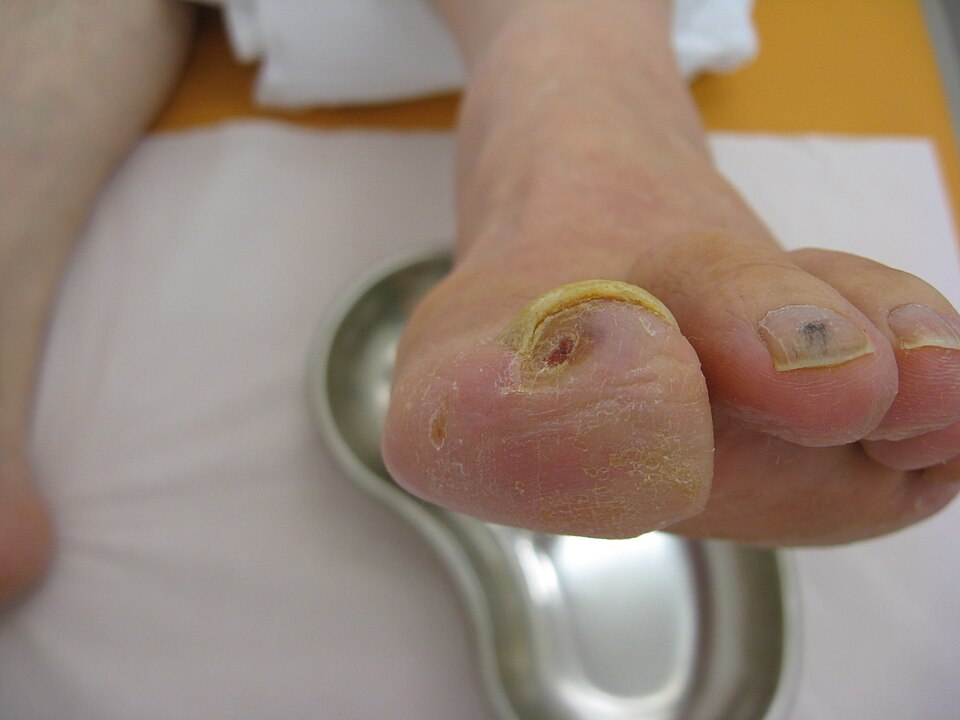
As disease worsens, the pain-free walking distance shortens until pain arrives at rest - a burning ache in the forefoot and toes, worst at night when the leg is flat in bed. Chronic ischemia leaves the skin shiny, hairless, and thin, with thickened nails and punched-out ulcers over the toes and pressure points, because starved tissue cannot maintain normal growth. Patients often report that dangling the leg over the side of the bed relieves rest pain, because gravity helps pull the meager blood flow downhill. If a plaque suddenly thromboses or a clot embolizes from the heart, the limb tips from chronic to acute limb ischemia - a surgical emergency whose hallmark is the six P's: pain, pallor, pulselessness, poikilothermia (the limb drifts to room temperature and feels cool), paresthesia, and paralysis. Paresthesia and paralysis are the late nerve-and-muscle findings; when they appear, the limb is close to being lost.
Venous disease: not enough blood getting out
Chronic venous insufficiency (CVI) is the mirror image. Here the arteries deliver blood fine, but the veins cannot return it. Damaged or incompetent valves - often after years of standing, obesity, pregnancy, or a prior clot - let blood flow backward and pool in the legs. This raises pressure in the veins and capillaries, a state called venous hypertension.
That backed-up pressure pushes fluid out into the tissues, producing chronic edema that is usually worst by the end of the day and improves overnight. Red blood cells also leak out and break down, depositing iron (hemosiderin), which stains the skin a permanent brownish discoloration around the lower leg and ankle - a change called hemosiderin staining. The same congested skin often turns red, itchy, scaly, and weepy, an eczema-like inflammation named stasis dermatitis. Over time the skin becomes leathery and tight (lipodermatosclerosis). Because the tissue is congested and poorly nourished, it breaks down into venous ulcers, classically over the inner ankle (the medial malleolus), where venous pressure is highest.
Telling an arterial ulcer from a venous ulcer
Ulcers are where students most often mix these up, so match the wound to its mechanism. An arterial ulcer forms because tissue is starved, so it appears where flow is poorest and looks dead. A venous ulcer forms because tissue is congested and weepy, so it appears where pooling is worst and looks wet.
| Feature | Arterial ulcer | Venous ulcer |
|---|---|---|
| Usual location | Toes, heels, bony points, lateral foot | Around the medial (inner) ankle |
| Wound edges | Well-defined, punched-out, deep | Irregular, shallow, sloping |
| Wound bed | Pale, gray, or dry; little drainage | Ruddy/beefy red; heavy drainage |
| Surrounding skin | Cool, pale, shiny, hairless | Warm, brown-stained, edematous |
| Pain | Severe, worse with elevation | Mild-moderate, better with elevation |
| Pulses | Diminished or absent | Usually present |
DVT, PE, and one word on aneurysm
A deep vein thrombosis (DVT) is a clot in a deep leg vein, favored by Virchow's triad: venous stasis, vessel-wall injury, and hypercoagulability (immobility, surgery, and estrogen are common real-world triggers). The leg is often warm, red, swollen, and tender. The danger is not the leg itself - it is that a piece can break loose, travel to the lungs, and lodge as a pulmonary embolism (PE), causing sudden shortness of breath, pleuritic chest pain, and a drop in oxygen. That is why we never vigorously massage a suspected DVT. Separately, an abdominal aortic aneurysm (AAA) is a balloon-like weakening of the aortic wall that may present as a pulsatile abdominal mass and can be fatal if it ruptures. Explore the arterial-versus-venous figure below to see these patterns side by side.
- PAD starves tissue of inflow: intermittent claudication (pain with walking, relieved by rest) plus a limb that is cool, pale, pulseless, and painful - worse with elevation.
- Chronic venous insufficiency is an outflow/pooling problem: edema, brownish hemosiderin staining, and ulcers around the medial ankle - better with elevation.
- Arterial ulcers are punched-out, dry, and on toes/pressure points; venous ulcers are shallow, wet, and around the inner ankle.
- DVT (Virchow's triad) can throw a clot to the lungs as a life-threatening pulmonary embolism - do not massage the leg.
- An abdominal aortic aneurysm is a weakened, ballooning aorta that can be fatal if it ruptures.
The chapter in one breath
- Blood pressure is cardiac output times systemic vascular resistance; hypertension (usually ≥130/80) is the silent, mostly asymptomatic disease that thickens vessels and overworks the heart until it damages the brain, kidneys, and eyes.
- Coronary artery disease is atherosclerotic plaque starving the myocardium; when a plaque ruptures and a clot fully blocks flow, the muscle downstream infarcts, giving the crushing chest pain, elevated troponin, and ST changes of an MI.
- Left-sided heart failure backs blood up into the lungs, so the classic signs are pulmonary: crackles, dyspnea, orthopnea, and frothy sputum from pulmonary edema.
- Right-sided heart failure backs blood up into the body, so you see peripheral congestion: dependent edema, jugular venous distention, ascites, and a congested, tender liver.
- Valve disorders come in two flavors: stenosis (a stiff, narrow valve the heart must strain to push through) and regurgitation (a leaky valve that lets blood flow backward), both forcing the chamber behind them to hypertrophy or dilate over time.
- The EKG tracks the electrical wave through the heart, where the P wave is atrial depolarization, the QRS is ventricular depolarization, and the T wave is ventricular repolarization.
- Atrial fibrillation is the most common serious arrhythmia, an irregularly irregular quiver that pools blood in the atria and throws clots, which is why these patients need anticoagulation to prevent stroke.
- Lethal rhythms demand instant action: ventricular fibrillation and pulseless ventricular tachycardia are shockable (defibrillate plus CPR), while true asystole is not shocked but treated with CPR and epinephrine.
- Peripheral vascular disease splits by side: arterial insufficiency gives pain with walking (claudication), cool pale pulseless feet, and painful distal wounds, while venous insufficiency gives warm swollen legs, brownish skin, and weepy medial-ankle ulcers.
ReferenceWords worth knowing
A quick reference for the terms in this chapter — skim it now, or circle back whenever a word trips you up.
- Angina
- Angina is chest pain or pressure that happens when a section of heart muscle temporarily gets less oxygen-rich blood than it needs, usually because a coronary artery is narrowed by plaque.
- Atherosclerosis
- Atherosclerosis is the slow buildup of fatty, cholesterol-rich plaque inside artery walls that stiffens and narrows the vessel and can eventually block blood flow.
- Atrial fibrillation
- Atrial fibrillation is a fast, disorganized quivering of the atria instead of a coordinated squeeze, which pools blood in the atria and raises the risk of clots and stroke.
- BNP (B-type natriuretic peptide)
- BNP is a hormone released by stretched, overworked heart muscle, so a high blood level often signals that the heart is failing to keep up with its fluid load.
- Cardiac output
- Cardiac output is the amount of blood the heart pumps out each minute, calculated as heart rate multiplied by stroke volume (usually about 4 to 8 L/min in adults).
- Claudication
- Claudication is cramping leg pain that comes on with walking and eases with rest, caused by narrowed arteries that can't deliver enough blood to exercising muscles.
- Ejection fraction
- Ejection fraction is the percentage of blood in a filled ventricle that gets pumped out with each beat, normally around 55 to 70 percent.
- Embolus
- An embolus is a clot, plaque fragment, air bubble, or other mass that travels through the bloodstream until it lodges in and blocks a smaller vessel downstream.
- Heart failure
- Heart failure is the condition in which the heart can no longer pump strongly enough or fill well enough to meet the body's needs, causing fluid backup and fatigue.
- Hypertension
- Hypertension is persistently high blood pressure (usually 130/80 mmHg or above) that forces the heart to work harder and gradually damages vessel walls and organs.
- Myocardial infarction
- Myocardial infarction, or heart attack, is the death of heart muscle caused when a coronary artery is fully blocked and that region of tissue is starved of oxygen.
- RAAS (renin-angiotensin-aldosterone system)
- RAAS is a hormone cascade that raises blood pressure by tightening blood vessels and telling the kidneys to hold onto salt and water when blood flow drops.
- Regurgitation
- Regurgitation is the backward leaking of blood through a heart valve that doesn't close tightly, forcing the heart to pump extra to make up for what flows the wrong way.
- Stenosis
- Stenosis is the abnormal narrowing or stiffening of a heart valve or vessel that restricts forward blood flow and makes the heart push harder to move blood through.
- Systemic vascular resistance
- Systemic vascular resistance is the total opposition to blood flow created by the tone (constriction or dilation) of the body's arteries, a major driver of blood pressure.
- Troponin
- Troponin is a protein released into the blood when heart muscle cells are injured, so a rising level is a key marker used to confirm a myocardial infarction.
DrillFlashcards for this unit
These are the Week 9 cards — the same deck as the hub, pulled in here so you can review the moment you finish reading. Tap the card to flip it; hit Shuffle to mix them up. To grade yourself and track mastery across the whole deck, open the full flashcards page.