Digestive: GI, Liver & Pancreas
The digestive tract is a single long tube with some powerful accessory organs, and trouble anywhere along it shows up fast. This chapter runs from the reflux and ulcers of the upper GI down through inflammatory bowel disease and obstruction, then into the liver, gallbladder, and pancreas — and the jaundice that signals something is backing up.
By the end of this chapter you can
- Explain GERD and peptic ulcer disease and their dangers.
- Contrast Crohn disease with ulcerative colitis.
- Recognize bowel obstruction and tell an upper from a lower GI bleed.
- Describe cirrhosis and its consequences (portal hypertension, varices, ascites, encephalopathy).
- Explain gallstones, pancreatitis, and the three types of jaundice.
Section 1Upper GI: GERD & peptic ulcers
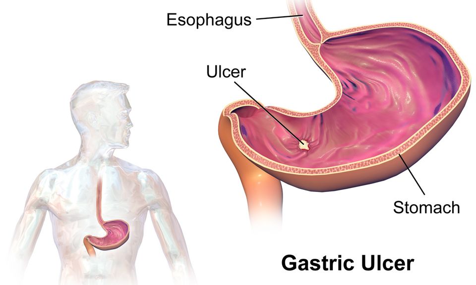
Your stomach makes acid on purpose. Special parietal cells in the stomach lining pump out hydrochloric acid (HCl) to start digesting food and to kill swallowed bacteria. That acid is powerful enough to burn skin, so the body has to protect itself in two ways. First, a muscular ring at the bottom of the esophagus called the lower esophageal sphincter (LES) stays tightly closed to keep acid down in the stomach where it belongs. Second, the stomach and duodenum are coated in a thick layer of mucus and bicarbonate that acts like a chemical raincoat, keeping acid from touching the delicate cells underneath. Trouble in the upper GI tract almost always comes down to one of these two defenses failing.
GERD: when the sphincter leaks
Gastroesophageal reflux disease (GERD) happens when the LES is weak or relaxes at the wrong time, letting stomach acid splash backward into the esophagus. The esophagus has no mucus raincoat, so the acid irritates its lining. That is why the classic symptom is heartburn (pyrosis) - a burning behind the breastbone, often worse after big meals or when lying down (gravity is no longer helping keep acid down). Patients may also describe regurgitation of sour fluid, a chronic cough, or a hoarse voice from acid reaching the throat.
Occasional reflux is normal; GERD is when it happens often enough to cause symptoms or damage. The real long-term worry is what repeated acid exposure does to the esophageal lining. Over years, the normal squamous cells can transform into intestinal-type columnar cells - a change called Barrett esophagus. This is the body adapting to survive the acid, but it is a form of metaplasia that raises the risk of esophageal adenocarcinoma. That is why chronic GERD is monitored, not just medicated.
Peptic ulcer disease: when the raincoat wears through
A peptic ulcer is an open sore that erodes through the protective mucosa into the wall of the stomach or duodenum. The two big culprits both attack the mucus defense. First is Helicobacter pylori, a spiral bacterium that burrows into the mucus layer and triggers inflammation that weakens it. Second is chronic use of NSAIDs (like ibuprofen and aspirin). NSAIDs block prostaglandins - the very chemicals that tell the stomach to make protective mucus and bicarbonate. Take away the prostaglandins and you take away the raincoat, so acid digests the tissue itself. Together, H. pylori and NSAIDs cause the large majority of peptic ulcers.
Gastric vs. duodenal: read the pain
Where the ulcer sits changes the timing of the pain, and this is a favorite exam point. The trick is to think about where the food goes.
| Feature | Gastric ulcer | Duodenal ulcer |
|---|---|---|
| Location | In the stomach lining | In the first part of the small intestine |
| Pain timing | Often worse with food (usually 30 min after eating) | Often relieved by food, then returns 2-3 hours later |
| Classic clue | May avoid eating, can lose weight | Night pain that eating settles; may gain weight |
| Typical cause lean | More often NSAID-related | More often H. pylori-related |
The logic: in a gastric ulcer, food entering the stomach means more acid production right on top of the sore, so eating hurts. In a duodenal ulcer, food buffers the acid and stimulates the stomach to close its outlet briefly, so eating soothes the pain until the meal moves through.
When an ulcer turns dangerous
Ulcers matter because they can go deeper. Two complications are emergencies. Bleeding occurs when the ulcer erodes into a blood vessel. Watch for hematemesis (vomiting blood, sometimes coffee-ground colored from digested blood) and melena (black, tarry, foul stool - blood that has traveled the whole gut). Slow chronic bleeding shows up as iron-deficiency anemia and fatigue. The second is perforation: the ulcer bores all the way through the wall, spilling acid and gut contents into the abdominal cavity. This causes sudden, severe pain and a rigid, board-like abdomen - a sign of peritonitis and a surgical emergency.
Take a look at the interactive figure below to see how a weak sphincter lets acid climb back into the esophagus.
- GERD is acid refluxing past a weak LES; chronic exposure can drive Barrett esophagus and raise esophageal cancer risk.
- Peptic ulcers form when the mucus defense fails - usually from H. pylori or NSAIDs (which block protective prostaglandins).
- Gastric ulcer pain is often worse with food; duodenal ulcer pain is often relieved by food and returns hours later.
- The dangerous complications are bleeding (hematemesis, melena, anemia) and perforation (sudden pain, rigid board-like abdomen, peritonitis).
Section 2Inflammatory bowel disease
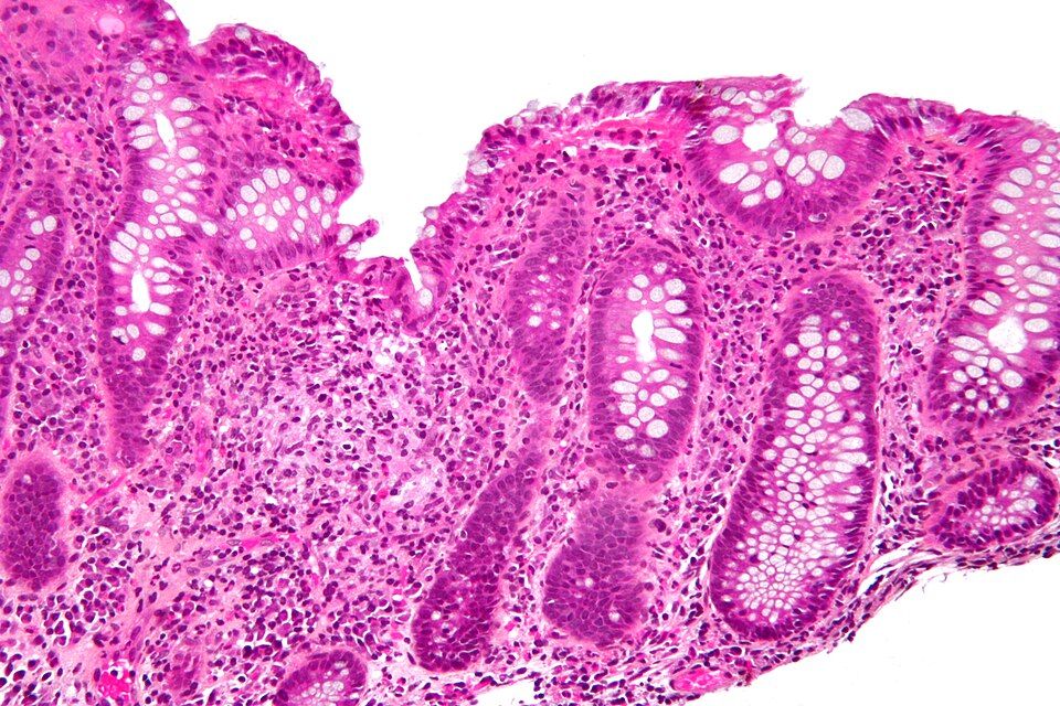
Your gut is a long tube that runs from your mouth to your anus, and its inner lining has a hard job: pull nutrients out of food while keeping trillions of bacteria on the inside of the tube and out of your bloodstream. That lining, the mucosa, sits on top of your immune system like a screen door, letting the good stuff through and holding the line against invaders. Normally your immune cells and your gut bacteria live in a quiet truce. In inflammatory bowel disease (IBD), that truce breaks down. The immune system starts attacking the bowel wall itself, causing chronic, immune-mediated inflammation that flares and settles, flares and settles, for the rest of a person's life.
Two diseases, one family
IBD is an umbrella term for two related conditions: Crohn disease and ulcerative colitis (UC). They share a lot. Both are chronic and relapsing, meaning they cycle between flares (active inflammation, misery) and remission (things calm down). Both involve an immune system that overreacts to the normal gut, driven by a mix of genetics and environmental triggers. And both can produce systemic misery beyond the belly, including fatigue, weight loss, and low-grade fever, because widespread inflammation is a whole-body event. What sets them apart is where they attack and how deep they go.
Crohn disease: patchy and deep
Crohn can strike anywhere from the mouth to the anus, though it loves the terminal ileum (the tail end of the small intestine) and the colon. Its signature is skip lesions: patches of angry, inflamed bowel sitting right next to perfectly healthy tissue, like a moth-eaten pattern. Crucially, the inflammation is transmural, meaning it burrows through all the layers of the bowel wall, not just the surface.
That depth explains Crohn's worst complications. When inflammation tunnels all the way through the wall, it can carve an abnormal connection between two structures, called a fistula (for example, bowel-to-skin or bowel-to-bladder). Chronic transmural damage also scars and narrows the tube, producing strictures that can block the bowel. Because the small intestine does most nutrient absorption, Crohn often causes malabsorption, weight loss, and vitamin deficiencies. The diarrhea is frequently non-bloody, and patients may have right-lower-quadrant pain that mimics appendicitis.
Ulcerative colitis: shallow but continuous
UC is more predictable in its footprint. It is limited to the colon only, it almost always starts in the rectum, and it spreads continuously upward with no skip areas, one unbroken zone of inflammation. It is also shallow: the damage stays in the mucosa (and submucosa), sparing the deeper muscle layers. Because it doesn't go transmural, UC rarely causes fistulas or strictures.
But that inflamed, ulcerated mucosal surface bleeds easily, so the hallmark symptom is bloody diarrhea mixed with mucus, often with crampy lower-abdominal pain and a constant urge to go (tenesmus). Losing blood and fluid stool many times a day can leave patients anemic and dehydrated. A feared emergency is toxic megacolon, where the colon dilates and can perforate. Long-standing UC also raises the risk of colorectal cancer, so patients need ongoing colonoscopy surveillance.
Side by side
| Feature | Crohn disease | Ulcerative colitis |
|---|---|---|
| Location | Mouth to anus (often terminal ileum) | Colon only, starts in rectum |
| Pattern | Skip lesions (patchy) | Continuous |
| Depth | Transmural (all layers) | Mucosa/submucosa only |
| Classic stool | Diarrhea, often non-bloody | Bloody diarrhea with mucus |
| Complications | Fistulas, strictures, malabsorption | Toxic megacolon, higher colon cancer risk |
IBD is not IBS
One last distinction that trips people up because the letters look alike. IBD is structural, real, visible inflammation and tissue damage you can see on scope and biopsy; IBS (irritable bowel syndrome) is functional, meaning the gut hurts and misbehaves but the tissue itself looks normal. Explore the interactive figure below to compare the Crohn and UC patterns side by side.
- IBD is chronic, immune-mediated inflammation of the bowel that relapses in flares and settles into remission; it splits into Crohn disease and ulcerative colitis.
- Crohn can hit anywhere mouth-to-anus with skip lesions and transmural inflammation, so it causes fistulas, strictures, and malabsorption.
- Ulcerative colitis stays in the colon only, spreads continuously from the rectum, and stays mucosal, so its hallmark is bloody diarrhea.
- Deep vs shallow explains the complications: Crohn tunnels through the wall (fistulas), UC ulcerates the surface (bleeding, toxic megacolon, cancer risk).
- Do not confuse IBD (structural inflammation, visible damage) with IBS (a functional disorder with normal-looking tissue).
Section 3Bowel obstruction & GI bleeding
Picture your intestines as a long, muscular hose, roughly 20 feet of it, that never stops working. Normally the gut moves its contents forward with rhythmic squeezing waves called peristalsis, driven by smooth muscle and coordinated by the enteric nervous system (the gut's own built-in wiring). Food, fluid, gas, and eventually stool all travel one direction: down and out. When that forward flow stops, everything backs up behind the blockage, and the trouble begins.
Mechanical obstruction vs paralytic ileus
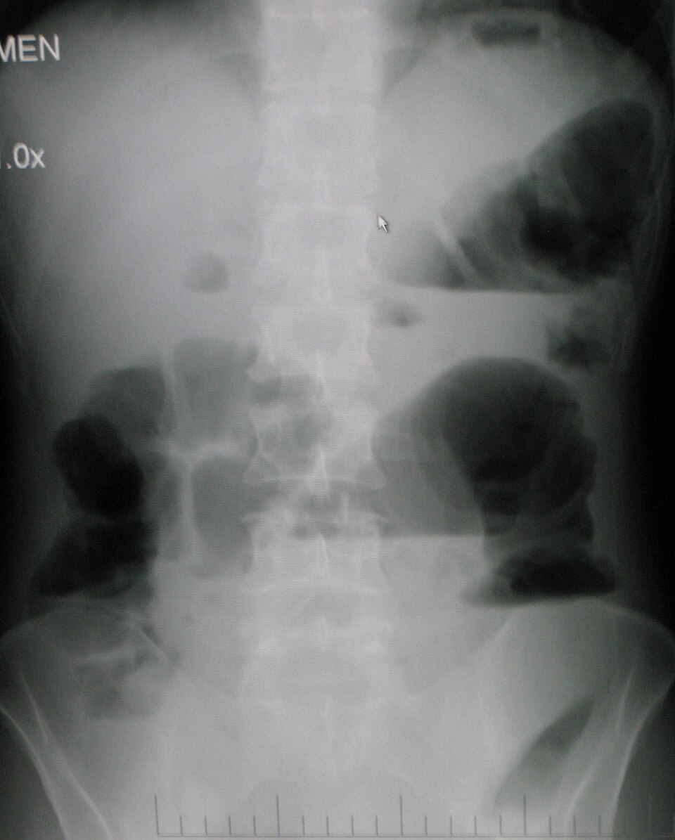
There are two very different reasons the highway can shut down. In a mechanical obstruction, something physically plugs the lumen. The most common culprits are adhesions (bands of internal scar tissue, usually from previous abdominal surgery), a hernia (a loop of bowel trapped where it pokes through the abdominal wall), and tumors (especially in the colon). The muscle still wants to push, so early on it squeezes harder against the wall it cannot pass.
In a paralytic ileus, nothing is physically blocking the tube, but the muscle simply stops moving. Peristalsis goes quiet. This happens after abdominal surgery (the gut is famously sluggish for a day or two), with peritonitis, with electrolyte problems (especially hypokalemia, since potassium is needed for smooth-muscle contraction), and with opioids, which slow the gut down. The distinction matters enormously: mechanical blockages often need a procedure to relieve them, while an ileus usually needs the underlying cause fixed and the bowel rested.
| Feature | Mechanical obstruction | Paralytic ileus |
|---|---|---|
| Problem | Physical blockage of the lumen | Gut muscle stops contracting |
| Common causes | Adhesions, hernia, tumor | Post-op state, hypokalemia, opioids, peritonitis |
| Bowel sounds (early) | High-pitched, hyperactive, tinkling | Diminished to absent from the start |
| Pain pattern | Crampy, wave-like (colicky) | Steady, diffuse discomfort |
| Usual fix | Often surgical or decompression | Treat the cause, rest the bowel |
The cardinal signs and why they happen
No matter the cause, obstruction produces a classic quartet. Abdominal distension develops because fluid and swallowed air pile up behind the block and the bowel balloons outward. Vomiting follows as the backed-up contents have nowhere to go but up; a low (distal) blockage often produces foul, feculent vomiting, while a high (proximal) small-bowel block causes earlier, more forceful emesis. Obstipation, the complete failure to pass any stool or gas, is a telling sign that nothing is getting through.
Bowel sounds tell a story over time. Early in a mechanical obstruction, listen for high-pitched, hyperactive, tinkling sounds as the muscle strains against the blockage. As hours pass and the muscle fatigues, or as an ileus sets in, those sounds fade to absent. The classic teaching: sounds go high-pitched then silent.
Why obstruction is an emergency
Trapped bowel does not just hurt, it dies. Fluid pours into the distended loop (third-spacing), dropping circulating volume and risking hypovolemic shock plus metabolic imbalances from all that lost fluid and electrolytes. Worse, if the swelling squeezes off the bowel's blood supply, the tissue becomes strangulated, turns ischemic, and can perforate, spilling gut contents into the abdomen and triggering life-threatening peritonitis and sepsis. Fever, rising heart rate, and sudden severe pain signal that a simple blockage has become a surgical emergency.
GI bleeding: upper vs lower
Bleeding anywhere in the digestive tract is graded by where it starts, because the appearance of the blood is a built-in clue. An upper GI bleed comes from above the ligament of Treitz, meaning the esophagus, stomach, or duodenum, and is often caused by peptic ulcers or esophageal varices. It shows up as hematemesis (vomited blood, sometimes coffee-ground in appearance once stomach acid has partly digested it) and melena (black, tarry, sticky stool). The stool is black because hemoglobin is chemically broken down during its long trip through the gut.
A lower GI bleed arises in the jejunum, ileum, or colon, commonly from diverticulosis, angiodysplasia, hemorrhoids, or colorectal cancer. Because the blood exits quickly without time to be digested, it appears as hematochezia: bright red or maroon blood mixed with or coating the stool.
Why a major bleed is an emergency
A significant GI bleed can quietly drain enough blood to cause hypovolemic shock before the source is even found. Watch for tachycardia, falling blood pressure, orthostatic dizziness, pallor, and a dropping hemoglobin and hematocrit. Early vital-sign changes often precede a visible drop in labs, so trending them and securing IV access is a nursing priority.
- Mechanical obstruction (adhesions, hernia, tumor) physically plugs the bowel, while paralytic ileus means the gut muscle simply stops moving.
- The cardinal signs are distension, vomiting, obstipation (no stool or gas), and bowel sounds that go high-pitched then absent.
- Upper GI bleed = hematemesis and melena (black, tarry, digested blood); lower GI bleed = hematochezia (bright red or maroon).
- Obstruction can progress to strangulation, perforation, and peritonitis; a major bleed can cause hypovolemic shock, so both are true emergencies.
- Monitor trending vital signs, look for tachycardia and hypotension, and watch for third-spacing and electrolyte loss.
Section 4The liver: hepatitis & cirrhosis
Think of your liver as the body's busiest chemistry lab. It sits under your right rib cage and quietly runs hundreds of jobs at once. Three of those jobs matter most for understanding what goes wrong. First, it is the body's detox center: it filters drugs, alcohol, and metabolic waste out of the blood, and it converts toxic ammonia (a byproduct of protein breakdown) into urea so the kidneys can excrete it. Second, it is a factory: it manufactures most of the body's clotting factors and albumin, the protein that holds fluid inside blood vessels. Third, it processes bilirubin, the yellow-orange pigment left over when old red blood cells are recycled, packaging it so it can leave the body in bile and stool. Hold onto those three jobs, because nearly every symptom of liver disease traces back to one of them failing.
Hepatitis: when the liver gets inflamed
Hepatitis simply means inflammation of the liver. The most common causes are viral, but alcohol and certain drugs (notably acetaminophen in overdose) can do the same damage. The three viral players behave differently, and that difference drives nursing care.
| Feature | Hepatitis A | Hepatitis B & C |
|---|---|---|
| How it spreads | Fecal-oral (contaminated food/water) | Blood & body fluids (needles, sex, birth) |
| Course | Acute, self-limiting; usually full recovery | Often becomes chronic; can smolder for years |
| Long-term risk | Rarely lasting damage | Cirrhosis & liver cancer |
| Prevention | Vaccine; hand hygiene | Hep B vaccine; no Hep C vaccine yet |
During acute hepatitis, patients often feel fatigued and nauseated, and many turn jaundiced (yellow skin and eyes) because the inflamed liver cannot keep up with bilirubin processing. Alcoholic and drug-induced hepatitis follow the same theme: the toxin injures liver cells (hepatocytes) faster than they can heal.
Cirrhosis: scarring that does not undo
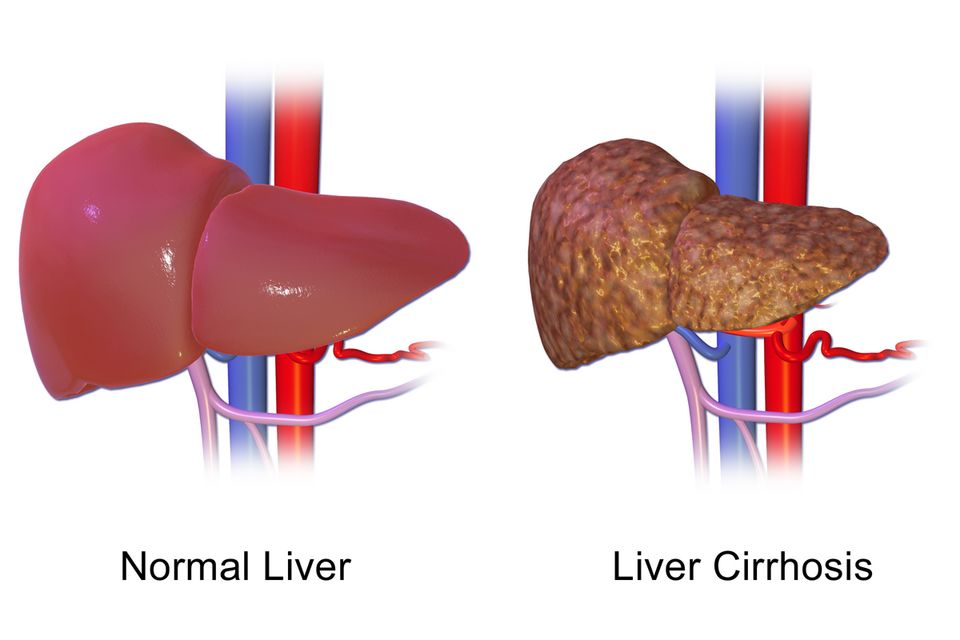
When injury to the liver continues long enough, healing itself becomes the problem. Instead of regenerating normal tissue, the liver lays down fibrosis (scar tissue). Cirrhosis is the end-stage result: the once-soft organ becomes hard, nodular, and shrunken. Scar tissue does not do liver work, and it does not let blood flow through normally. Both of those facts explain the cascade of complications that follow.
Portal hypertension and its downstream chaos
Normally, blood from the intestines drains through the portal vein into the liver. Cirrhotic scar tissue squeezes those channels, so pressure backs up, a state called portal hypertension. High pressure in this system forces blood to find detours through smaller veins that were never built for the load. Where those veins swell, we get problems:
- Esophageal varices are ballooned, fragile veins at the base of the esophagus. They can rupture and cause sudden, massive, life-threatening bleeding.
- Ascites is fluid pooling in the abdomen. Two forces drive it: the high portal pressure pushes fluid out of vessels, and the failing liver makes too little albumin to hold fluid in. Low albumin means low oncotic pressure, so water leaks into the belly.
Why liver failure causes bleeding and jaundice
Remember the factory job. A failing liver stops making enough clotting factors, so patients bruise and bleed easily, and lab tests show a prolonged PT/INR. Pair that with fragile varices, and bleeding becomes a true emergency. Remember the bilirubin job, too: as the liver falters, bilirubin builds up in the blood and stains the skin and sclera yellow, the classic jaundice.
Hepatic encephalopathy: the toxin job fails
When the detox center goes down, ammonia from protein digestion is no longer converted to urea. It accumulates and crosses into the brain, causing hepatic encephalopathy: confusion, personality changes, and a flapping tremor of the hands called asterixis. Nurses often watch for early confusion as a warning sign, and the drug lactulose is given to trap ammonia in the gut and pull it out in stool.
- The liver detoxifies (converting ammonia to urea), builds clotting factors and albumin, and processes bilirubin — remember these jobs and the symptoms make sense.
- Hepatitis is liver inflammation; Hep A spreads fecal-oral and resolves, while Hep B and C spread through blood, often turn chronic, and lead to cirrhosis.
- Cirrhosis is irreversible scarring; the scar tissue blocks blood flow and causes portal hypertension, driving esophageal varices and ascites.
- Liver failure causes bleeding (too few clotting factors, high PT/INR) and jaundice (bilirubin buildup).
- Rising ammonia causes hepatic encephalopathy (confusion, asterixis), typically treated with lactulose.
Section 5Gallbladder & pancreas
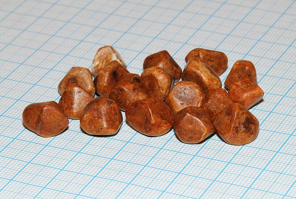
Your gallbladder and pancreas are two quiet helpers that sit just under your liver and stomach. Normally, the gallbladder is a small green sac that stores bile (a soap-like fluid the liver makes to break up fat). When you eat a greasy meal, a hormone called cholecystokinin (CCK) tells the gallbladder to squeeze, sending bile down the common bile duct into the small intestine. The pancreas, meanwhile, pours powerful digestive enzymes down its own pancreatic duct, which usually joins the bile duct right before both empty into the duodenum. Those enzymes are supposed to stay switched off until they reach the gut. Trouble starts when a stone plugs the plumbing, or when those enzymes turn on too early.
Gallstones and cholecystitis
When bile has too much cholesterol or sits too long, it can harden into gallstones (cholelithiasis). Many stones are silent. But if a stone lodges in the gallbladder's neck or the cystic duct, the gallbladder keeps squeezing against a locked door, becomes trapped, inflamed, and infected. This is acute cholecystitis. Because fatty food is the trigger for gallbladder contraction, the classic story is right-upper-quadrant (RUQ) pain 30 minutes to a few hours after a fatty meal, often radiating to the right shoulder or scapula, plus nausea and vomiting. The pain can be severe and cramping (biliary colic).
A bedside clue is Murphy's sign. You place your hand under the right rib margin and ask the patient to take a deep breath. As the inflamed gallbladder descends onto your fingers, the patient catches their breath and stops from pain. A positive Murphy's sign points strongly to cholecystitis. Think of the classic risk profile as the 4 F's: female, forty, fertile, and overweight.
When a stone blocks the ducts
A stone that escapes the gallbladder can travel and wedge deeper. If it blocks the common bile duct (choledocholithiasis), bile backs up. Bilirubin spills into the blood, so the skin and eyes turn yellow (jaundice), the urine darkens, and stools turn clay-pale because bile pigment no longer reaches the gut. Trapped, stagnant bile invites infection of the whole biliary tree (cholangitis).
Here is the dangerous overlap: because the bile duct and pancreatic duct usually share a final channel, one stone can dam both. If it blocks where the pancreatic duct drains, enzymes cannot escape, pressure rises, and the pancreas is set up to attack itself.
Acute pancreatitis: the organ that digests itself
Acute pancreatitis happens when those digestive enzymes activate inside the pancreas instead of the intestine. Trypsin switches on prematurely and triggers the rest, so the gland begins to autodigest its own tissue (autodigestion). The result is intense inflammation, swelling, and sometimes bleeding.
The hallmark is severe, constant epigastric pain that bores straight through to the back. Patients often feel worse lying flat and better leaning forward. Nausea and vomiting are common, and the belly may be tender and distended. On labs, lipase rises sharply (usually more than three times the upper normal limit) and stays elevated longer than amylase, which makes lipase the more specific marker.
What causes it
By far the two leading causes are gallstones and alcohol (a helpful memory hook is "I GET SMASHED," where the first culprits are Idiopathic, Gallstones, and Ethanol). Gallstones block the shared duct; alcohol is directly toxic to pancreatic cells and thickens secretions.
| Feature | Cholecystitis | Acute pancreatitis |
|---|---|---|
| Core problem | Blocked, inflamed gallbladder | Pancreas digesting itself |
| Pain location | Right upper quadrant, to right shoulder | Epigastric, boring to the back |
| Trigger | Fatty meal | Alcohol or a gallstone |
| Key exam/lab | Positive Murphy's sign | High lipase |
- Gallstones can block bile flow; if the neck or cystic duct is trapped, the gallbladder inflames (cholecystitis) with RUQ pain after fatty meals and a positive Murphy's sign.
- A stone in the common bile duct causes jaundice, dark urine, and pale stools; because the ducts share a channel, one stone can block the pancreatic duct too.
- Acute pancreatitis is autodigestion: enzymes activate early and the pancreas attacks itself, causing severe epigastric pain that bores to the back.
- Lipase is the key lab, usually more than triple the upper limit, and is more specific than amylase.
- The top two causes of pancreatitis are gallstones and alcohol.
Section 6Jaundice & bilirubin
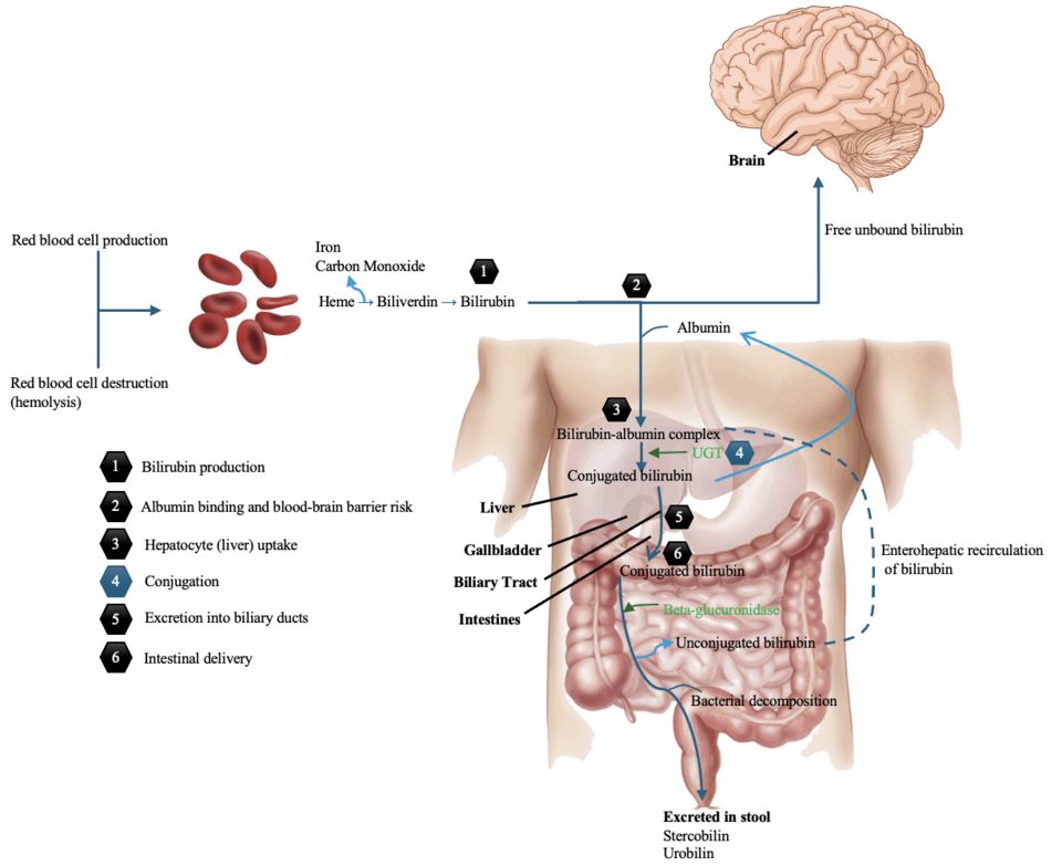
Every day, your body quietly retires about 1% of its red blood cells, mostly because they have worn out after roughly 120 days in circulation. When a red cell breaks down, the hemoglobin inside it is dismantled. The heme portion becomes a yellow-orange pigment called bilirubin. This first form is unconjugated (indirect) bilirubin, and it has a problem: it is fat-loving and not water-soluble, so it cannot simply be flushed out in urine. Instead it rides through the blood attached to albumin and is delivered to the liver.
How the liver handles bilirubin
The liver is the cleanup station. Liver cells (hepatocytes) take up unconjugated bilirubin and attach sugar molecules to it using an enzyme called UDP-glucuronyl transferase. This step is conjugation, and it converts the pigment into conjugated (direct) bilirubin, which is now water-soluble. The liver secretes conjugated bilirubin into bile, which drains through the bile ducts into the small intestine. There, gut bacteria change it into urobilinogen and then stercobilin, the pigment that gives stool its normal brown color. A small amount is reabsorbed and leaves in urine as urobilin, giving urine its yellow tint.
Jaundice (also called icterus) is the yellowing of skin, mucous membranes, and the sclera of the eyes. It appears when serum bilirubin rises above roughly 2 to 3 mg/dL. Because bilirubin loves fatty tissue and elastin-rich tissue, the sclera and skin show it first. Jaundice is never a disease by itself; it is a signal that one of the steps above has broken down. To find the break, nurses group jaundice by where in the pathway the problem sits.
The three types of jaundice
Prehepatic (hemolytic) jaundice happens before the liver, when red cells are destroyed faster than the liver can keep up. Causes include hemolytic anemias, sickle cell crisis, and transfusion reactions. The liver is healthy but overwhelmed, so unconjugated bilirubin piles up in the blood.
Hepatic (intrahepatic) jaundice comes from the liver itself being sick. In hepatitis, cirrhosis, or drug injury, damaged hepatocytes cannot conjugate and excrete bilirubin efficiently. Both unconjugated and conjugated forms often rise.
Posthepatic (obstructive) jaundice happens after the liver, when bile cannot drain. A gallstone in the common bile duct or a pancreatic head tumor blocks the flow. Conjugated bilirubin, already water-soluble, backs up and spills into the blood.
| Feature | Prehepatic (hemolytic) | Posthepatic (obstructive) |
|---|---|---|
| Problem | Too many red cells breaking down | Blocked bile drainage |
| Main bilirubin type | Unconjugated (indirect) | Conjugated (direct) |
| Urine color | Usually normal | Dark (tea-colored) |
| Stool color | Normal or dark | Pale/clay-colored |
| Itching (pruritus) | Uncommon | Common |
Why urine darkens and stool turns pale
This clue is one of the most useful at the bedside, and it makes sense once you follow the plumbing. In obstruction, conjugated bilirubin cannot reach the intestine, so no stercobilin is made and stool loses its brown pigment, turning pale or clay-colored. Meanwhile the water-soluble conjugated bilirubin backs up into the blood, and because the kidneys can filter this water-soluble form, it appears in urine, turning it dark, tea-colored. Bile salts also build up under the skin, often causing intense pruritus (itching).
Newborn jaundice in one line
Physiologic jaundice of the newborn is common and usually harmless: a baby's immature liver has not yet made enough UDP-glucuronyl transferase to conjugate bilirubin quickly, so unconjugated bilirubin rises for a few days and then falls on its own. It needs no treatment unless the level crosses the hour-specific threshold for the infant's age and risk factors, at which point phototherapy is started.
An interactive figure below lets you explore how rising bilirubin turns tissues yellow.
- Bilirubin is a pigment from broken-down red cells; the liver conjugates it (making it water-soluble) and excretes it in bile.
- Jaundice appears when serum bilirubin exceeds about 2 to 3 mg/dL and is classified as prehepatic (hemolysis, unconjugated), hepatic (liver disease, both), or posthepatic (obstruction, conjugated).
- Obstruction causes dark urine (water-soluble conjugated bilirubin spills into urine) and pale stool (no stercobilin reaches the gut), often with pruritus.
- Hemolytic jaundice usually leaves urine and stool color normal because the excess bilirubin is unconjugated and cannot enter urine.
- Newborn physiologic jaundice reflects an immature liver enzyme system and resolves on its own; phototherapy is added only when the bilirubin crosses the hour-specific threshold, and untreated extremes risk kernicterus.
The chapter in one breath
- GERD happens when a weak lower esophageal sphincter lets acidic stomach contents wash back up, burning the esophageal lining and causing heartburn, regurgitation, and (over time) Barrett esophagus from chronic acid injury.
- Peptic ulcers are breaks in the stomach or duodenal wall, usually from H. pylori infection or NSAIDs stripping the protective mucus barrier, so acid digests the tissue itself; watch for coffee-ground emesis or melena signaling a bleed.
- Crohn disease causes patchy, full-thickness (transmural) inflammation anywhere mouth-to-anus with skip lesions and fistulas, while ulcerative colitis is continuous, mucosa-only inflammation limited to the colon with bloody diarrhea.
- A bowel obstruction blocks intestinal flow, backing up gas and fluid to cause distension, vomiting, and often high-pitched then absent bowel sounds; strangulation cuts off blood supply and threatens ischemic necrosis.
- A GI bleed from an upper source shows as black tarry melena or hematemesis, while a lower source shows bright red hematochezia; either can drop hemoglobin and drive the patient toward hypovolemic shock.
- Hepatitis is liver inflammation (viral A-E, alcohol, or toxins) that impairs the liver's jobs, and repeated injury drives scarring into cirrhosis, where fibrosis raises portal hypertension and causes ascites, esophageal varices, and bleeding from lost clotting factors.
- Gallstones form when bile is oversaturated with cholesterol; a stone lodged in the cystic duct triggers cholecystitis with RUQ pain worsening after fatty meals, and a stone blocking the shared channel at the ampulla obstructs pancreatic drainage, so enzymes back up under pressure inside the gland and activate prematurely.
- Pancreatitis is auto-digestion of the pancreas by its own activated enzymes (usually from gallstones or alcohol), producing severe epigastric pain radiating to the back with sharply elevated lipase and amylase.
- Jaundice is yellow skin and sclera from bilirubin buildup, and its type points to the problem: unconjugated from excess hemolysis or an immature/failing liver, versus conjugated from blocked bile flow that also gives dark urine and clay-colored stools.
ReferenceWords worth knowing
A quick reference for the terms in this chapter — skim it now, or circle back whenever a word trips you up.
- Ascites
- The buildup of protein-rich fluid inside the peritoneal cavity (the belly), usually driven by portal hypertension and low blood albumin, which makes the abdomen swell and feel tight.
- Barrett esophagus
- A change in the lower esophageal lining from normal squamous cells to intestinal-type columnar cells (metaplasia) after years of acid reflux, which raises the risk of esophageal cancer.
- Bilirubin
- The yellow pigment made when the body breaks down old red blood cells; when it builds up in the blood it causes jaundice and turns the skin and whites of the eyes yellow.
- Cholecystitis
- Inflammation of the gallbladder, usually because a gallstone blocks the cystic duct, causing right-upper-quadrant pain, fever, and often a positive Murphy sign.
- Cirrhosis
- Permanent scarring (fibrosis) of the liver that replaces working tissue with hard nodules, so the liver slowly loses its ability to filter blood, make proteins, and process toxins.
- Crohn disease
- A chronic inflammatory bowel disease that can inflame any part of the GI tract from mouth to anus in patchy skip lesions, with inflammation going through the full thickness of the bowel wall (transmural).
- GERD
- Gastroesophageal reflux disease, the frequent backflow of stomach acid into the esophagus because the lower esophageal sphincter is weak or relaxes too easily, causing heartburn and regurgitation.
- Hematochezia
- The passage of bright red or maroon blood through the rectum, usually signaling bleeding low in the GI tract (colon, rectum, or anus) or a very rapid upper GI bleed.
- Hepatic encephalopathy
- A decline in brain function, ranging from confusion to coma, that happens when a failing liver can no longer clear ammonia and other toxins from the blood so they reach the brain.
- H. pylori
- Helicobacter pylori, a spiral-shaped bacterium that burrows into the stomach's protective mucus layer and is the leading cause of peptic ulcers and chronic gastritis.
- Ileus
- A temporary loss of the normal muscular wave motion (peristalsis) of the intestine, often after abdominal surgery, so contents stop moving even though nothing is physically blocking them.
- Melena
- Black, tarry, foul-smelling stool that forms when blood from an upper GI bleed is digested on its way through the gut, turning the hemoglobin dark.
- Pancreatitis
- Inflammation of the pancreas that occurs when its digestive enzymes activate too early and begin to digest the organ itself, usually triggered by gallstones or alcohol and causing severe epigastric pain that radiates to the back.
- Peptic ulcer
- An open sore in the lining of the stomach or duodenum where acid and pepsin have eroded through the protective mucosa, most often caused by H. pylori or NSAID use.
- Portal hypertension
- Abnormally high blood pressure in the portal venous system feeding the liver, usually from cirrhosis backing up blood flow, which drives ascites, esophageal varices, and an enlarged spleen.
- Ulcerative colitis
- A chronic inflammatory bowel disease limited to the colon and rectum that inflames only the innermost lining (mucosa) in one continuous stretch, causing bloody diarrhea and urgency.
DrillFlashcards for this unit
These are the Week 13 cards — the same deck as the hub, pulled in here so you can review the moment you finish reading. Tap the card to flip it; hit Shuffle to mix them up. To grade yourself and track mastery across the whole deck, open the full flashcards page.