Pathophysiology
Foundations: The Cell, ABGs & Genetics
Before any organ can fail, something goes wrong at the level of the cell, the fluids around it, or the genes that built it. This opening chapter lays the foundation the rest of the book stands on — how cells cope and die, how water and electrolytes stay in balance, how the body defends its pH, and how traits and disorders pass down through families.
By the end of this chapter you can
- Tell reversible from irreversible cell injury, and name the five cellular adaptations.
- Contrast apoptosis with necrosis, and recognize the main necrosis types.
- Explain how fluid moves between compartments and the four ways edema forms.
- Predict the effects of sodium, potassium, and calcium imbalances.
- Interpret a basic ABG — is it respiratory or metabolic, and is it compensating?
- Use inheritance patterns and a Punnett square to predict risk, and recognize trisomy 21 and the sex-chromosome disorders.
Section 1Cells: injury, adaptation & death
Every cell in your body is a tiny, hard-working machine, and like any machine it needs fuel and it needs to get rid of waste. Two supplies matter most: oxygen and glucose. Cells burn glucose with oxygen in the mitochondria to make ATP, the energy currency that runs every pump, every repair, and every heartbeat's worth of work. Take away the oxygen and the whole factory starts to fail. That is why the single most common cause of cell injury is a lack of oxygen.
When the fuel runs out: hypoxia and ischemia
Hypoxia means low oxygen reaching the tissue. Ischemia is a specific, more dangerous flavor of it: blood flow itself is blocked, so the cell loses not only oxygen but also glucose and its ability to wash away waste. Without ATP, the sodium-potassium pump quits, sodium and water rush in, and the cell swells (cellular edema, the earliest visible change). If oxygen returns quickly, the cell recovers. This is reversible injury. But if the insult drags on, mitochondria rupture, calcium floods in, enzymes start digesting the cell from the inside, and the damage becomes irreversible.
Free radicals and oxidative damage
Injury also comes from unstable molecules called free radicals (reactive oxygen species) that carry an unpaired electron and grab electrons from anything nearby, damaging membranes, proteins, and DNA. This oxidative stress underlies radiation injury, reperfusion damage, and aging. The body defends itself with antioxidants and enzymes, but when radicals outrun the defenses, the cell is harmed.
Five ways cells adapt
Before cells give up, they try to adjust to chronic stress. There are five classic adaptations:
- Atrophy - shrinking. A leg in a cast for weeks loses muscle bulk from disuse.
- Hypertrophy - cells get bigger. The heart wall thickens against high blood pressure; a weightlifter's muscles enlarge.
- Hyperplasia - more cells. The uterus grows a thicker lining each cycle under hormones; the liver regenerates.
- Metaplasia - one mature cell type is swapped for a tougher one. In a smoker's airway, delicate ciliated cells become flat squamous cells; in the reflux-damaged lower esophagus the change runs the other way, as the squamous lining is replaced by columnar cells (Barrett esophagus).
- Dysplasia - disordered, abnormal-looking cells. This is not a normal adaptation; it signals trouble and can be a precancerous change, as on an abnormal Pap smear.
Two ways cells die
When adaptation fails, cells die by one of two very different routes. Apoptosis is programmed, tidy cell death - the cell shrinks, packages itself into neat fragments, and is quietly cleared without spilling its contents. It is normal and useful (it sculpts fingers in the womb and removes worn-out cells). Necrosis is messy, unplanned death from injury - the cell bursts, spills its enzymes, and provokes inflammation in the surrounding tissue.
| Feature | Apoptosis | Necrosis |
|---|---|---|
| Trigger | Programmed, physiologic | Injury (usually ischemia, toxin) |
| Energy | Requires ATP | ATP depleted |
| Membrane | Stays intact | Ruptures, spills contents |
| Inflammation | None | Yes, often marked |
Necrosis comes in patterns worth knowing:
- Coagulative - firm, preserved outline; the classic look after an ischemic infarct (heart, kidney).
- Liquefactive - tissue melts into liquid; typical of the brain after a stroke and of bacterial abscesses.
- Caseous - a crumbly, cheese-like center; the hallmark of tuberculosis.
Signs of chronic stress inside the cell
Cells under long-term strain often show accumulations - substances that pile up because the cell cannot process or export them, such as fat in liver cells (steatosis) or brown lipofuscin, the "wear-and-tear" pigment of aging. Cells and dead tissue may also undergo calcification, laying down calcium salts. When it happens in already-damaged or dead tissue while the blood calcium is normal, it is dystrophic calcification; when a high blood calcium drives deposits into otherwise-normal tissue, it is metastatic. Both are quiet fingerprints of injury the body has been fighting for a long time.
The five adaptations can be hard to keep straight in words - explore the interactive figure below to see atrophy, hypertrophy, hyperplasia, metaplasia, and dysplasia side by side.
- Cells need a steady supply of oxygen and glucose to make ATP; hypoxia and ischemia are the most common causes of injury, and ischemia is more dangerous because it also cuts off glucose and waste removal.
- Injury is reversible (cell swelling from failed pumps) until mitochondria rupture and calcium floods in, at which point it becomes irreversible; free radicals add oxidative damage.
- The five adaptations are atrophy (shrink), hypertrophy (bigger cells), hyperplasia (more cells), metaplasia (swapped cell type), and dysplasia (disordered, often precancerous).
- Apoptosis is tidy, programmed, and non-inflammatory; necrosis is messy and provokes inflammation, with coagulative (infarct), liquefactive (brain/abscess), and caseous (TB) patterns.
- Chronic cell stress leaves footprints - abnormal accumulations (fat, lipofuscin) and calcification.
Section 2Fluid balance & edema
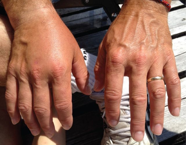
Your body is mostly water, and where that water sits matters just as much as how much you have. In a healthy adult, water makes up roughly 60% of body weight. A handy memory trick is the 60-40-20 rule: about 60% of body weight is total water, about 40% sits inside cells, and about 20% sits outside them.
The fluid compartments
Think of two big rooms. The intracellular fluid (ICF) compartment is all the water inside your cells, and it holds the majority, roughly two-thirds of body water. The extracellular fluid (ECF) compartment is everything outside the cells, the remaining one-third. The ECF splits again into two parts: the interstitial fluid that bathes the spaces between cells, and the intravascular fluid, which is the plasma inside your blood vessels. Fluid is always shifting between these spaces, and keeping it balanced is what keeps cells alive and blood pressure steady.
Osmosis and tonicity
Water follows solute. Osmosis is the movement of water across a semipermeable membrane from an area of lower solute concentration to higher, chasing the particles it cannot easily follow. Tonicity describes how a solution affects cell volume, and it is one of the highest-yield concepts you will use at the bedside when hanging IV fluids.
| Solution | What it does to the cell | Why |
|---|---|---|
| Isotonic (e.g., 0.9% normal saline) | No net change, cell stays the same | Solute concentration matches the cell, so water does not shift |
| Hypotonic (e.g., 0.45% saline) | Cell swells, may burst (lyse) | Outside is more dilute, so water rushes into the cell |
| Hypertonic (e.g., 3% saline) | Cell shrinks (crenates) | Outside is more concentrated, so water is pulled out of the cell |
Starling forces: the tug-of-war at the capillary
At every capillary, two opposing pressures decide whether fluid leaks out or gets pulled back in. This push-pull is described by the Starling forces. Hydrostatic pressure is the outward push of fluid against the vessel wall, essentially the pressure of blood being pumped, and it drives fluid out into the tissues. Oncotic pressure (also called colloid osmotic pressure) is the inward pull created mostly by albumin, the large protein that stays in the bloodstream and draws water back in. Normally, fluid pushes out at the arterial end and gets reabsorbed at the venous end, with the lymphatic system mopping up the small leftover. When this balance tips, fluid piles up in the tissues, and that is edema.
The four mechanisms of edema
Edema is not one problem, it is any disruption of that Starling balance or the drainage system. Four mechanisms cause it:
- Increased hydrostatic pressure. Too much outward push forces fluid into tissues. This is classic in heart failure (blood backs up) and venous obstruction like a DVT, causing dependent swelling in the ankles and feet.
- Decreased plasma oncotic pressure. Low albumin means a weak inward pull, so fluid stays out. Seen in cirrhosis, nephrotic syndrome, and severe malnutrition.
- Lymphatic obstruction. If the lymph channels that normally drain leftover fluid are blocked, protein-rich fluid backs up as lymphedema, often after lymph node removal or radiation in cancer treatment.
- Sodium and water retention. Where sodium goes, water follows. When the kidneys hold onto sodium (often driven by aldosterone or renal failure), the extra volume raises hydrostatic pressure and expands the ECF, causing generalized swelling.
Third spacing
Third spacing is fluid that shifts out of the vessels and interstitium and gets trapped in a space where it does no useful work, such as the abdomen (ascites), the pleural cavity, or the bowel wall after surgery or in sepsis. This matters clinically because that fluid is lost to circulation: the patient can look puffy and swollen while actually being intravascularly dry, with a low blood pressure, rising heart rate, and poor urine output. You may be giving fluids to someone who appears fluid-overloaded, which is why assessing perfusion, not just appearance, is essential. Explore the compartments and these shifts in the interactive figure below.
- Total body water is roughly 60% of weight, split about 40% intracellular and 20% extracellular (interstitial + intravascular), the 60-40-20 rule.
- Tonicity predicts cell volume: hypotonic swells cells, hypertonic shrinks them, isotonic leaves them unchanged.
- Starling forces balance outward hydrostatic push against inward oncotic (albumin) pull; tipping that balance causes edema.
- Edema arises from four mechanisms: high hydrostatic pressure, low oncotic pressure (low albumin), lymphatic obstruction, or sodium/water retention.
- Third spacing traps fluid where it cannot circulate, so a patient can look edematous yet be intravascularly depleted, judge by perfusion and vitals.
Section 3The electrolytes that matter most
Think of electrolytes as the tiny charged particles dissolved in your body's water that make nerves fire, muscles squeeze, and the heart beat on rhythm. Because they carry an electrical charge, even small shifts in their concentration change how excitable your cells are. The body works hard to keep them in a narrow range, and when it can't, the nervous system and the heart are usually the first to complain. Let's walk through the big three, plus a quiet helper.
Sodium: the master of water balance
Sodium (normal roughly 135 to 145 mEq/L) is the main electrolyte outside your cells, and where sodium goes, water follows. That single rule explains almost every symptom. Because sodium controls where water sits, sodium problems are really water problems, and the brain feels them most because it is packed inside a rigid skull.
In hyponatremia (low sodium), the blood outside cells becomes dilute, so water moves into cells by osmosis and they swell. In brain cells this means cerebral edema, which is why patients get headache, confusion, lethargy, and in severe or rapid cases, seizures. In hypernatremia (high sodium), the outside is too salty, so water is pulled out of cells and they shrink. The brain literally dehydrates, causing thirst, restlessness, irritability, and altered mental status. Notice the pattern: sodium imbalances are neurologic because they move water in and out of neurons.
Potassium: the heart's electrolyte
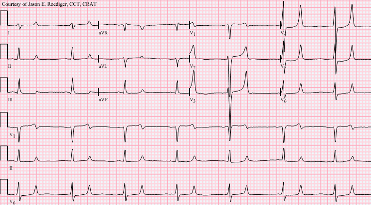
Potassium (normal about 3.5 to 5.0 mEq/L) is the major electrolyte inside cells, and it sets the electrical resting membrane potential that lets heart muscle depolarize and repolarize on cue. This is why potassium is often called the most dangerous electrolyte: both too little and too much can stop the heart, and the danger shows up on the ECG.
In hypokalemia, cardiac cells become sluggish to repolarize, producing flattened T waves, ST depression, and U waves, along with muscle weakness and risk of lethal arrhythmias. In hyperkalemia, cells depolarize too easily at first, giving the classic peaked (tall, tented) T waves, then a widening QRS, and finally a sine-wave pattern that can slide into ventricular fibrillation or asystole. Because both extremes are arrhythmogenic, potassium sits at the top of the emergency list.
| Feature | Hypokalemia | Hyperkalemia |
|---|---|---|
| Level | < 3.5 mEq/L | > 5.0 mEq/L |
| ECG | Flat T waves, U waves, ST depression | Peaked T waves, wide QRS |
| Muscle | Weakness, cramps, ileus | Weakness, later paralysis |
| Main danger | Fatal arrhythmia | Fatal arrhythmia / arrest |
Calcium: nerves, muscles, and PTH
Calcium (total normal roughly 8.5 to 10.5 mg/dL) stabilizes nerve and muscle membranes and is guarded by parathyroid hormone (PTH), which raises calcium by pulling it from bone, saving it in the kidney, and activating vitamin D. Here the rule flips: low calcium makes cells more excitable.
In hypocalcemia, membranes become twitchy and fire too easily, causing tetany, muscle cramps, tingling, and the two bedside signs: Trousseau sign (carpal spasm when a blood-pressure cuff is inflated) and Chvostek sign (facial twitch when you tap the cheek over the facial nerve). In hypercalcemia, often from too much PTH or from cancer, membranes are over-stabilized, so things slow down: "stones, bones, groans, and psychiatric moans" means kidney stones, bone pain, constipation and abdominal pain, and lethargy or confusion.
Magnesium: the quiet partner
Magnesium (normal about 1.7 to 2.4 mg/dL, which is roughly 1.4 to 2.0 mEq/L) tends to travel with potassium and calcium. Low magnesium makes potassium hard to replace and worsens low calcium, so you often cannot fix a stubborn hypokalemia until you correct the magnesium first. Think of it as the electrolyte that keeps the other two honest.
Which imbalances are true emergencies? The ones that threaten the heart or brain fastest: severe hyperkalemia and hypokalemia (lethal arrhythmias within minutes), symptomatic hyponatremia with seizures (brain swelling), and severe hypocalcemia that can cause laryngospasm. Explore the interactive figure below to see how each imbalance shifts.
- Sodium controls water, so its imbalances are neurologic: low sodium swells brain cells (edema, seizures), high sodium shrinks them (thirst, confusion) - and both must be corrected slowly.
- Potassium is the most dangerous electrolyte because both hypo- and hyperkalemia cause lethal arrhythmias; watch the ECG for flat T/U waves (low) versus peaked T waves and wide QRS (high).
- Calcium reverses the rule - low calcium raises excitability, causing tetany with positive Trousseau and Chvostek signs; PTH is the main regulator.
- Magnesium tracks with potassium and calcium; a low magnesium often blocks successful potassium replacement.
- True emergencies are the ones hitting the heart or brain fastest: severe hyper/hypokalemia and symptomatic hyponatremia.
Section 4Acid-base balance & the ABG
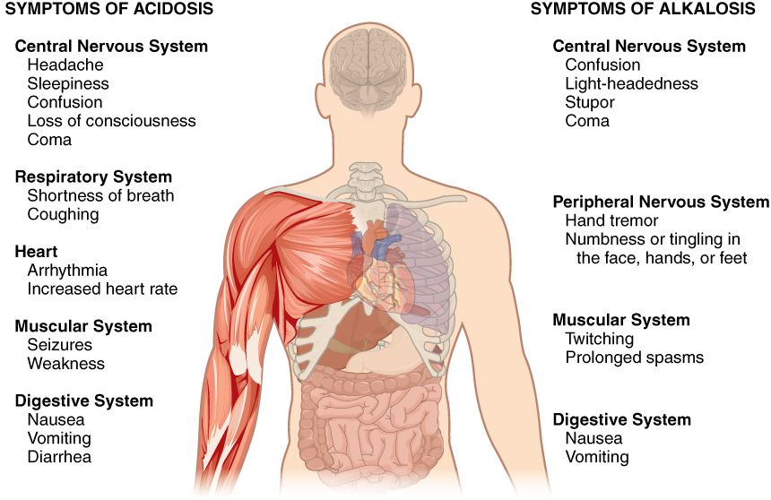
Your blood is fussy about one thing above almost all others: its pH. Every enzyme, every heartbeat, every nerve signal depends on the blood staying in a narrow window of 7.35 to 7.45. That is only barely on the alkaline (basic) side of neutral, and your body guards it obsessively. Slip below 7.35 and we call it acidosis; drift above 7.45 and it is alkalosis. Push much past 6.8 or 7.8 in either direction and cells simply stop working. So the body runs two teams whose whole job is defending that pH.
Two defenders: the lungs (fast) and the kidneys (slow)
Think of pH as a seesaw between an acid and a base. The acid side is carbon dioxide (CO2), a waste gas that dissolves into carbonic acid. The base side is bicarbonate (HCO3), the buffer that soaks up extra acid. The lungs control the CO2 side and they are fast - breathe harder and you blow off CO2 (less acid) within minutes; breathe slower and CO2 builds up. The kidneys control the bicarbonate side and they are slow, taking hours to days to hold onto or dump HCO3. Fast lungs, slow kidneys - remember that and half of ABGs make sense.
The four primary disorders
Every simple acid-base problem is a story about one of those two chemicals going wrong. If the trouble starts with CO2 (a breathing problem), it is respiratory. If it starts with HCO3 (a metabolic or kidney problem), it is metabolic.
| Disorder | Primary change | Classic cause |
|---|---|---|
| Respiratory acidosis | CO2 high (retained acid) | COPD, hypoventilation, opioid overdose |
| Respiratory alkalosis | CO2 low (blown off) | Hyperventilation, anxiety, early sepsis |
| Metabolic acidosis | HCO3 low (acid excess) | DKA, shock/lactic acid, diarrhea |
| Metabolic alkalosis | HCO3 high | Vomiting, NG suction, diuretics |
Reading an ABG in three steps
You do not need to memorize a chart - just walk down three numbers in order. Standard normals are pH 7.35-7.45, PaCO2 35-45 mmHg, and HCO3 22-26 mEq/L.
- Look at the pH first. Below 7.35 = acidosis. Above 7.45 = alkalosis. This tells you which direction the body is actually in.
- Look at the CO2. Above 45 is acidic; below 35 is alkalotic (because CO2 is an acid). If the CO2 matches the pH direction, the lungs are the culprit - it is respiratory.
- Look at the HCO3. Below 22 is acidic; above 26 is alkalotic. If the HCO3 matches the pH direction, the kidneys/metabolism are the culprit - it is metabolic.
Whichever value moves the same way as the pH is your primary problem. Example: pH 7.28 (acidosis) with CO2 60 (acidic) - the CO2 explains it, so this is respiratory acidosis, often from a COPD patient who is retaining CO2.
Compensation: the other team pitches in
Here is the elegant part. When one system fails, the other one tries to drag the pH back toward normal. If the lungs cause an acidosis, the kidneys slowly retain bicarbonate to buffer it. If the kidneys (metabolic) cause an acidosis, the lungs speed up to blow off CO2 - those deep, sighing Kussmaul respirations in DKA are exactly this rescue in action. The compensating system pushes its own number in the same direction as the primary problem. So in a compensated respiratory acidosis you will see high CO2 (the problem) and high HCO3 (the kidney rescue).
Watch the pH to judge how far compensation has gone. If pH is back inside 7.35-7.45, it is fully compensated. If pH is still abnormal but the second value has clearly shifted to help, it is partially compensated. If only the primary system is off and the other has not budged, it is uncompensated. Remember: compensation nudges pH toward normal but rarely overshoots it - the body does not overcorrect past neutral.
Tie it to real patients and it sticks: the vomiting patient loses stomach acid and swings alkalotic (metabolic alkalosis); the panicking, hyperventilating patient blows off CO2 (respiratory alkalosis); the septic or hemorrhaging patient in shock pours out lactic acid (metabolic acidosis); the sedated post-op patient who is barely breathing retains CO2 (respiratory acidosis). Explore the step-by-step ABG interpreter below to practice walking those three numbers yourself.
- Normal blood pH is 7.35-7.45; below is acidosis, above is alkalosis. The lungs defend it fast via CO2, the kidneys slowly via bicarbonate.
- CO2 problems are respiratory; HCO3 problems are metabolic. High CO2 or low HCO3 = acidosis; low CO2 or high HCO3 = alkalosis.
- Read an ABG in order: pH, then CO2, then HCO3. Whichever value moves the same direction as the pH is the primary problem.
- Compensation is the opposite system shifting to pull pH back toward normal (kidneys retain HCO3 for a respiratory acidosis; lungs blow off CO2 in DKA). It rarely fully overshoots normal.
- Classic causes: DKA and shock (metabolic acidosis), COPD/hypoventilation (respiratory acidosis), vomiting (metabolic alkalosis), hyperventilation (respiratory alkalosis).
Section 5Genes & inheritance
Every cell in your body carries a full instruction manual written in DNA. Think of DNA as a long string of just four chemical letters (A, T, C, G). A gene is one meaningful sentence in that manual, and each gene tells the cell how to build one protein. Proteins do almost everything: they form structures, carry oxygen, speed up reactions as enzymes, and open and close channels. The flow is simple to remember: DNA is copied (transcribed) into mRNA, and mRNA is read (translated) into a protein. When the recipe is spelled correctly, the protein works.
What a mutation actually does
A mutation is a spelling change in a gene. Sometimes it is harmless. Other times it swaps one letter and changes one amino acid, so the protein folds wrong or stops working. In sickle cell disease, a single letter change turns normal hemoglobin into a sticky version that warps red cells into a sickle shape. So the whole chain matters: a tiny DNA typo can cascade into a missing or broken protein, and a broken protein shows up as disease. This is why genetic conditions are so consistent within a family - everyone inherits the same misspelled recipe.
Autosomal dominant vs autosomal recessive
You inherit two copies of most genes - one from each parent (these live on the autosomes, chromosomes 1 through 22). Whether a condition appears depends on how many broken copies it takes.
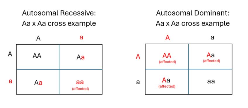
In autosomal dominant inheritance, one faulty copy is enough to show the trait, so it typically appears in every generation and affects males and females equally. Huntington disease is the classic example. In autosomal recessive inheritance, you need two faulty copies to be affected. People with just one copy are healthy carriers - they carry the gene silently and can pass it on. Cystic fibrosis and sickle cell disease follow this pattern, which is why they often seem to skip generations and appear when two carriers have a child.
| Feature | Autosomal dominant | Autosomal recessive |
|---|---|---|
| Copies needed to show | One | Two |
| Family pattern | Usually every generation | Often skips generations |
| Carriers | Rare concept - one copy shows | Yes - one copy is a silent carrier |
| Examples | Huntington disease | Cystic fibrosis, sickle cell |
X-linked recessive: why males are hit harder
Some genes sit on the X chromosome. Females are XX and males are XY. In X-linked recessive conditions like hemophilia, the faulty gene is on the X. A female has a backup X, so one healthy copy usually covers for the broken one - she is typically a carrier. A male has only one X and no second copy to compensate, so a single faulty gene shows. That is why X-linked recessive disorders affect males far more often. A carrier mother has a 50 percent chance of passing the affected X to each son.
Predicting the odds with a Punnett square
A Punnett square is a simple grid that lays out each parent's possible gene copies and shows the chance of each combination in their children. Use capital letters for dominant and lowercase for recessive. For two carriers of a recessive condition (both Aa), the grid gives four boxes: AA, Aa, Aa, aa. That means about a 25 percent chance of an affected child (aa), a 50 percent chance of a carrier, and a 25 percent chance of neither. Each pregnancy is an independent roll - the odds reset every time. You can explore the patterns and squares in the interactive figure below.
Penetrance and expressivity
Penetrance is whether a gene shows at all - the percentage of people with the gene who actually develop the trait. Expressivity is how strongly it shows - how mild or severe the same gene looks from person to person.
- DNA holds the recipe; a gene builds a protein; a mutation is a spelling change that can break that protein and cause disease.
- Autosomal dominant needs one copy and appears every generation (Huntington); autosomal recessive needs two copies, with healthy carriers (cystic fibrosis, sickle cell).
- X-linked recessive conditions (hemophilia) mostly affect males because they have only one X and no backup copy.
- A Punnett square predicts offspring odds; two recessive carriers have about a 25 percent chance of an affected child each pregnancy.
- Penetrance is whether the gene shows; expressivity is how strongly it shows.
Section 6Chromosomal & congenital disorders
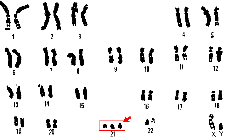
Every cell in your body normally carries 46 chromosomes arranged in 23 pairs: 22 pairs of autosomes plus one pair of sex chromosomes (XX in females, XY in males). You inherit exactly half from each parent. This tidy sorting happens during meiosis, the special cell division that makes eggs and sperm. When meiosis works, each gamete ends up with exactly 23 chromosomes, and fertilization restores the full set of 46. Chromosomal disorders happen when that sorting goes wrong, or when an outside exposure damages a developing embryo. Explore the interactive figure below to see how an extra chromosome reshapes the picture.
Nondisjunction: chromosomes that refuse to separate
The most common mistake is nondisjunction - a pair of chromosomes fails to pull apart during meiosis. One gamete ends up with an extra copy, and the other is left short. If that gamete joins in fertilization, the embryo carries the wrong count, a state called aneuploidy. Getting three copies of a chromosome instead of two is a trisomy; having only one copy is a monosomy. Because chromosomes are packed with genes, even one extra or missing chromosome throws off the dose of hundreds of proteins - which is why these disorders affect so many body systems at once. Nondisjunction becomes more likely with advancing maternal age, which is why the risk of trisomies climbs after about age 35.
Trisomy 21 and its cousins
Trisomy 21 (Down syndrome) is the most common surviving autosomal trisomy - three copies of chromosome 21. Classic features usually include intellectual disability, a flat facial profile with upslanting palpebral fissures, a single palmar (simian) crease, and hypotonia (low muscle tone). Because that extra genetic material affects many systems, congenital heart defects (often an atrioventricular septal defect) are common, along with a higher risk of leukemia and early Alzheimer-type changes. Two rarer, far more severe trisomies are worth knowing: trisomy 18 (Edwards syndrome) and trisomy 13 (Patau syndrome). Both usually cause profound impairment and multiple organ malformations, and most affected infants do not survive past infancy.
When the sex chromosomes are off
Nondisjunction can also hit the sex chromosomes. Turner syndrome (45,X) is a monosomy - a female with only one X. It usually presents with short stature, a webbed neck, ovarian dysgenesis (nonfunctioning ovaries causing infertility and absent puberty without hormone therapy), and often coarctation of the aorta. Klinefelter syndrome (47,XXY) is a male with an extra X. It often shows up around puberty with tall stature and small firm testes: the seminiferous tubules scar over and germ cells are lost, so the semen has no sperm (azoospermia) and the man is infertile. In parallel, the Leydig cells fail, and that reduced testosterone is what produces the gynecomastia and delayed or incomplete puberty. Testosterone replacement fixes the hormonal features but does not restore fertility, because the tubules themselves are gone. Notice the pattern: an extra sex chromosome tends to be better tolerated than an extra autosome, largely because the body naturally silences extra X chromosomes.
| Feature | Turner (45,X) | Klinefelter (47,XXY) |
|---|---|---|
| Karyotype | One X, missing a sex chromosome | Two X plus one Y |
| Phenotype | Female | Male |
| Stature | Short | Tall |
| Classic clue | Webbed neck, coarctation | Small testes, gynecomastia |
| Fertility | Usually infertile (ovarian dysgenesis) | Usually infertile (testicular dysgenesis, azoospermia); testosterone also low |
Teratogens: when the environment is the culprit
Not every congenital disorder is genetic. A teratogen is any agent that disrupts normal fetal development - a drug, infection, chemical, or radiation. Timing matters enormously. The embryonic period (roughly weeks 3 to 8), when organs are first forming, is the window of greatest vulnerability; an exposure then can cause major structural defects, while later exposure tends to affect growth and function. A key cluster to memorize is the TORCH infections: Toxoplasmosis, Other (including syphilis), Rubella, Cytomegalovirus, and Herpes simplex. These maternal infections can cross the placenta and cause malformations, growth restriction, hearing loss, and neurologic damage. Alcohol is one of the most preventable teratogens; prenatal exposure can produce fetal alcohol spectrum disorder, with characteristic facial features, growth deficits, and lifelong cognitive impairment. There is no known safe amount.
Multifactorial disorders
Finally, many common conditions - neural tube defects, cleft lip and palate, congenital heart disease - are multifactorial, arising from a combination of several genes plus environmental influences rather than a single cause.
- Nondisjunction - chromosomes failing to separate in meiosis - causes trisomies (extra copy) and monosomies (missing copy).
- Trisomy 21 (Down syndrome) is the most common autosomal trisomy; trisomy 18 (Edwards) and 13 (Patau) are rarer and usually fatal in infancy.
- Sex-chromosome disorders include Turner (45,X), a short-statured infertile female, and Klinefelter (47,XXY), a tall infertile male.
- Teratogens - including the TORCH infections and alcohol - do the most structural harm during the embryonic period (weeks 3 to 8).
- Multifactorial disorders arise from combined genetic and environmental causes.
The chapter in one breath
- Cellular adaptation is the cell's survival plan under stress, running through five modes: atrophy (shrink), hypertrophy (enlarge), hyperplasia (multiply), metaplasia (swap to a tougher cell type), and dysplasia (disordered growth that can precede cancer).
- When stress outpaces adaptation you get reversible injury (cell swelling from failing Na+/K+ pumps), and if hypoxia and ATP loss persist it tips into irreversible injury and cell death.
- Apoptosis is tidy, programmed, energy-using single-cell suicide with no inflammation, while necrosis is messy, accidental death where cells rupture, spill their contents, and trigger inflammation.
- Body water splits into intracellular (about two-thirds) and extracellular compartments, and edema happens when fluid leaks into tissues from high hydrostatic pressure, low albumin (oncotic pull), blocked lymphatics, or sodium and water retention.
- The must-know electrolytes are sodium (water balance and nerve firing), potassium (the cardiac rhythm ion, dangerous high or low), calcium (bones, clotting, muscle contraction), and magnesium (the quiet cofactor that stabilizes nerves and heart).
- Acid-base balance is a tug-of-war between the lungs (fast, blowing off CO2) and kidneys (slow, adjusting HCO3-), keeping arterial pH in the tight 7.35-7.45 window.
- Read an ABG by checking pH first (acid vs alkaline), then CO2 for a respiratory cause and HCO3- for a metabolic cause, then decide if the other system is compensating to nudge pH back toward normal.
- Inheritance patterns follow the pedigree: autosomal dominant (one copy shows, every generation), autosomal recessive (needs two copies, carriers hidden), and X-linked recessive (hits males hardest, like hemophilia).
- Chromosomal disorders come from wrong chromosome numbers, classically trisomy 21 (Down syndrome) from nondisjunction, while many congenital defects like neural tube defects arise from gene-environment hits (for example, low folate) during development.
ReferenceWords worth knowing
A quick reference for the terms in this chapter — skim it now, or circle back whenever a word trips you up.
- Apoptosis
- Apoptosis is orderly, programmed cell death in which a cell shrinks and neatly packages itself for removal without spilling its contents, so no inflammation results.
- Atrophy
- Atrophy is a decrease in the size of cells (and therefore the whole tissue or organ) in response to disuse, denervation, aging, or reduced blood supply, as when a casted limb shrinks.
- Autosomal recessive
- Autosomal recessive describes an inheritance pattern in which a person must receive two copies of a mutated gene (one from each parent) on a non-sex chromosome to show the disease, so unaffected carriers can silently pass it on.
- Compensation (acid-base)
- Compensation is the body's attempt to restore normal blood pH by using the system that is not primarily broken, so the lungs adjust CO₂ for a metabolic problem and the kidneys adjust bicarbonate for a respiratory problem.
- Dysplasia
- Dysplasia is disordered growth in which cells vary in size, shape, and organization; it is not cancer but is often a warning change that can precede it.
- Edema
- Edema is the buildup of excess fluid in the interstitial space (the tissue between cells), producing visible swelling and sometimes pitting when pressed.
- Hyperplasia
- Hyperplasia is an increase in the number of cells in a tissue due to increased cell division, such as the endometrium thickening under hormonal stimulation.
- Hypertrophy
- Hypertrophy is an increase in the size of individual cells (without adding new cells), enlarging the organ, as when heart muscle thickens against high blood pressure.
- Hyponatremia
- Hyponatremia is a lower-than-normal blood sodium level (usually below about 135 mEq/L) that often reflects too much water relative to sodium and can cause confusion or seizures.
- Hypoxia
- Hypoxia is an inadequate supply of oxygen reaching the tissues, which starves cells of the fuel they need to make energy.
- Ischemia
- Ischemia is reduced blood flow to a tissue that limits delivery of both oxygen and nutrients while also letting wastes build up, and it is a common cause of hypoxia.
- Metaplasia
- Metaplasia is the reversible replacement of one mature cell type by another, tougher one under stress (such as airway cells changing under chronic cigarette smoke); the swap withstands the stress but loses the original tissue's specialized function and can precede dysplasia.
- Necrosis
- Necrosis is disorderly, unplanned cell death from injury in which cells swell and burst, spilling their contents and triggering inflammation.
- Nondisjunction
- Nondisjunction is the failure of chromosomes to separate properly during cell division, leaving a cell with an extra or missing chromosome, as in trisomy 21 (Down syndrome).
- Oncotic pressure
- Oncotic pressure is the inward pull that plasma proteins (mainly albumin) exert to keep fluid inside blood vessels; when it drops, fluid leaks out and edema forms.
- Tonicity
- Tonicity is how a solution's solute concentration affects a cell's water movement, causing the cell to swell in a hypotonic solution, shrink in a hypertonic one, or stay unchanged in an isotonic one.
DrillFlashcards for this unit
These are the Week 1 cards — the same deck as the hub, pulled in here so you can review the moment you finish reading. Tap the card to flip it; hit Shuffle to mix them up. To grade yourself and track mastery across the whole deck, open the full flashcards page.
Check yourself
Immunity, Inflammation & Hypersensitivity
The immune system is the body’s standing army — mostly invisible, until it wins too slowly, overreacts, or turns on its own side. This chapter follows the whole arc: who the invaders are, how the innate and adaptive defenses answer them, what inflammation costs, and what happens when immunity misfires as allergy, autoimmunity, or immunodeficiency.
By the end of this chapter you can
- Name the major classes of pathogens and how each causes disease.
- Contrast innate with adaptive immunity, and humoral with cell-mediated defense.
- Match each of the five antibody classes to its signature role.
- Explain the cardinal signs of inflammation by their mechanisms.
- Tell the four hypersensitivity types apart with a classic example of each.
- Describe how autoimmunity and immunodeficiency (including HIV/AIDS) arise.
Section 1The invaders: types of pathogens
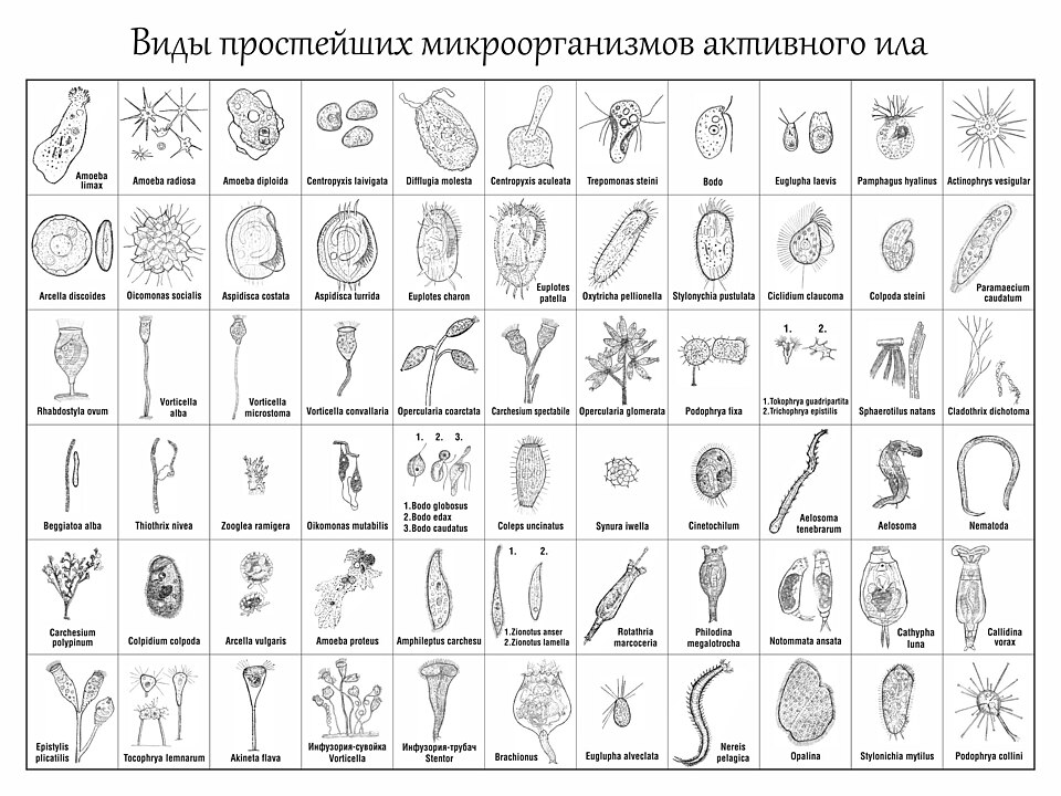
Right now, as you read this, you are covered and colonized by trillions of microorganisms, and that is completely normal. We call these resident microbes your normal flora (or microbiota). They live on your skin, in your mouth, and especially in your gut, and they earn their keep: they crowd out troublemakers, help digest food, and even make some vitamins (like vitamin K). Most of the time you and your flora have a peaceful, mutually helpful relationship. A pathogen is different. It is any organism that can cause disease, either by invading tissue, releasing toxins, or triggering an immune response that harms you. Some organisms are opportunistic: harmless in their usual spot, but dangerous when they get somewhere they do not belong (for example, gut bacteria entering the bloodstream after surgery). Understanding what kind of invader you are dealing with is the first step, because the type of organism decides the treatment.
Bacteria: tiny, but they play dirty
Bacteria are single-celled organisms with their own machinery to live and reproduce, which is why many can grow on their own outside our cells. Nurses sort them first by shape (round cocci, rod-shaped bacilli) and, crucially, by the Gram stain. In plain terms, gram-positive bacteria have a thick peptidoglycan cell wall (and no outer membrane) that traps and holds the purple stain, while gram-negative bacteria have a thin wall hidden under an extra outer membrane, so they lose the purple and take a pink counterstain. That difference is not just cosmetic. Gram-negative bacteria carry endotoxin (lipopolysaccharide, LPS) in that outer membrane; when the bacteria die and break apart, LPS floods out and can drive high fever, dangerously low blood pressure, and septic shock. Bacteria harm us in two broad ways: by invasion (physically spreading into and destroying tissue) and by releasing toxins. Exotoxins are proteins some bacteria secrete that damage cells at a distance (think of the toxins behind tetanus or C. difficile colitis), while endotoxin comes from the gram-negative wall itself.
Viruses: hijackers that cannot live alone
Viruses are not really cells at all. They are genetic material (DNA or RNA) wrapped in a protein coat, and they are obligate intracellular parasites meaning they must get inside one of your living cells to do anything. A virus attaches to a host cell, enters it and releases its genes inside, and then hijacks the cell's own ribosomes and enzymes to mass-produce new virus copies. The cell is turned into a factory and is often destroyed in the process. Because viruses hide inside your cells and borrow your machinery, antibiotics do nothing against them, and treatment is harder to target without harming the host.
Fungi, parasites, and a word on prions
Fungi (yeasts like Candida and molds) are more complex cells that often turn pathogenic when the immune system is weakened or when normal flora is wiped out by antibiotics. Protozoa are single-celled parasites (for example, the Plasmodium that causes malaria), while helminths are the multicellular parasitic worms. Finally, prions are the strange outliers: misfolded proteins, with no DNA or RNA at all, that cause other proteins to misfold, leading to fatal brain diseases.
Matching the treatment to the invader
Here is the payoff: naming the class of organism guides the whole plan. Antibiotics target features only bacteria have (like the cell wall), so they are useless against viruses. Giving them anyway breeds antibiotic resistance and can trigger a C. difficile infection by wiping out protective flora.
| Pathogen | Key trait | Treatment class |
|---|---|---|
| Bacteria | Own cells; toxins & invasion | Antibiotics |
| Viruses | Hijack host cells | Antivirals |
| Fungi | Yeasts & molds | Antifungals |
| Parasites | Protozoa & worms | Antiparasitics |
Explore the major classes of pathogens in the figure below to see how these families line up side by side.
- Normal flora usually protect us; a pathogen is any organism that causes disease, and opportunists turn harmful in the wrong place or when defenses drop.
- Bacteria harm us by invasion and toxins; gram-negative endotoxin (LPS) can drive fever and septic shock, while the Gram stain guides early therapy.
- Viruses are obligate intracellular parasites that hijack host cells to replicate, so antibiotics do not work on them.
- Fungi, protozoa, and helminths each need their own drug class; prions are misfolded proteins with no nucleic acid.
- Matching drug to organism (antibiotic vs antiviral vs antifungal) matters, and misusing antibiotics breeds resistance and can cause C. difficile.
Section 2The body’s defenses
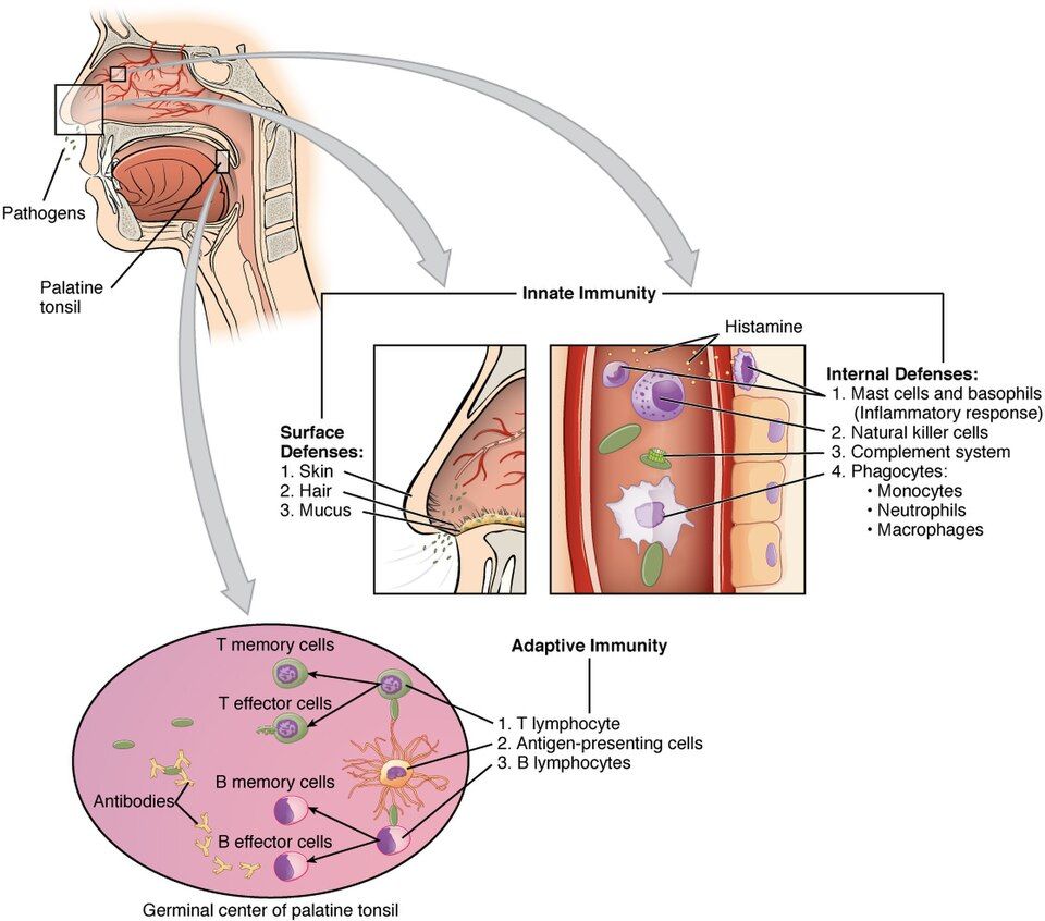
Every second of every day, your body is fending off bacteria, viruses, and other invaders without you ever noticing. It does this with two teams that work together: a fast, general-purpose crew called innate immunity, and a slower but far more precise crew called adaptive immunity. Understanding how these two teams operate is the foundation for everything you will learn about infection, inflammation, and hypersensitivity.
Innate immunity: fast, but nonspecific
Innate immunity is the defense you are born with. It responds within minutes to hours, but it does not care exactly who the invader is: it treats most threats the same way. Think of it as the building's locks, alarms, and security guards.
- Physical and chemical barriers come first. Intact skin is a wall invaders cannot easily cross, and mucous membranes lining the airways and gut trap microbes so cilia and secretions can sweep them out. Stomach acid and lysozyme in tears add chemical defense. This is why a break in the skin, like an IV site or a pressure injury, is a real infection risk.
- Phagocytes are cells that eat invaders. Neutrophils arrive first and fast (they dominate the early response and are the main cell in pus), while macrophages arrive a bit later, engulf debris, and clean up. This eating process is called phagocytosis.
- Complement is a cascade of plasma proteins that punches holes in microbes, tags them for phagocytes (opsonization), and amplifies inflammation.
- Natural killer (NK) cells destroy your own cells that have turned dangerous, especially virus-infected and some tumor cells.
- Fever is a whole-body innate response. Chemical messengers called pyrogens reset the hypothalamus to a higher temperature, which slows microbial growth and speeds up immune cells. This is why a fever, though uncomfortable, is often a sign the defenses are working.
Adaptive immunity: specific, with memory
Adaptive immunity is slower to start (days on a first exposure) but it is specific: it targets one particular invader, and it remembers. It has two branches.
- Humoral immunity is run by B cells. When activated, B cells become plasma cells that pump out antibodies (immunoglobulins) into the blood and fluids. Antibodies neutralize toxins, clump microbes together, and flag them for destruction. This branch is especially good against invaders outside your cells, like bacteria in the bloodstream.
- Cell-mediated immunity is run by T cells and handles threats inside cells, such as viruses, and abnormal cells.
The T-cell coordinators and killers
Two T-cell types are worth knowing by name. Helper T cells (CD4) are the coordinators: they release cytokines that activate B cells, cytotoxic T cells, and macrophages. Because HIV attacks CD4 cells, a falling CD4 count leaves the whole system leaderless, which is why HIV is so devastating. Cytotoxic T cells (CD8) are the killers: they directly destroy virus-infected and cancerous cells.
Active vs passive immunity
Immunity can be earned or borrowed. In active immunity your own body mounts the response and builds memory, either by getting sick or by vaccination (a safe preview of the invader). It is slow to develop but long-lasting. In passive immunity you receive ready-made antibodies from someone else, so protection is immediate but temporary, because you never made memory cells. Examples include antibodies crossing the placenta to the fetus and antibodies delivered in breast milk.
| Feature | Active immunity | Passive immunity |
|---|---|---|
| Source | Your own immune response | Borrowed antibodies |
| Onset | Slow (days to weeks) | Immediate |
| Duration | Long-lasting (memory) | Short (no memory) |
| Examples | Infection, vaccines | Placenta, breast milk, antibody injections |
Primary vs secondary response
The primary response is your body's first meeting with an invader. It is slow, often taking a week or more to build up antibodies, which is why you can feel sick during that first encounter. But the immune system files away memory cells. On a repeat exposure, the secondary (memory) response is faster and stronger, producing far more antibodies far more quickly, so you may never feel sick at all. This is exactly why vaccines and booster doses work: they safely create memory so the real invader triggers a rapid secondary response. Explore the innate-versus-adaptive figure below to see how these teams fit together.
- Innate immunity (barriers, neutrophils and macrophages, complement, NK cells, fever) is fast but nonspecific; adaptive immunity is slower but specific with memory.
- Adaptive immunity has two branches: humoral (B cells make antibodies, best against extracellular threats) and cell-mediated (T cells, best against intracellular threats).
- Helper T cells (CD4) coordinate the response; cytotoxic T cells (CD8) kill infected and abnormal cells.
- Active immunity (infection or vaccine) is self-made and long-lasting; passive immunity (placenta, breast milk, antibody injections) is borrowed, immediate, and temporary.
- The secondary (memory) response is faster and stronger than the primary response, which is the basis for vaccines and boosters.
Section 3Antibodies (immunoglobulins)
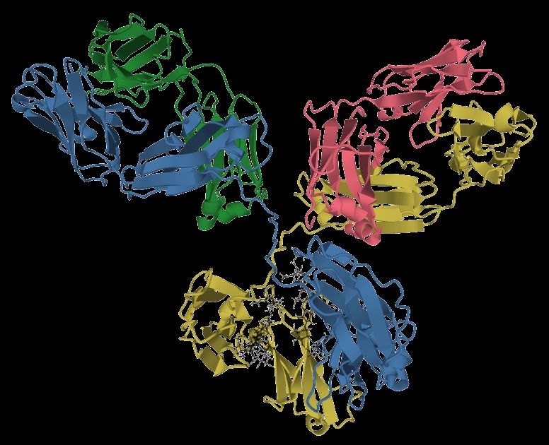
Your immune system faces a huge problem: how do you recognize millions of different germs, when each one looks a little different? The answer is antibodies, also called immunoglobulins (Ig). Think of an antibody as a tiny Y-shaped protein tag that your body custom-builds to match one specific target. The tips of the Y are the variable region, shaped to lock onto a particular antigen (a molecular fingerprint on a microbe). The stem of the Y, the constant region, is the handle that the rest of the immune system grabs.
Antibodies are made by plasma cells, which are mature B lymphocytes that have been activated by their antigen. Once released, an antibody does three main jobs. First, it tags the invader so that phagocytes recognize and eat it, a process called opsonization. Second, it can neutralize a threat by physically coating a virus or a toxin so it can no longer attach to your cells. Third, it can activate complement, a cascade of blood proteins that punches holes in bacterial membranes and amplifies inflammation. In short: tag, neutralize, and activate.
The five classes and their signature jobs
All antibodies share that Y shape, but the constant-region handle comes in five flavors, and each flavor sends the antibody to a different job and a different place in the body.
- IgG is the most abundant antibody in blood and the workhorse of the secondary (memory) response. It is the only class that crosses the placenta, and it is excellent at opsonization, neutralization, and activating complement.
- IgM is the first to appear in a new infection and the largest antibody, because it circulates as a pentamer (five Y units joined together). Those ten binding arms make it a powerful early complement activator.
- IgA guards the mucosal surfaces, the wet linings of the gut, lungs, and airways, and it is concentrated in secretions like saliva, tears, and breast milk, giving a nursing newborn ready-made protection.
- IgE drives allergy and type I hypersensitivity and defends against parasites. It binds mast cells and basophils; when antigen cross-links it, those cells release histamine.
- IgD mostly sits on the surface of naive B cells as part of the B-cell receptor, helping them get activated.
Reading antibody levels: recent versus past infection
Because IgM and IgG appear on different timelines, a lab panel can tell a story about when an exposure happened. When you meet an antigen for the first time, IgM rises first, usually within days, then fades over a few weeks. IgG rises later and stays elevated for months to years, providing lasting immunity. Nurses use this pattern constantly.
| Feature | IgM elevated | IgG elevated |
|---|---|---|
| What it suggests | Recent or current infection | Past infection or immunity |
| Timing | Appears first, fades in weeks | Appears later, persists long-term |
| Response type | Primary response | Secondary / memory response |
| Classic example | Acute hepatitis or rubella | Prior vaccination or old exposure |
So IgM positive with IgG negative usually points to a fresh infection, while IgG positive with IgM negative usually means the person was infected or vaccinated in the past and is now immune.
Why placenta-crossing IgG protects a newborn
A newborn's own immune system is still learning and cannot make strong antibody responses yet. Because IgG crosses the placenta during pregnancy, the mother essentially loans her baby a full library of antibodies. This passive immunity shields the infant for roughly the first several months of life while their own B cells mature. IgA in breast milk adds a second layer of protection right at the gut lining. The trade-off is that this borrowed protection is temporary, which is exactly why the infant vaccination schedule begins before it fully wears off.
The interactive figure below lets you explore the five classes side by side.
- Antibodies (immunoglobulins) are Y-shaped proteins from plasma cells that tag, neutralize, and activate complement against a specific antigen.
- Five classes, memory hook GAMED: IgG (most abundant, crosses placenta), IgA (mucosa and breast milk), IgM (first and largest, pentamer), IgE (allergy and parasites), IgD (B-cell receptor).
- IgM rises first and fades, signaling a recent infection; IgG rises later and persists, signaling past infection or immunity.
- Maternal IgG crosses the placenta to give the newborn temporary passive immunity for the first months of life.
Section 4Inflammation & healing
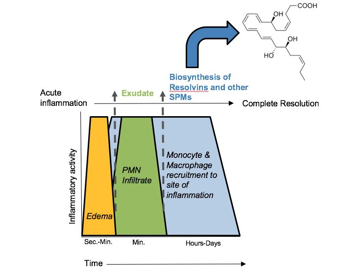
Think of inflammation as your body's first-responder team. It is not the same as infection. Infection means microbes have invaded; inflammation is the response, and it fires whether the trigger is bacteria, a splinter, a burn, a sprain, or dying tissue. In healthy tissue, capillaries run quietly and white blood cells patrol the bloodstream. The moment cells are injured, that calm is interrupted on purpose: the goal is to contain the threat, clear the debris, and set up repair. Understanding the response means understanding a handful of chemical mediators and what they do to blood vessels.
The five cardinal signs and why each happens
Injured cells and nearby mast cells release histamine, along with prostaglandins, bradykinin, and cytokines. These mediators drive two vascular changes that explain every classic sign.
- Redness (rubor) and heat (calor) come from vasodilation - the arterioles widen, so more warm blood floods the area.
- Swelling (tumor) comes from increased capillary permeability. The vessel walls loosen and let protein-rich fluid leak into the tissue, forming edema.
- Pain (dolor) comes from bradykinin and prostaglandins stimulating nerve endings, plus pressure from the swelling.
- Loss of function (functio laesa) is the sum of pain and swelling - a swollen, aching joint simply will not move well.
Meanwhile, neutrophils are the first white cells to arrive, squeezing out of vessels (diapedesis) to begin phagocytosis of pathogens and debris.
Acute versus chronic inflammation
Acute inflammation is fast and self-limiting - hours to a few days - and ideally ends in full healing. Chronic inflammation lingers for weeks, months, or years, usually because the trigger never clears (persistent infection, autoimmune disease, ongoing irritation). The cell cast changes too: neutrophils give way to macrophages and lymphocytes. The cost to the tissue is real - chronic inflammation drives fibrosis (scarring), tissue destruction, and sometimes granuloma formation, so the organ slowly loses working cells and gains stiff, non-functional scar.
Exudates and the systemic response
The fluid that leaks out is called exudate. Serous exudate is thin and watery (think of a blister); purulent exudate is thick pus, full of dead neutrophils and microbes, and signals infection. Inflammation is not only local. Cytokines such as IL-1 and IL-6 travel to the brain and liver, producing systemic signs: fever (a raised hypothalamic set-point), leukocytosis (elevated WBC count), and rising acute-phase reactants - a climbing CRP (a true acute-phase protein), and a rising ESR (an indirect marker driven by fibrinogen), are lab clues that inflammation is active somewhere in the body.
Healing: resolution versus repair
Two outcomes are possible. Resolution is the best case: the tissue was only mildly injured and the cells can regenerate, so it returns to normal. Repair with scar (fibrosis) happens when damage is extensive or the cells cannot regenerate - fibroblasts lay down collagen and granulation tissue fills the gap, leaving a scar that holds but does not fully function.
| Feature | Primary intention | Secondary intention |
|---|---|---|
| Wound edges | Clean, approximated (sutured) | Wide, gaping, cannot be closed |
| Tissue loss | Minimal | Significant |
| Healing time | Fast | Slower |
| Scar | Small | Larger, more granulation tissue |
| Infection risk | Lower | Higher |
Why anti-inflammatory drugs work
NSAIDs (ibuprofen, aspirin) block the COX enzymes that make prostaglandins, so they blunt pain, fever, and swelling but leave much of the immune response intact. Corticosteroids (prednisone) act much higher upstream, suppressing the genes for many mediators at once - they are far more powerful but also more immunosuppressive, which is why they raise infection risk and slow healing. Explore the acute inflammatory response and its cardinal signs in the figure below.
- The five cardinal signs (redness, heat, swelling, pain, loss of function) all trace back to vasodilation and increased capillary permeability driven by histamine and prostaglandins.
- Acute inflammation is short and usually heals; chronic inflammation persists and costs tissue through fibrosis and destruction.
- Systemic signs of inflammation include fever, leukocytosis, and elevated CRP/ESR; purulent exudate suggests infection.
- Healing is either resolution (back to normal) or repair with scar; clean wounds heal by primary intention, gaping wounds by secondary.
- NSAIDs block prostaglandins (COX); corticosteroids suppress mediators broadly and can mask signs of infection.
Section 5When immunity overreacts: hypersensitivity
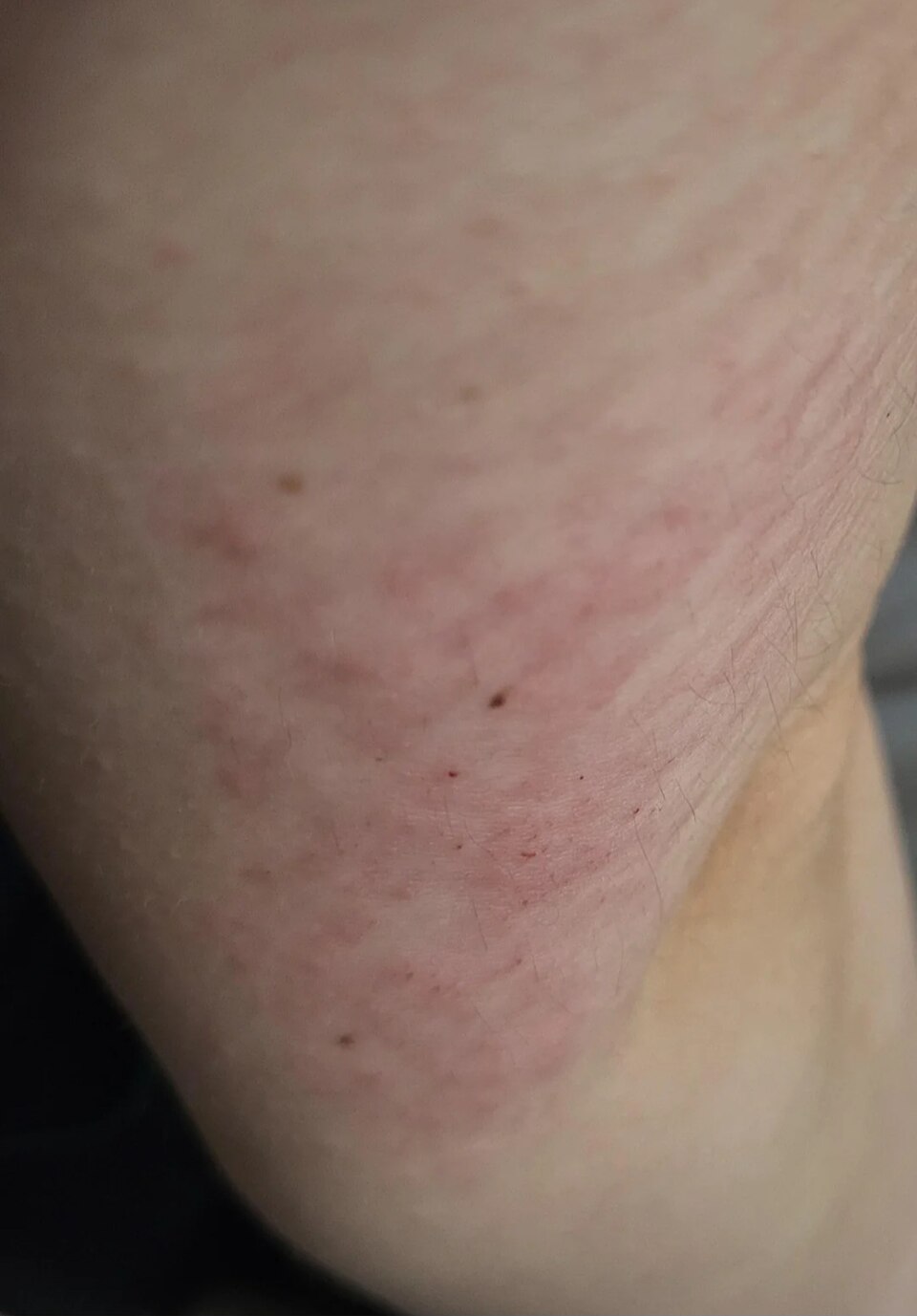
Your immune system is built to protect you, but sometimes it protects too hard. Hypersensitivity means the immune response is exaggerated or misdirected, so the defense itself becomes the source of tissue damage. To understand this, remember how normal immunity works. Your body makes antibodies (immunoglobulins) that tag foreign invaders, and specialized T cells that recognize threats directly. Normally these tools attack germs and leave your own cells alone. In hypersensitivity, that precision breaks down. The immune system reacts to something harmless (pollen, a food, a medication), targets your own cells, or leaves inflammatory debris behind that damages healthy tissue.
Clinicians sort these overreactions into four classic types, and a simple memory device keeps them straight: ACID - Anaphylactic, Cytotoxic, Immune-complex, Delayed. The first three are driven by antibodies; only the fourth is not.
Type I - Immediate (IgE-mediated): allergy and anaphylaxis
This is the classic allergy. On first exposure, the body makes IgE antibodies against an otherwise harmless allergen and parks them on the surface of mast cells and basophils (this silent step is called sensitization). On the next exposure, the allergen bridges those IgE molecules, and the mast cells degranulate, dumping histamine and other mediators within minutes. Histamine causes vasodilation (redness, warmth, low blood pressure), increased capillary permeability (swelling, hives), and bronchoconstriction (wheezing). A local reaction is hay fever or hives; a whole-body reaction is anaphylaxis, where airway swelling and vasodilatory shock can kill quickly. This is why epinephrine is the first-line rescue - it reverses bronchoconstriction and vasodilation fast.
Type II - Cytotoxic (antibody against cells)
Here antibodies (usually IgG or IgM) bind directly to antigens on the surface of specific cells, marking them for destruction by complement and phagocytes. The classic example is a mismatched blood transfusion: give type A blood to a type B patient and the recipient's preformed antibodies attack the donor red cells, causing rapid hemolysis, fever, flank pain, and dark urine. Another example is hemolytic disease of the newborn, where an Rh-negative mother makes anti-Rh antibodies that cross the placenta and destroy an Rh-positive baby's red cells.
Type III - Immune-complex mediated
In Type III, antibody and antigen clump together into immune complexes that circulate and then deposit in tissues - joints, skin, kidney glomeruli, blood vessel walls. Once lodged, they activate complement and trigger inflammation right where they settle. This is the mechanism behind systemic lupus erythematosus (SLE) and serum sickness (a reaction to foreign proteins, such as certain antivenoms). The key difference from Type II: the antibody is not aimed at a fixed cell, it is floating debris that inflames wherever it lands. Timing is broad rather than fixed - a localized Arthus reaction appears within about 4 to 12 hours, classic serum sickness takes 7 to 14 days after the foreign protein is given (fever, rash, joint pain), and SLE is lifelong and relapsing. So a reaction showing up a week after antivenom or antithymocyte globulin is still Type III.
Type IV - Delayed (T-cell mediated)
Type IV is the odd one out - no antibody at all. Instead, sensitized T lymphocytes recognize the antigen and recruit macrophages, and because this cell-driven process takes time, the reaction peaks at 48 to 72 hours. Read a TB skin test (PPD) at 48-72 hours for exactly this reason - the firm raised induration is delayed T-cell inflammation. Other examples include contact dermatitis (poison ivy, nickel) and transplant (graft) rejection.
| Type | Mediator | Timing | Classic example |
|---|---|---|---|
| I - Anaphylactic | IgE, mast cells, histamine | Seconds to minutes | Anaphylaxis, hay fever |
| II - Cytotoxic | IgG/IgM against cell surface | Minutes to hours | Transfusion reaction, HDN |
| III - Immune-complex | Circulating antibody-antigen complexes | Hours to weeks | SLE, serum sickness |
| IV - Delayed | T cells (no antibody) | 48-72 hours | TB skin test, poison ivy |
Explore the four types side by side in the interactive figure below.
- ACID orders the four types: Anaphylactic (I), Cytotoxic (II), Immune-complex (III), Delayed (IV).
- Types I, II, and III are antibody-driven; Type IV is T-cell driven with no antibody.
- Type I needs prior sensitization - the severe reaction hits on re-exposure, and epinephrine is the anaphylaxis rescue.
- Type II targets specific cells (transfusion reaction, hemolytic disease of the newborn); Type III deposits floating complexes that inflame tissues (SLE, serum sickness).
- Type IV is delayed 48-72 hours - the reason a TB skin test is read days later.
Section 6Autoimmunity & immunodeficiency
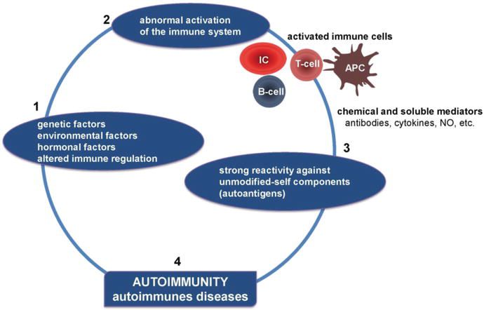
Your immune system has an almost impossible job: it must attack anything foreign - bacteria, viruses, cancer cells - while leaving you completely alone. The ability to tell "self" from "non-self" is called self-tolerance, and it is built during immune development. As lymphocytes mature, any that react strongly against your own tissues are normally deleted or switched off (a process of central and peripheral tolerance). When that filtering breaks down, the immune system turns its weapons inward. This section covers what happens when tolerance fails (autoimmunity) and, on the flip side, when the immune system is too weak to defend at all (immunodeficiency).
When self-tolerance breaks down: autoimmunity
In autoimmune disease, the immune system loses self-tolerance and begins producing autoantibodies and self-reactive T cells that treat normal tissue like an invader. The result is chronic inflammation and tissue destruction at whatever the immune system targets. The trigger is usually a mix of genetic susceptibility (often certain HLA types) plus an environmental push such as an infection, hormones, or drugs. A helpful way to learn these diseases is to ask one question: what tissue is being attacked? Explore the interactive figure below to see how tolerance breaks down step by step.
| Disease | Target attacked | Classic result |
|---|---|---|
| SLE (lupus) | Many tissues (multi-system) - skin, joints, kidneys, blood | Butterfly rash, joint pain, lupus nephritis; ANA usually positive |
| Rheumatoid arthritis | Synovial joints (the joint lining) | Symmetric, warm, swollen joints; morning stiffness |
| Multiple sclerosis | Myelin around CNS nerves | Slowed nerve signals - weakness, vision changes, numbness |
| Type 1 diabetes | Beta cells of the pancreas | No insulin made - high blood glucose |
| Hashimoto thyroiditis | Thyroid (destroys it) | Hypothyroidism - fatigue, cold, weight gain |
| Graves disease | Thyroid (over-stimulates it) | Hyperthyroidism - heat intolerance, weight loss, exophthalmos |
Notice that Hashimoto and Graves hit the same organ with opposite outcomes. In Hashimoto, autoantibodies and T cells slowly destroy thyroid tissue, so hormone output falls. In Graves, the autoantibody actually mimics TSH and switches the gland on constantly, flooding the body with thyroid hormone. Same target, opposite mechanism - a favorite exam contrast.
The opposite problem: immunodeficiency
Sometimes the danger is not an overactive immune system but an underactive one. In immunodeficiency, part of the immune defense is missing or weakened, so ordinary germs become dangerous. There are two broad types:
- Primary (congenital) - the person is born with a genetic defect in immune development. These are usually rare and often show up in infancy with repeated, severe, or unusual infections.
- Acquired (secondary) - the immune system develops normally but is later damaged by something else: infection (HIV), chemotherapy, long-term steroids or other immunosuppressants, malnutrition, or aging. These are far more common.
HIV/AIDS and the CD4 count
The key example of acquired immunodeficiency is HIV (human immunodeficiency virus). HIV specifically infects and destroys CD4+ T-helper cells - the "quarterback" cells that coordinate nearly the entire immune response. As the virus replicates, the CD4 count steadily falls. A normal CD4 count is usually around 500 to 1,500 cells/mm³. When it drops below about 200 cells/mm³ (or an AIDS-defining opportunistic infection appears), the diagnosis becomes AIDS. At that point the body can no longer fend off opportunistic infections - organisms like Pneumocystis pneumonia that a healthy immune system would easily control. So the CD4 count is essentially a scoreboard of how much immune defense remains.
When inflammation goes body-wide: SIRS and sepsis
Inflammation is usually local and protective. But when an infection spills the inflammatory response into the whole bloodstream, the reaction becomes dysregulated and dangerous. SIRS (systemic inflammatory response syndrome) describes this body-wide inflammatory storm - often marked by abnormal temperature, high heart rate, fast breathing, and abnormal white-cell count. When infection triggers this body-wide response, it is called sepsis - and current definitions reserve that word for infection plus new organ dysfunction, not infection alone. Here the same mediators meant to fight germs cause widespread vasodilation and leaky vessels, dropping blood pressure and starving organs of oxygen - which can progress to septic shock and organ failure. It is a sobering reminder that too much immune activity can be as deadly as too little.
Why immunosuppressed patients are so fragile
Whether the cause is HIV, chemotherapy, transplant anti-rejection drugs, or steroids, an immunosuppressed patient has fewer or weaker defenders. Germs that would barely register in a healthy person can cause overwhelming infection, and the usual warning signs may be muted - a patient with neutropenia - an absolute neutrophil count below about 1,500/mm3, and severe below 500/mm3 - may not mount a fever or form pus, so infection hides until it is advanced. This is why nursing care emphasizes strict hand hygiene, neutropenic precautions, and treating even a low-grade fever as a possible emergency. Note the wording: it is the neutrophil count, not the total white-cell count, that sets the risk - a patient can carry a normal or even high WBC and still be dangerously neutropenic.
- Autoimmunity is a loss of self-tolerance - the immune system attacks the body's own tissue; learn each disease by asking what tissue is targeted (SLE = multi-system, RA = joints, MS = myelin, T1DM = beta cells, Hashimoto/Graves = thyroid).
- Hashimoto destroys the thyroid (hypothyroid) while Graves over-stimulates it (hyperthyroid) - same organ, opposite effect.
- Immunodeficiency is either primary (born with it) or acquired; HIV is the key acquired example, destroying CD4+ helper cells, and a CD4 count below ~200 cells/mm³ defines AIDS.
- SIRS/sepsis is a dangerous, body-wide inflammatory response to infection that can cause vasodilation, low blood pressure, and septic shock.
- Immunosuppressed patients catch infections easily and may show blunted signs (little fever or pus), so any fever is treated urgently.
The chapter in one breath
- Pathogens differ by structure and treatment: bacteria (cell wall, antibiotics), viruses (need a host cell, antivirals), fungi, protozoa/parasites, and misfolded-protein prions each demand a different defense and drug.
- Innate immunity is the fast, nonspecific first responder you are born with (skin, phagocytes, complement, fever) while adaptive immunity is slower but specific and remembers the invader through B cells (antibodies) and T cells.
- The five immunoglobulin classes split the work: IgG (most abundant, crosses the placenta), IgM (first responder, biggest), IgA (guards mucous membranes and breast milk), IgE (allergy and parasites), and IgD (B-cell surface receptor).
- Inflammation is the tissue-level alarm with five cardinal signs: redness (rubor), heat (calor), swelling (tumor), pain (dolor), and loss of function, all driven by vasodilation and increased capillary permeability from histamine and other mediators.
- The four hypersensitivity reactions run I (immediate, IgE-mediated, anaphylaxis), II (cytotoxic, antibody attacks cells, as in transfusion reactions), III (immune-complex deposition, as in lupus), and IV (delayed, T-cell-mediated, as in TB skin test and contact dermatitis).
- Autoimmunity is a loss of self-tolerance where the immune system attacks the body's own tissue, producing diseases like rheumatoid arthritis, systemic lupus erythematosus, and type 1 diabetes.
- Immunodeficiency is a weakened or missing defense that can be primary (congenital) or secondary (acquired from chemo, steroids, or malnutrition), leaving the patient prone to opportunistic infection.
- HIV is a retrovirus that destroys CD4+ helper T cells, crippling adaptive immunity; when CD4 counts fall below 200 cells/mm³ or opportunistic infections appear, the illness has progressed to AIDS.
ReferenceWords worth knowing
A quick reference for the terms in this chapter — skim it now, or circle back whenever a word trips you up.
- Anaphylaxis
- A severe, rapid, whole-body type I hypersensitivity reaction in which mass release of histamine causes widespread vasodilation, bronchoconstriction, and dangerously low blood pressure that can kill within minutes if untreated.
- Antibody
- A Y-shaped protein (also called an immunoglobulin) made by plasma cells that binds one specific antigen to tag it for destruction, neutralize it, or trigger other immune defenses.
- Antibody titer
- A lab measurement of how much of a specific antibody is in the blood, used to check immunity from vaccination or past infection or to track an active immune response.
- Antigen
- Any molecule, usually a protein or sugar on a germ or foreign cell, that the immune system recognizes as "not self" and responds to by making antibodies or activating T cells.
- Adaptive immunity
- The slower, learned arm of defense that targets a specific antigen and builds memory, so the next exposure to that same invader is met faster and harder.
- Autoimmunity
- A breakdown of self-tolerance in which the immune system mistakes the body's own healthy tissues for a threat and attacks them, as in lupus or rheumatoid arthritis.
- Cell-mediated immunity
- The arm of adaptive immunity driven by T lymphocytes rather than antibodies, which directly kills virus-infected or cancerous cells and coordinates other immune responses.
- Complement
- A cascade of blood proteins that, once triggered, punches holes in microbe membranes, coats germs to mark them for eating, and amplifies inflammation.
- Cytokine
- A small signaling protein (such as an interleukin or interferon) that immune cells release to communicate, calling in reinforcements and directing the strength and type of the response.
- Exudate
- The protein-rich fluid, cells, and debris that leak from blood vessels into inflamed tissue, ranging from watery (serous) to pus-filled (purulent) depending on the cause.
- Humoral immunity
- The arm of adaptive immunity carried out by B lymphocytes and the antibodies they secrete, which circulate in body fluids to neutralize pathogens and toxins.
- Immunodeficiency
- A weakened or missing part of the immune system, whether inherited or acquired (as in HIV or chemotherapy), that leaves a person prone to frequent or unusual infections.
- Inflammation
- The body's protective response to injury or infection, producing the classic redness, heat, swelling, and pain as blood vessels widen and immune cells rush to the site.
- Innate immunity
- The fast, nonspecific first line of defense present from birth, including skin barriers, phagocytes, and inflammation, that attacks any invader without needing prior exposure.
- Pathogen
- Any disease-causing microorganism, such as a bacterium, virus, fungus, or parasite, that can invade the body and trigger an immune response.
- Phagocyte
- A "cell-eating" white blood cell, such as a neutrophil or macrophage, that engulfs and digests germs, dead cells, and debris to clear infection.
DrillFlashcards for this unit
These are the Week 2 cards — the same deck as the hub, pulled in here so you can review the moment you finish reading. Tap the card to flip it; hit Shuffle to mix them up. To grade yourself and track mastery across the whole deck, open the full flashcards page.
Check yourself
Oncology: How Cancer Works
Cancer is not one disease but a process — a single cell that stops obeying the body’s rules and starts dividing on its own. This chapter follows that process from the first mutation to a spreading tumor: how a normal cell turns malignant, how we tell benign from dangerous, how cancer travels, what sets it off, and how it is graded, staged, and treated.
By the end of this chapter you can
- Explain how a normal cell becomes cancer through mutated oncogenes and tumor-suppressor genes.
- Contrast benign and malignant tumors, and use the -oma / carcinoma / sarcoma naming.
- Describe how cancer invades, and the routes and common sites of metastasis.
- Name major carcinogens and the roles of p53 and BRCA.
- Tell grading from staging (TNM), and recognize tumor lysis syndrome.
Section 1From a normal cell to cancer
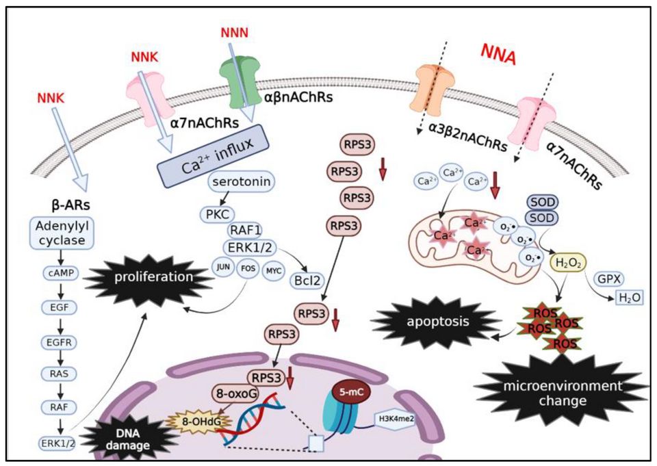
Every cell in your body lives by a simple rule: divide only when you are told to, and stop when the job is done. A healthy cell reads chemical signals from its neighbors, copies its DNA carefully, checks that copy for errors, and either keeps going or quietly retires. That balance between growing and stopping is controlled by genes. When those control genes get damaged, the balance tips, and a normal cell can slowly turn into a cancer cell. Cancer is not an invader from outside. It is one of your own cells that has lost its instructions.
The accelerator and the brakes
Think of cell division like driving a car. Two sets of genes keep it under control. Proto-oncogenes are the accelerator: they normally tell the cell when it is time to grow and divide. Tumor-suppressor genes are the brakes: they slow division, fix damaged DNA, or order a broken cell to self-destruct. In a normal cell these work together, so growth happens only when it should.
Mutations break this system in two different ways. When a proto-oncogene mutates, it becomes an oncogene a stuck accelerator that screams "grow!" even with no signal. This is a gain of function, and usually only one copy of the gene needs to change. When a tumor-suppressor gene mutates, the brakes fail. This is a loss of function, and typically both copies must be knocked out before protection is lost. The famous tumor suppressor p53 (often called the "guardian of the genome") normally halts a damaged cell and triggers apoptosis; when p53 fails, damaged cells survive and keep dividing.
The multistep journey: how tissue changes over time
Cancer does not appear overnight. Tissue drifts through recognizable stages as mutations accumulate:
- Hyperplasia too many cells, but they still look normal and stay organized.
- Dysplasia cells start looking abnormal disordered shapes, sizes, and arrangement. This is pre-cancerous but still reversible.
- Carcinoma in situ cells look fully cancerous but stay contained above the basement membrane. Nothing has spread yet, so cure rates are high.
- Invasive cancer cells break through the basement membrane into surrounding tissue and can travel to distant sites (metastasis).
What makes a cancer cell dangerous: the hallmarks
Researchers describe cancer through shared "hallmark" behaviors. In plain terms, cancer cells learn to:
- Divide without permission the stuck-accelerator, failed-brakes problem, so they multiply even when the body signals stop.
- Evade apoptosis normal cells with broken DNA are told to die (programmed cell death). Cancer cells silence that signal often by disabling p53 so damaged cells refuse to die.
- Grow their own blood supply a process called angiogenesis. Once a tumor grows past roughly 1 to 2 mm, it starves for oxygen, so it releases signals that recruit new blood vessels to feed itself and, later, to give cancer cells a highway to spread.
Tumor is not the same as cancer
Students mix these up constantly. A tumor (or neoplasm) is simply a lump an abnormal mass of cells. Cancer means the tumor is malignant: it invades and can spread. A benign tumor, like a lipoma, is a lump that stays put.
| Feature | Benign tumor | Malignant tumor (cancer) |
|---|---|---|
| Growth | Usually slow | Often rapid |
| Border | Well-defined, often encapsulated | Irregular, invades nearby tissue |
| Cell appearance | Looks like normal tissue (well-differentiated) | Abnormal, disorganized (poorly differentiated) |
| Spread (metastasis) | No | Yes via blood or lymph |
| Danger | Usually harmless unless pressing on something | Life-threatening if untreated |
Explore the figure below to watch a cell move from normal, through adaptation, into dysplasia the same stepwise drift described here.
- Proto-oncogenes are the accelerator and tumor-suppressor genes are the brakes; cancer needs a stuck accelerator (oncogene, one hit) and failed brakes (tumor suppressor, usually two hits).
- Cancer develops in steps: hyperplasia to dysplasia to carcinoma in situ to invasive cancer, as mutations pile up over time.
- Hallmark behaviors: uncontrolled division, evading apoptosis (often by disabling p53), and angiogenesis growing its own blood supply.
- A tumor is any lump; only a malignant tumor is cancer, defined by its ability to invade and metastasize.
- Invasion is marked by breaking through the basement membrane the line between contained and spreading disease.
Section 2Benign vs malignant
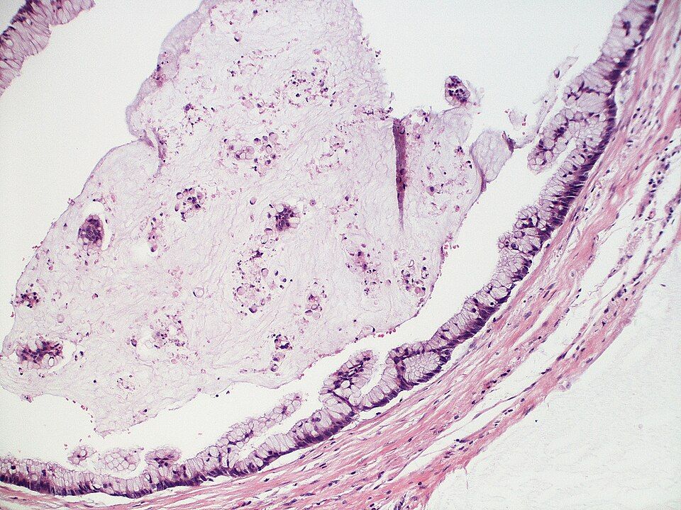
Your body makes new cells constantly. Skin, gut lining, blood cells, and more are always being replaced, and normally this happens under tight control: cells divide only when signaled, mature into a specific job, and stop dividing when the tissue is repaired. This maturing process is called differentiation. A well-differentiated cell looks and acts like the tissue it came from. A neoplasm (literally "new growth") is a lump of cells that has escaped normal growth controls and keeps dividing when it should not. The big clinical question is always the same: is this growth benign or malignant?
Benign growths: slow, walled-off, and well-behaved
A benign tumor grows slowly and stays put. Because it grows gradually, the surrounding tissue often has time to form a fibrous capsule around it, so the mass is encapsulated and can usually be shelled out cleanly by a surgeon. Its cells are well-differentiated, meaning they still closely resemble the normal tissue of origin and often still do that tissue's job. Benign tumors do not invade nearby structures and do not metastasize (spread to distant sites). Under the microscope the cells look orderly, with normal-sized nuclei and few dividing cells (low mitotic rate).
Malignant growths: fast, invasive, and mobile
Malignant tumors (true cancer) behave the opposite way. They grow quickly, so they rarely form a tidy capsule; instead they send finger-like projections that invade and destroy surrounding tissue, which is why the cut surface can look crab-like (the origin of the word "cancer"). Their cells are poorly differentiated or undifferentiated, meaning they no longer look like or act like the parent tissue. Most importantly, malignant cells can break away, travel through blood or lymph, and seed new tumors elsewhere. That spread is metastasis, and it is the feature that makes cancer so dangerous and so hard to cure.
| Feature | Benign | Malignant |
|---|---|---|
| Growth rate | Slow | Rapid, often erratic |
| Border | Encapsulated, well-defined | Invasive, poorly defined |
| Differentiation | Well-differentiated | Poorly differentiated to anaplastic |
| Metastasis | Never | Common |
| Recurrence after removal | Rare | Common |
How tumors are named
Naming follows a pattern worth memorizing. The suffix -oma usually signals a benign tumor named for its tissue of origin: a lipoma is a benign fat tumor, an adenoma a benign glandular tumor, a leiomyoma a benign smooth-muscle tumor (a uterine "fibroid"). Malignant tumors get one of two labels based on their tissue of origin: carcinomas from epithelial tissue, sarcomas from connective (mesenchymal) tissue. A carcinoma arises from epithelial tissue (skin, gland, and organ linings) and is by far the most common category, including adenocarcinoma of the colon or breast. A sarcoma arises from connective tissue such as bone (osteosarcoma), cartilage, muscle, or fat. A few dangerous "-oma" names break the rule, notably melanoma and lymphoma, which are malignant despite the friendly suffix.
Differentiation and anaplasia
Pathologists describe cancers by how far the cells have drifted from normal. Grading measures differentiation. A well-differentiated (low-grade) cancer still resembles its origin and usually behaves less aggressively. Anaplasia is the extreme opposite: cells so primitive and disorganized that they barely resemble any tissue. Anaplastic cells show pleomorphism (variable size and shape), large dark nuclei with a high nucleus-to-cytoplasm ratio, and abundant, sometimes abnormal, mitoses. In general, the less differentiated the cell, the more aggressive the tumor and the worse the prognosis.
When "benign" still hurts you
Benign does not always mean harmless. A tumor that would be trivial elsewhere can be life-threatening because of where it sits. A benign meningioma in the skull has nowhere to expand; as it grows it raises intracranial pressure and compresses brain tissue, causing headaches, seizures, or neurologic deficits. A benign adenoma can secrete hormones (for example, insulin from a pancreatic tumor, causing dangerous hypoglycemia), and a benign mass can obstruct a hollow organ or press on a vital vessel. Location, secretion, and compression, not just the label, drive the risk.
- Benign tumors are slow, encapsulated, well-differentiated, and stay put; malignant tumors are fast, invasive, poorly differentiated, and metastasize.
- Metastasis is the defining danger of malignancy and the main reason cancer is hard to cure.
- Naming: -oma usually means benign, carcinoma means malignant epithelial, sarcoma means malignant connective tissue; watch the exceptions melanoma and lymphoma.
- Differentiation drives grade; anaplasia (primitive, pleomorphic, high mitotic cells) signals the most aggressive tumors.
- A benign tumor can still be deadly because of its location, hormone secretion, or compression, classically a meningioma raising intracranial pressure.
Section 3How cancer grows and spreads
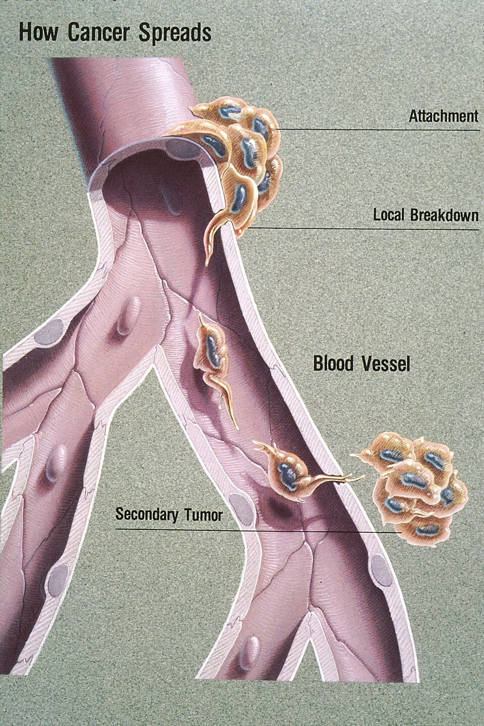
Normally, your tissues are beautifully organized. Cells stay in their own neighborhood, held in place by a thin, tough sheet called the basement membrane that separates the surface layer (epithelium) from the deeper connective tissue below. Cells also respect their neighbors: they stop dividing when they touch (contact inhibition), they anchor down where they belong, and they call for new blood vessels only when the body genuinely needs them. Cancer is what happens when a cell breaks every one of these rules at once, and understanding how it grows and travels is really a story about a cell that has forgotten how to stay put. Explore it below as we walk through each step.
Local invasion: breaking through the basement membrane
A tumor usually starts as a well-behaved lump that stays on its own side of the basement membrane. As long as it has not crossed that barrier, we call it carcinoma in situ ("in place") - and this is often curable, because it has not gone anywhere yet. The dangerous turning point is local invasion, when cancer cells secrete enzymes called matrix metalloproteinases (MMPs) that literally digest the basement membrane and the surrounding extracellular matrix. Once that fence is dissolved, the cells lose their normal stickiness (they down-regulate E-cadherin, the "glue" protein) and crawl into the deeper tissue. This is the moment a cancer becomes invasive, and it is why pathologists care so much about whether the basement membrane is intact.
Metastasis: how cancer travels to distant sites
Metastasis is the spread of cancer from the original (primary) tumor to a distant site. It is the single biggest reason cancer becomes life-threatening. There are three main routes.
- Lymphatic spread - cells enter lymphatic vessels and travel to nearby lymph nodes first. This is the classic route for carcinomas (epithelial cancers like breast and colon). It is why nurses palpate lymph nodes and why the sentinel lymph node - the first node draining a tumor - is biopsied to check whether spread has begun.
- Hematogenous spread - cells invade blood vessels and ride the bloodstream to far-off organs. This is the favored route for sarcomas (connective-tissue cancers) and explains distant organ metastases.
- Seeding of body cavities - cells shed off the tumor surface and scatter across a cavity lining, like peritoneal seeding in ovarian cancer. This can cause malignant ascites (fluid buildup) or pleural effusions.
| Feature | Lymphatic spread | Hematogenous spread |
|---|---|---|
| Typical cancer type | Carcinomas (epithelial) | Sarcomas (connective tissue) |
| First stop | Regional lymph nodes | Distant organs |
| Clinical clue | Enlarged, firm nodes | Masses in lung, liver, bone, brain |
| Assessed by | Sentinel node biopsy | CT/PET imaging, bone scan |
Where cancer tends to land
Metastases are not random. Cells often lodge in the first capillary bed they reach, and certain organs also provide fertile "soil" for tumor cells to settle - a concept called the seed-and-soil hypothesis. The four most common landing sites are lung, liver, bone, and brain. The lung and liver catch a large share of circulating cells because they act as biological filters with dense capillary networks (the liver receives portal blood; the lungs filter all venous return). Bone metastases are common in breast and prostate cancer and often cause deep pain, pathologic fractures, and high calcium levels (hypercalcemia). Brain metastases can raise intracranial pressure and cause headaches, seizures, or new neurologic deficits.
Angiogenesis: the tumor builds its own supply lines
A clump of cancer cells can only grow to about 1-2 mm before the cells in the center start starving - they are too far from any blood vessel to get oxygen and nutrients by diffusion. To keep growing, the tumor pulls off a striking trick called angiogenesis: it releases chemical signals, especially vascular endothelial growth factor (VEGF), that coax nearby blood vessels to sprout new branches straight into the tumor. This "angiogenic switch" gives the tumor its own supply line for continued growth and, unfortunately, an on-ramp into the bloodstream for metastasis. These new vessels are usually leaky and disorganized, which contributes to swelling and makes drug delivery unpredictable. Because VEGF is so central, several cancer drugs (anti-angiogenic agents like bevacizumab) work by blocking it to try to starve the tumor.
- Cancer becomes invasive when it uses enzymes (MMPs) to break through the basement membrane; before that, carcinoma in situ is often curable.
- Metastasis spreads through three routes: lymphatic (typical of carcinomas), hematogenous/bloodstream (typical of sarcomas), and seeding of body cavities.
- The most common landing sites are lung, liver, bone, and brain; new bone pain in a cancer patient is a red flag for metastasis.
- Angiogenesis, driven by VEGF, lets a tumor recruit its own blood supply to grow past 1-2 mm and also opens a route for cells to escape and spread.
Section 4What causes cancer
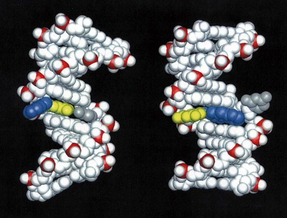
Every day, your body makes billions of new cells. Old cells wear out, get injured, or reach the end of their lifespan, so a healthy cell divides to make a fresh copy of itself. This is normally a tightly controlled process. Before a cell divides, it copies its entire DNA, then pauses at built-in checkpoints to proofread that copy. If the DNA looks damaged, the cell either fixes it or, if the damage is too severe, deliberately kills itself through a tidy self-destruct program called apoptosis. Cancer begins when this careful system breaks down and a cell starts dividing on its own schedule, ignoring the stop signals, and refusing to die.
So the real question is: what damages the DNA and the control genes badly enough to let a cell go rogue? The answer is usually a slow buildup of mutations (permanent changes in the DNA sequence) hitting the specific genes that run the cell cycle.
Carcinogens: the things that damage DNA
A carcinogen is any agent that causes cancer, usually by damaging DNA directly or by forcing cells to divide so fast that copy errors pile up. They fall into a few broad groups:
- Chemical — the classic example is tobacco smoke, which carries dozens of DNA-damaging compounds. Others include asbestos, benzene, and certain dyes. Many act at the point of contact (asbestos in the lungs, dyes in the bladder), but some, like benzene, are absorbed and cause blood cancers such as leukemia.
- Radiation — ultraviolet (UV) light from the sun damages skin-cell DNA and drives melanoma and other skin cancers. Ionizing radiation (X-rays, radon) does the same internally.
- Viral and microbial — some infections insert their own genes or cause chronic irritation. HPV (cervical and oropharyngeal cancers), EBV (some lymphomas), hepatitis B (liver cancer), and the bacterium H. pylori (stomach cancer) are the ones to know.
Oncogenes and tumor-suppressor genes
Two families of genes normally keep cell division in check, and cancer usually involves damage to both. Think of driving a car: you need a working gas pedal and working brakes.
Proto-oncogenes are the gas pedal. They tell a cell when it is time to grow and divide. When a proto-oncogene mutates and gets stuck in the "on" position, it becomes an oncogene that floors the accelerator, driving nonstop division.
Tumor-suppressor genes are the brakes. They slow division, run the DNA proofreading checkpoints, and trigger apoptosis when a cell is too damaged. The most famous is p53, nicknamed the guardian of the genome because it halts the cell cycle to allow repair, and orders apoptosis when repair fails. When p53 is knocked out, damaged cells sail through checkpoints and accumulate even more mutations. The BRCA1 and BRCA2 genes are also tumor suppressors; they help repair broken DNA, so when they fail, breast and ovarian tissue lose a key safety net.
| Feature | Oncogene | Tumor-suppressor gene |
|---|---|---|
| Normal job | Promotes cell growth (gas pedal) | Restrains growth, repairs DNA, triggers apoptosis (brakes) |
| Effect of mutation | Gain of function - stuck ON | Loss of function - brakes fail |
| Copies needed to cause trouble | Usually one mutated copy is enough | Usually both copies must be lost |
| Examples | Mutated RAS, HER2 | p53, BRCA1/2, RB |
Inherited vs acquired mutations, and why some cancers run in families
Most cancers come from acquired (somatic) mutations - damage that builds up in one person's cells over a lifetime from carcinogens, random copying errors, and aging. These are not passed to children, which is why the majority of cancers are sporadic.
A smaller group involves inherited (germline) mutations, present in every cell from birth because they came from a parent's egg or sperm. Someone who inherits one broken copy of a tumor-suppressor gene like BRCA1 only needs to lose the second copy for that safety net to fail - they are already halfway there. That head start is why these cancers appear earlier in life, often in both breasts or in multiple family members. This is the mechanism behind familial patterns and the basis for genetic counseling.
Chronic inflammation and failed immune surveillance
Two background factors quietly raise the odds. First, chronic inflammation (as in long-standing hepatitis, H. pylori gastritis, or inflammatory bowel disease) keeps tissue in a constant cycle of injury and repair. Every extra round of division is another chance for a mutation to slip in. Second, your immune system normally patrols for abnormal cells and destroys them - a process called immune surveillance. Cancer cells that learn to hide from or exhaust these immune patrols escape destruction, which is why immunosuppressed patients often have higher cancer rates.
- Cancer starts when DNA damage disables the normal controls on cell division, letting a cell divide without limit and dodge apoptosis.
- Carcinogens - chemical (tobacco), radiation (UV), and viral/microbial (HPV, EBV, hepatitis B, H. pylori) - cause cancer largely by damaging DNA; many are preventable through vaccines and lifestyle.
- Oncogenes are a stuck gas pedal (gain of function); tumor suppressors like p53 (guardian of the genome) and BRCA1/2 are failed brakes (loss of function).
- Most mutations are acquired over a lifetime; inherited germline mutations give a head start toward cancer, explaining earlier onset and familial clustering.
- Chronic inflammation and failed immune surveillance are contributors that let mutated cells accumulate and escape destruction.
Section 5Grading, staging & living with cancer
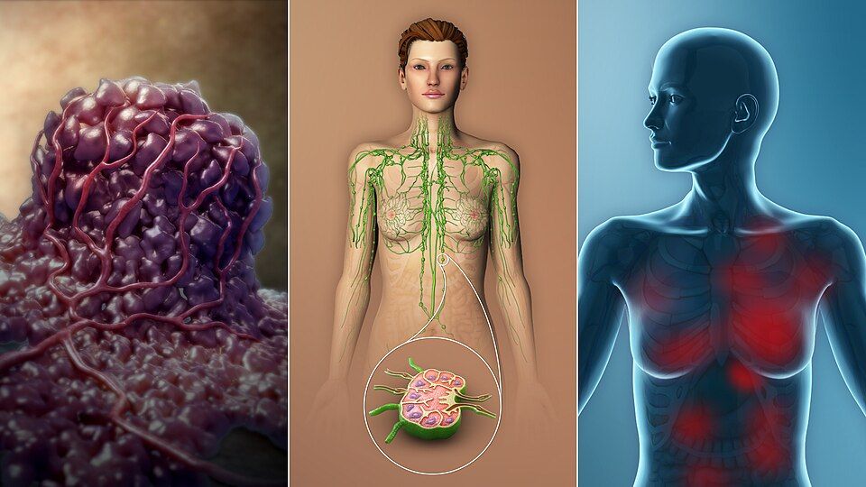
Your cells normally follow strict rules. They divide only when told to, they mature into a specific job (a skin cell, a colon cell), and they stay in their own neighborhood. Cancer breaks all three rules: the cells divide without permission, they fail to mature, and they invade and travel. Once a tumor exists, two of the first questions a care team asks are how ugly do these cells look under the microscope? and how far has this spread? Those are two different questions, and mixing them up is one of the most common student errors.
Grading vs staging
Grading is about appearance. A pathologist looks at the tumor cells and asks how much they still resemble the normal tissue they came from. Well-differentiated (low-grade) cells still look and behave a lot like the parent tissue and tend to grow slowly. Poorly differentiated or anaplastic (high-grade) cells look wild and disorganized and usually behave more aggressively. Grading is usually reported as G1 through G4.
Staging is about spread - how big the tumor is and how far it has traveled. The most widely used framework is the TNM system: T for the size and extent of the primary tumor, N for how many regional lymph nodes are involved, and M for distant metastasis (spread to far organs). Higher numbers mean more advanced disease, and staging drives treatment decisions and prognosis more powerfully than grade does.
| Feature | Grading | Staging |
|---|---|---|
| Question it answers | How abnormal do the cells look? | How far has it spread? |
| How it is measured | Differentiation under the microscope | TNM: Tumor, Nodes, Metastasis |
| Typical scale | G1 (well-differentiated) to G4 (anaplastic) | Stage I to Stage IV |
| Main use | Predicts aggressiveness | Guides treatment and overall prognosis |
The CAUTION warning signs
The American Cancer Society mnemonic CAUTION lists classic early clues that should prompt evaluation. They matter because early detection usually means a lower stage and better outcomes:
- C - Change in bowel or bladder habits
- A - A sore that does not heal
- U - Unusual bleeding or discharge
- T - Thickening or a lump (breast or elsewhere)
- I - Indigestion or difficulty swallowing
- O - Obvious change in a wart or mole
- N - Nagging cough or hoarseness
When the tumor causes trouble from a distance
Paraneoplastic syndromes happen when a tumor secretes hormones or hormone-like substances, causing symptoms far from the tumor itself - classic examples are SIADH (small cell lung cancer making ADH, driving down sodium) and hypercalcemia (from PTH-related peptide or bone breakdown).
Tumor lysis syndrome (TLS) is an oncologic emergency. When large numbers of cancer cells are destroyed quickly - often right after chemotherapy starts - they dump their contents into the blood. The result is high potassium (hyperkalemia), high phosphate, high uric acid, and low calcium, which can trigger fatal arrhythmias and acute kidney injury. Nurses watch labs closely and push hydration.
The four treatment pillars
Most cancer care combines four approaches. Surgery removes the tumor. Radiation damages the DNA of cells in a targeted area. Chemotherapy uses systemic drugs to kill dividing cells throughout the body. Immunotherapy retrains or unleashes the patient's own immune system to recognize and attack cancer.
Here is the key mechanism behind chemo's side effects: most chemotherapy targets rapidly dividing cells, because that is what cancer does. But some healthy tissues also divide fast, so they take collateral damage. That is exactly why chemo predictably causes hair loss (alopecia) from fast-dividing hair follicles, nausea, vomiting, and mouth sores (mucositis) from the fast-turnover gut lining, and bone marrow suppression (myelosuppression) - dropping white cells, red cells, and platelets. The low white count (neutropenia) is the scary one, because it leaves the patient wide open to infection.
- Grade describes how abnormal cells look (differentiation, G1-G4); stage describes how far cancer has spread using TNM (Tumor, Nodes, Metastasis).
- The CAUTION mnemonic flags early warning signs that warrant evaluation and can catch cancer at a lower, more treatable stage.
- Paraneoplastic syndromes (like SIADH and hypercalcemia) cause distant hormone-driven symptoms; tumor lysis syndrome is an emergency with hyperkalemia, hyperphosphatemia, hyperuricemia, and hypocalcemia.
- The four pillars are surgery, radiation, chemotherapy, and immunotherapy.
- Chemo harms fast-dividing normal cells too, causing alopecia, mucositis/GI upset, and myelosuppression - watch for neutropenic fever.
The chapter in one breath
- Carcinogenesis is a stepwise loss of control: a normal cell mutates until it ignores stop signals, dodges apoptosis, and divides on its own, becoming a clone of rogue cells.
- Cancer arises when proto-oncogenes mutate into oncogenes stuck in the on position (accelerator) and tumor suppressor genes like p53 fail (brakes), so growth and DNA-repair checkpoints stop working.
- Benign tumors are usually slow, encapsulated, well-differentiated, and stay put; malignant tumors grow fast, lack a capsule, look abnormal, and spread.
- Invasion is local spread as cancer cells release enzymes to chew through basement membrane and infiltrate nearby tissue, which is why margins matter in surgery.
- Metastasis is distant spread through blood or lymph to form secondary tumors; common landing sites are lung, liver, bone, and brain, and it signals advanced disease.
- Causes are multifactorial carcinogens and risk factors: tobacco, radiation, UV light, certain chemicals, oncogenic viruses (HPV, hepatitis, EBV), chronic inflammation, and inherited mutations (like BRCA).
- Grading describes how abnormal the cells look under the microscope (differentiation), while staging (often TNM: Tumor size, Nodes, Metastasis) describes how far it has spread and drives prognosis.
- Treatment follows a few core principles: surgery removes local disease, radiation targets a region, and chemotherapy works systemically by hitting rapidly dividing cells, often combined for better cure rates.
- Chemotherapy is not selective, so it also harms fast-turnover normal cells, explaining the classic toxicities: bone marrow suppression, hair loss, nausea, and mucositis.
ReferenceWords worth knowing
A quick reference for the terms in this chapter — skim it now, or circle back whenever a word trips you up.
- Anaplasia
- Anaplasia is the extreme loss of a cell's normal appearance and organization, where cancer cells look so wild and disorganized (with big, irregular nuclei and chaotic mitoses) that you often can't tell what tissue they came from, marking a highly malignant tumor.
- Angiogenesis
- Angiogenesis is the growing of new blood vessels, which a tumor hijacks by releasing signals (like VEGF) to build its own blood supply so it can keep feeding and expanding beyond a couple of millimeters.
- Benign
- A benign tumor is a growth that stays put, grows slowly, is usually well-differentiated and encapsulated, and does not invade nearby tissue or spread to distant sites, though it can still cause harm by pressing on nearby structures.
- Carcinogen
- A carcinogen is any agent that can cause cancer by damaging DNA or promoting abnormal growth, such as tobacco smoke, UV and ionizing radiation, certain chemicals, or oncogenic viruses like HPV.
- Carcinoma
- A carcinoma is a cancer that arises from epithelial cells, meaning the cells that line and cover body surfaces and glands (skin, breast, lung, colon, prostate), and it is the most common category of cancer.
- Carcinoma in situ
- Carcinoma in situ is an early cancer whose malignant cells are still confined to their original layer and have not yet broken through the basement membrane to invade, so it is often curable if caught and removed at this stage.
- Differentiation
- Differentiation is how closely a cell resembles the mature, specialized tissue it came from; well-differentiated cells look and behave almost normally, while poorly differentiated ones look primitive and tend to be more aggressive.
- Dysplasia
- Dysplasia is disordered but still reversible cell growth where cells vary in size, shape, and organization, a pre-cancerous change that can progress to cancer if the trigger continues.
- Malignant
- A malignant tumor is a cancerous growth that invades surrounding tissue, tends to be poorly differentiated, and can spread (metastasize) to distant parts of the body, making it life-threatening if untreated.
- Metastasis
- Metastasis is the spread of cancer cells from the original (primary) tumor to distant sites through the blood or lymphatic system, where they seed new (secondary) tumors, and it is the main reason cancer becomes deadly.
- Neoplasm
- A neoplasm, literally "new growth," is a mass of cells that keep dividing beyond normal limits and serve no useful purpose, and it may be either benign or malignant.
- Oncogene
- An oncogene is a mutated or overactive version of a normal growth-promoting gene (a proto-oncogene) that, once switched on, pushes a cell to divide relentlessly, acting like a gas pedal stuck to the floor.
- Paraneoplastic syndrome
- A paraneoplastic syndrome is a set of symptoms caused by substances (like hormones or cytokines) a tumor secretes rather than by the tumor's physical bulk, so a patient might show effects (such as low sodium or high calcium) far from the actual cancer site.
- Sarcoma
- A sarcoma is a cancer that arises from connective and supportive tissues, such as bone, cartilage, muscle, fat, or blood vessels, and it is far less common than carcinoma.
- TNM staging
- TNM staging is the standard system for describing how far a cancer has progressed by rating three things: T for the size and extent of the primary tumor, N for spread to regional lymph nodes, and M for distant metastasis.
- Tumor-suppressor gene
- A tumor-suppressor gene (such as p53 or BRCA) normally slows cell division, repairs DNA, or triggers a damaged cell to self-destruct, so when it is lost or inactivated the cell's brakes fail and cancer can develop.
DrillFlashcards for this unit
These are the Week 3 cards — the same deck as the hub, pulled in here so you can review the moment you finish reading. Tap the card to flip it; hit Shuffle to mix them up. To grade yourself and track mastery across the whole deck, open the full flashcards page.
Check yourself
Pain
Pain is the body’s alarm system — usually protective, sometimes broken, and always personal. This chapter traces a pain signal from the nerve ending that detects damage all the way to the brain that feels it, sorts pain into its types, explains why rubbing a bump helps, and covers how nurses assess and safely relieve it.
By the end of this chapter you can
- Trace the pain pathway through transduction, transmission, perception, and modulation.
- Distinguish somatic, visceral, and neuropathic pain, and acute from chronic.
- Explain the gate control theory and the body’s own pain-relief system.
- Predict referred-pain patterns and assess pain in patients who cannot self-report.
- Apply the WHO analgesic ladder and name the key risks of opioids.
Section 1Nociception & the pain pathway
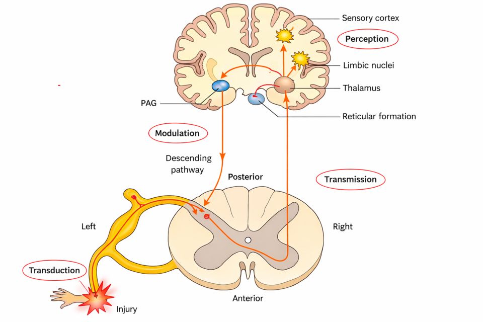
Pain feels like a problem, but in most cases it is your body's alarm system doing exactly what it should. When tissue is threatened or damaged, specialized nerve endings fire and send a warning racing toward your brain: stop, protect this spot, something is wrong. Understanding how that signal is born, how it travels, and how the body can turn the volume up or down is the foundation for everything you will do in pain management.
Nociceptors: the sensors that detect damage
The journey starts with nociceptors - free nerve endings scattered through the skin, muscles, joints, and internal organs. Think of them as tiny smoke detectors. They do not respond to gentle touch; they wake up only when a stimulus is intense enough to signal real or potential tissue damage. That trigger can be mechanical (a cut or crush), thermal (a hot stove), or chemical. When cells are injured they spill out a soup of substances - prostaglandins, bradykinin, histamine, substance P, and hydrogen ions - and this inflammatory soup lowers the firing threshold so the nociceptors become easier to set off. This is why a sunburned shoulder screams at a light pat: the sensors have been sensitized.
The four steps of nociception
Textbooks break the pain process into four linked steps. Learn them in order, because nursing interventions target specific steps.
- Transduction - the noxious stimulus is converted into an electrical signal at the nociceptor. This is where NSAIDs work, blocking prostaglandin production so the sensors stay quieter.
- Transmission - the electrical impulse travels along peripheral nerve fibers to the dorsal horn of the spinal cord, then upward to the brain. Local anesthetics interrupt this step.
- Perception - the brain (cortex and limbic system) becomes consciously aware of the pain and gives it meaning and emotion. This is where distraction, relaxation, and imagery help.
- Modulation - the nervous system turns the signal up or down. Descending pathways release the body's own endogenous opioids (endorphins, enkephalins) plus serotonin and norepinephrine, which dampen the message. Opioids and many antidepressants boost this natural braking system.
Two kinds of fibers, two kinds of pain
Not all pain messages travel at the same speed, and this explains a common experience: stub your toe and you feel a sharp jolt first, then a throbbing ache a moment later. Two different fibers are talking. A-delta fibers are lightly myelinated, so they conduct fast - they carry the sharp, bright, easy-to-pinpoint pain that makes you pull your hand away instantly. C fibers are unmyelinated and slow, carrying the dull, aching, burning, poorly localized pain that lingers afterward. Both fibers hand their signal off in the dorsal horn and cross to the opposite side, then ride the spinothalamic tract up to the thalamus, which relays it to the cortex for perception.
| Feature | A-delta fibers | C fibers |
|---|---|---|
| Myelin | Thinly myelinated | Unmyelinated |
| Speed | Fast | Slow |
| Quality of pain | Sharp, pricking | Dull, aching, burning |
| Localization | Well localized | Diffuse, hard to pinpoint |
| Timing | First (fast) pain | Second (slow) pain |
Why pain protects us - and what happens when it is gone
All of this machinery exists for one reason: survival. Pain forces you to withdraw from harm, to guard an injured limb, and to seek help. It is protective. That is exactly why losing the ability to feel pain is so dangerous. In diabetic peripheral neuropathy, chronic high blood glucose damages the small sensory nerves in the feet, so the nociceptors and their fibers can no longer sound the alarm. A patient may step on a tack, develop a blister from ill-fitting shoes, or walk on an open ulcer for days - completely unaware. Without the warning signal, minor injuries go unnoticed, become infected, and can progress to serious wounds or even amputation. The lesson for nursing practice is powerful: absent pain is not a blessing but a hazard, which is why we teach daily foot inspection and protective footwear for anyone with sensory loss.
Explore the pathway below to trace a single signal from the moment of injury, up the spinothalamic tract, all the way to conscious perception in the brain.
- Nociceptors are free nerve endings that detect mechanical, thermal, and chemical stimuli signaling tissue damage; injury releases prostaglandins and bradykinin that sensitize them.
- Nociception has four steps - transduction, transmission, perception, and modulation - and each is a distinct target for pain-relieving interventions.
- A-delta fibers carry fast, sharp, well-localized pain while C fibers carry slow, dull, aching pain; both ascend the spinothalamic tract to the brain.
- The body modulates pain by releasing endogenous opioids (endorphins, enkephalins) through descending pathways, which is why opioids and some antidepressants relieve pain.
- Pain is protective; when it is absent, as in diabetic neuropathy, unnoticed foot injuries can progress to ulcers, infection, and amputation - so protective foot care is essential.
Section 2The types of pain
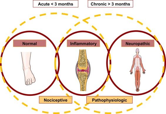
Pain is not a design flaw. In a healthy body, it is one of your best-working alarm systems. Specialized nerve endings called nociceptors sit in your skin, muscles, joints, and organs, quietly waiting. When something threatens to damage tissue, they fire, sending a signal up the spinal cord to the brain, which reads it as "that hurts, do something." This protective alarm makes you pull your hand off a hot stove before you even think about it. The trouble starts when the alarm rings for the wrong reasons, keeps ringing after the danger is gone, or wires itself into a permanent problem. Understanding what kind of pain you are dealing with is the first step, because different pain comes from different places and answers to different treatments.
Nociceptive pain: the normal alarm working as intended
Nociceptive pain is the pain of actual or threatened tissue damage picked up by nociceptors. It is the most familiar type, and it splits into two flavors depending on where those nerve endings live.
Somatic pain comes from the "body wall" tissues you can point to, such as skin, muscle, bone, and joints. These areas are densely supplied with nerves and mapped precisely in the brain, so somatic pain is usually sharp, throbbing, and well-localized. A sprained ankle or a surgical incision hurts right there, and the patient can put a finger on it.
Visceral pain comes from the internal organs such as the gut, bladder, and heart. These organs have fewer, more scattered nociceptors, and their signals travel on nerves that share spinal pathways with the skin. The result is pain that is usually dull, deep, cramping, and poorly localized, often described as "somewhere in my belly." Because of that shared wiring, visceral pain is frequently referred, meaning it is felt in a body-wall location away from the actual organ. The classic example is cardiac pain felt in the left arm or jaw. Visceral pain also tends to bring along nausea, sweating, and restlessness because it activates the autonomic nervous system.
| Feature | Somatic | Visceral |
|---|---|---|
| Source | Skin, muscle, bone, joints | Internal organs |
| Quality | Sharp, throbbing, aching | Dull, deep, cramping |
| Localization | Well-localized, easy to point to | Poorly localized, vague |
| Referred? | Rarely | Often (e.g., MI to arm/jaw) |
Neuropathic pain: the alarm system itself is damaged
Neuropathic pain is different in kind. Here the problem is not tissue damage sending a signal, but damage or dysfunction of the nerves that carry the signal. Injured nerves become abnormally excitable and fire on their own, so the alarm sounds without any real threat. Patients describe it in electrical, burning language, such as burning, shooting, stabbing, tingling, or "pins and needles." They may also have allodynia (pain from something that should not hurt, like a bedsheet) and hyperalgesia (an exaggerated response to a mildly painful stimulus). Common causes include diabetic peripheral neuropathy, phantom limb pain after amputation, and sciatica from a compressed nerve root. This matters clinically because ordinary painkillers often barely touch it.
Acute versus chronic: how long the alarm rings
A separate way to sort pain is by its time course. Acute pain is the protective alarm doing its job. It comes on with an identifiable injury, warns you, and usually resolves as the tissue heals, typically within days to a few weeks. It often carries visible sympathetic signs such as increased heart rate, elevated blood pressure, and sweating.
Chronic pain is pain that persists beyond about three months, or beyond the expected healing time. At this point the nervous system can undergo central sensitization, becoming more efficient at transmitting and amplifying pain, so the pain outlives its usefulness and becomes its own disease. Chronic pain frequently loses the obvious sympathetic signs and instead shows up as fatigue, depression, sleep disruption, and withdrawal. Treating it means looking beyond the original injury to the whole person.
Why the type steers the treatment
Matching the mechanism to the therapy is the whole point of classifying pain. Nociceptive pain generally responds to NSAIDs, acetaminophen, and, for severe cases, opioids. Neuropathic pain usually needs anticonvulsants or antidepressants that calm overactive nerves. Acute pain is treated aggressively and briefly to allow healing, while chronic pain calls for a multimodal plan that combines medications with physical therapy, psychological support, and non-drug strategies.
- Nociceptive pain reflects tissue damage: somatic is sharp and well-localized; visceral is dull, poorly localized, and often referred.
- Neuropathic pain comes from damaged nerves firing on their own, feels burning or shooting, and is seen in diabetic neuropathy, phantom limb, and sciatica.
- Acute pain is protective and resolves with healing; chronic pain lasts beyond ~3 months and becomes its own problem via central sensitization.
- The type of pain guides therapy: NSAIDs/opioids for nociceptive, anticonvulsants/antidepressants for neuropathic, and multimodal plans for chronic pain.
Section 3How pain is modulated
Here is something strange but true: pain is not a fixed signal traveling a one-way wire from your finger to your brain. The nervous system is constantly editing the pain message on its way up, turning the volume up or down before you ever feel it. This editing is called pain modulation, and it happens at every level, from the spinal cord to the brain. Understanding it explains a lot of everyday things, like why rubbing a stubbed toe helps, why a soldier or athlete can keep going despite a serious injury, and why some people hurt for years after the tissue has completely healed.
The gate control theory: crowding out the pain signal
In 1965, Melzack and Wall proposed the gate control theory, and it still guides nursing practice today. Picture a gate in the dorsal horn of the spinal cord where incoming signals compete for passage to the brain. Pain travels on small, slow fibers: thinly myelinated A-delta fibers (sharp, fast pain) and unmyelinated C fibers (dull, aching, burning pain). Touch, pressure, and vibration travel on large, fast A-beta fibers.
Here is the key idea: when you activate the big A-beta fibers, they excite inhibitory interneurons in the dorsal horn that partly close the gate on the pain fibers. The non-painful input literally crowds out some of the painful input. This is the mechanism behind rubbing a bumped elbow, applying heat or cold, massage, and TENS (transcutaneous electrical nerve stimulation), which delivers a mild buzzing current to flood those large fibers and dampen the pain message.
The brain talks back: endogenous opioids and descending inhibition
The gate is not controlled only from below. The brain sends signals downward to the spinal cord to quiet pain, a process called descending inhibition. Nuclei in the midbrain (the periaqueductal gray) and brainstem release neurotransmitters, including serotonin and norepinephrine, that suppress transmission in the dorsal horn. This is also why certain antidepressants (SNRIs, tricyclics) help chronic pain: they boost these descending pathways.
The body also makes its own opioids, called endogenous opioids. The main families are endorphins and enkephalins (plus dynorphins). They bind the same opioid receptors (mu, delta, kappa) that morphine does, blocking the release of pain neurotransmitters like substance P. This is the chemistry behind the calm after intense exercise, the reduced pain during a crisis, and part of why placebo relief is real, not imaginary.
| Feature | Gate closes (less pain) | Gate opens (more pain) |
|---|---|---|
| Fiber activity | Large A-beta touch fibers firing | A-delta and C pain fibers firing |
| From the brain | Descending inhibition active | Descending pathways weak or fatigued |
| Chemistry | Endorphins, enkephalins, serotonin, norepinephrine | Substance P, glutamate |
| State of mind | Relaxed, distracted, hopeful | Anxious, exhausted, focused on the pain |
| Nursing levers | Heat/cold, massage, TENS, distraction, opioids | Untreated anxiety, poor sleep, fear |
When the volume knob gets stuck: central sensitization and wind-up
Normally, modulation keeps pain proportional to injury. But when pain signaling continues intensely and repeatedly, the nervous system can start amplifying itself. Rapid, repeated firing of C fibers produces wind-up, where each stimulus triggers a progressively bigger response in dorsal horn neurons, often through overactivation of NMDA receptors by glutamate. Over time this settles into central sensitization: the spinal cord and brain become hyper-responsive, essentially turning up their own gain.
Two classic signs follow. In hyperalgesia, a painful stimulus hurts far more than it should. In allodynia, something that should not hurt at all, like a bedsheet, a light touch, or a breeze, is felt as pain. These are not exaggeration or drug-seeking; they are the fingerprint of a sensitized nervous system.
Why chronic pain outlasts the injury
This is the crux of chronic pain. Once central sensitization is established, the nervous system keeps generating pain even after the original tissue has healed. The alarm keeps ringing after the fire is out. Reasons include lasting changes in dorsal horn wiring, weakened descending inhibition, and remodeling in pain-processing brain regions. That is why chronic pain (usually defined as pain lasting more than about three months) is now understood as a disease of the nervous system in its own right, not simply ongoing tissue damage. It also explains why chronic pain often needs a broader plan, such as SNRIs or tricyclics, gabapentinoids, graded activity, cognitive strategies, and good sleep, rather than opioids alone, since the problem lives largely in the modulation system itself.
- Pain is modulated, not fixed: the spinal cord and brain turn the signal up or down before you feel it.
- Gate control: large A-beta touch fibers can close the spinal gate on A-delta and C pain fibers, which is why rubbing, heat/cold, and TENS help.
- The brain fights back: descending inhibition plus endogenous opioids (endorphins and enkephalins), serotonin, and norepinephrine suppress pain in the dorsal horn.
- Central sensitization and wind-up turn up the nervous system's own volume, producing hyperalgesia and allodynia.
- Chronic pain can persist after healing because the sensitized nervous system keeps sounding the alarm, so it often needs a multimodal, non-opioid-centered plan.
Section 4Referred pain & special patterns
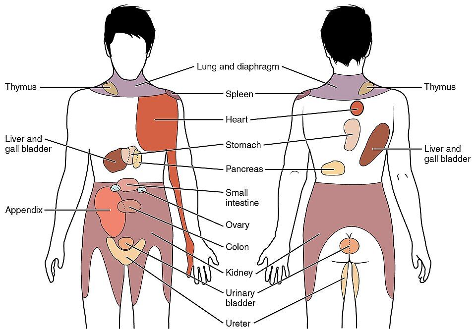
Normally, your brain is very good at telling you where something hurts. A nerve carries a pain signal from a specific spot, travels to a specific level of the spinal cord, and your brain reads the return address and points to the right place. This mapping is usually accurate for skin and muscle, which have dense, well-organized sensory nerves. But some pain does not follow the rules, and understanding why helps you catch dangerous problems and treat pain you cannot easily see.
Referred pain: the crossed-wire problem
Referred pain is pain felt in a body area away from the organ actually causing it. The classic example: a heart attack (myocardial infarction) often hurts in the left arm, jaw, neck, or shoulder rather than only the chest. Why? The nerves carrying pain from the heart enter the spinal cord at the same segments (roughly T1 to T4) as the nerves from the skin of the arm and chest wall. This is called convergence: visceral (organ) signals and somatic (skin) signals share the same second-order neurons on their way up. The brain has spent your whole life feeling your arm and almost never feeling your heart, so when a shared pathway fires, it guesses the familiar source and projects the pain outward to the skin.
Organs give vague, poorly localized pain because their sensory nerves are sparse. That is why visceral pain is often described as deep, dull, cramping, or squeezing, while somatic pain from skin or bone is sharp and easy to pinpoint. Knowing common referral patterns is a safety skill: diaphragm irritation refers to the shoulder tip, gallbladder pain to the right shoulder/scapula, and cardiac pain to the left arm or jaw.
Phantom limb pain
After an amputation, many people still feel the missing limb, and a large share feel pain in it, sometimes burning, cramping, or shooting. This is phantom limb pain. The limb is gone, but the nerves, spinal cord, and the brain's somatosensory map that once served it are still there. Cut nerve endings can form tangled neuromas that fire on their own, the spinal cord becomes hyperexcitable (central sensitization), and the brain's map reorganizes, sometimes misreading nearby signals as coming from the lost limb. It is real neuropathic pain, not imagination, and validating it matters. Treatments target the nervous system: neuropathic agents (gabapentin, certain antidepressants), and mirror therapy, which uses the reflection of the intact limb to help the brain remap.
Assessing pain when the person cannot tell you
Pain is whatever the person says it is, but some people cannot say. Infants, patients who are sedated or intubated, and people with advanced dementia or cognitive impairment need behavioral (observational) pain scales. You watch the body instead of asking a number.
| Population | Tool | What you observe |
|---|---|---|
| Nonverbal older adult / dementia | PAINAD | Breathing, vocalization, facial expression, body language, consolability |
| Critically ill / intubated adult | CPOT or BPS | Facial expression, muscle tension, ventilator compliance |
| Infant / young child | FLACC | Face, Legs, Activity, Cry, Consolability |
Look for grimacing, guarding, restlessness, moaning, or a rigid body. Also watch for physiologic clues, though these are less reliable over time: raised heart rate, blood pressure, and respiratory rate. Consider the situation too; if a procedure or condition would hurt you, assume it hurts them, and treat.
Why pain is under-treated in older adults
Older adults are frequently under-medicated, and it usually comes from stacked barriers rather than one cause:
- Communication gaps: dementia, aphasia, or delirium make self-report hard, so pain gets missed.
- False beliefs: both patients and staff may wrongly assume pain is a normal part of aging, so no one treats it.
- Stoicism and fear: older patients may under-report to avoid being a burden, or fear addiction and side effects.
- Prescriber caution: real worries about falls, confusion, kidney function, and drug interactions make clinicians hesitant, sometimes to the point of undertreatment.
Untreated pain is not benign; it drives delirium, poor sleep, immobility (raising risks of pneumonia, pressure injuries, and clots), depression, and slower recovery. The nursing goal is honest, proactive assessment using the right tool, then advocating for adequate, carefully titrated relief.
- Referred pain happens because visceral and somatic nerves converge at the same spinal segment; cardiac pain classically refers to the left arm, jaw, or shoulder.
- Learn common referral patterns and treat atypical presentations (jaw pain, nausea, dyspnea) as possible MI, especially in women, older adults, and people with diabetes.
- Phantom limb pain is real neuropathic pain from neuroma firing, central sensitization, and cortical remapping; mirror therapy and neuropathic agents help.
- When patients cannot self-report, use behavioral scales: FLACC (infants), PAINAD (dementia), CPOT/BPS (critically ill), watching face, body, and consolability.
- Pain in older adults is often under-treated due to communication barriers and false beliefs; untreated pain fuels delirium, immobility, and complications, so assess proactively and advocate.
Section 5Assessing & managing pain
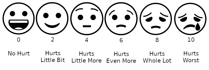
Pain is your body's alarm system, and it usually does an important job. When tissue is threatened or damaged, specialized nerve endings called nociceptors fire and send a signal up the spinal cord to the brain, where it is finally interpreted as "that hurts." This pathway (transduction, transmission, perception, and modulation) is protective. It makes you pull your hand off a hot stove and guard a sprained ankle so it can heal. The trouble is that the alarm is invisible. You cannot see it on a monitor or draw it in a tube, which is exactly why pain is one of the hardest things a nurse assesses.
Pain is whatever the patient says it is
The guiding rule in nursing is that pain is a subjective, self-reported experience. Margo McCaffery's classic definition still holds: pain is whatever the person says it is, existing whenever they say it does. Two people with the same surgery can report a 3 and a 9, and both are correct, because pain is shaped by nerves and by fear, culture, past experience, and mood. So we ask the patient rather than guessing.
To turn that report into something we can track, we use rating scales. The tool has to match the patient in front of you.
- 0-10 Numeric Rating Scale - for alert adults who can think in numbers. Zero is no pain, ten is the worst imaginable.
- Wong-Baker FACES - cartoon faces from smiling to crying, useful for young children (usually around age 3 and up) or anyone who struggles with numbers.
- FLACC - a behavioral score (Face, Legs, Activity, Cry, Consolability) for infants and preverbal children who cannot self-report.
- PAINAD - Pain Assessment in Advanced Dementia, which reads breathing, vocalization, facial expression, body language, and consolability in nonverbal older adults.
Whichever scale you pick, keep using the same one for that patient so the numbers are comparable, and pair the score with a fuller assessment such as OLDCARTS or PQRST (location, quality, radiation, timing, what makes it better or worse).
Climbing the WHO analgesic ladder
The World Health Organization (WHO) analgesic ladder is a simple, stepwise plan for matching drug strength to pain intensity. It was built for cancer pain but the logic guides much of pain management.
- Step 1 - mild pain: non-opioid analgesics such as acetaminophen or NSAIDs, with or without an adjuvant (a helper drug like an antidepressant or anticonvulsant for nerve pain).
- Step 2 - moderate pain: a weak opioid (for example codeine or tramadol) added to the non-opioid.
- Step 3 - severe pain: a strong opioid such as morphine, hydromorphone, or fentanyl, still combined with non-opioids and adjuvants.
Two ideas make the ladder work: use multimodal analgesia (drugs that attack pain by different mechanisms so you need less of any one), and dose around the clock for constant pain rather than waiting for it to spike.
Non-opioids vs opioids
Non-opioids work mostly at the site of injury. NSAIDs (ibuprofen, ketorolac, naproxen) block cyclooxygenase (COX) enzymes, so less prostaglandin is made and the nociceptors quiet down; this also fights inflammation but can irritate the stomach lining and stress the kidneys. Acetaminophen relieves pain and fever centrally with little anti-inflammatory effect and, importantly, no GI bleeding risk, but too much (usually more than about 4 grams a day) can cause hepatotoxicity. Opioids act differently: they bind mu receptors in the brain and spinal cord to dampen how pain is transmitted and perceived. They are powerful and have no organ-damaging ceiling, but they carry serious risks.
| Feature | Non-opioids | Opioids |
|---|---|---|
| Main site of action | Peripheral (injury site); acetaminophen more central | Central mu receptors (brain, spinal cord) |
| Dose ceiling | Yes - max daily dose to protect liver/GI/kidney | No true ceiling; titrate to effect |
| Key risks | GI bleed, kidney injury (NSAIDs); liver damage (acetaminophen) | Respiratory depression, constipation, sedation, tolerance, dependence |
| Best for | Mild pain, inflammation, fever | Moderate to severe pain |
The opioid risk you watch hardest is respiratory depression. Mu receptors in the brainstem blunt the drive to breathe, so an oversedated patient breathes slowly and shallowly before the rate ever crashes. Rising sedation is the early warning sign; check level of consciousness and respiratory rate, and keep naloxone (the opioid antagonist) available. Nearly every patient also gets constipation, because gut mu receptors slow motility, so a bowel regimen starts alongside the opioid. Over time patients may develop tolerance (needing more drug for the same relief) and physical dependence (withdrawal if stopped abruptly) - normal physiologic adaptations that are not the same as addiction, which is compulsive use despite harm.
Non-pharmacologic methods
Medication is not the whole plan. Non-drug measures work through the gate control theory - competing signals and top-down input can partly "close the gate" in the spinal cord and reduce how much pain reaches awareness. Cold numbs and slows nerve conduction and eases swelling in fresh injuries; heat relaxes muscle and boosts blood flow for stiffness and cramping. Positioning and support offload strained tissue, while distraction, guided imagery, music, and relaxation or slow breathing lower anxiety and muscle tension, which turns down the pain volume. These pair with drugs and often let you use lower doses.
- Pain is subjective and self-reported; believe the patient and match the scale to them - 0-10 for alert adults, FACES for children, FLACC for the preverbal, PAINAD for advanced dementia.
- The WHO ladder steps from non-opioids to strong opioids, using multimodal, around-the-clock dosing for constant pain.
- Non-opioids act mostly peripherally with organ-based dose ceilings (NSAIDs: GI/kidney; acetaminophen: liver), while opioids act centrally on mu receptors with no true ceiling.
- Watch opioids for respiratory depression - rising sedation is the earliest sign - and expect constipation, tolerance, and dependence; keep naloxone ready.
- Non-drug methods (heat/cold, positioning, distraction, relaxation) work via gate control and let you achieve comfort with lower drug doses.
The chapter in one breath
- Nociception has four steps — transduction (tissue damage releases prostaglandins, bradykinin, and substance P that fire the nerve ending), transmission up the spinal cord, perception in the brain, and modulation that dials the signal up or down.
- Fast, sharp, well-localized pain travels on myelinated A-delta fibers, while dull, aching, poorly-localized pain crawls along unmyelinated C fibers — which is why you feel a pinprick before the throb.
- Nociceptive pain comes from actual tissue injury (somatic = sharp/localized, visceral = deep/cramping), whereas neuropathic pain comes from nerve damage itself and is described as burning, shooting, or electric.
- The gate control theory explains why rubbing a bump helps — large A-beta touch fibers "close the gate" in the dorsal horn, blocking the smaller pain fibers, the same principle behind heat, massage, and TENS.
- Descending modulation from the brainstem releases the body's own endogenous opioids (endorphins, enkephalins) plus serotonin and norepinephrine to naturally quiet pain signals.
- Referred pain is felt away from its source because visceral and somatic nerves converge on the same spinal segment — classically cardiac pain radiating to the left arm or jaw.
- Pain is subjective, so "whatever the patient says it is" is the standard — use a validated tool (0–10 numeric, Wong-Baker FACES, or the behavioral FLACC/PAINAD for nonverbal patients) and a full OLDCARTS/PQRST assessment.
- Treat pain with a multimodal, stepwise plan (the WHO ladder), pairing non-opioids like acetaminophen and NSAIDs with adjuvants and opioids only as needed to hit the lowest effective dose.
- The most dangerous opioid side effect is respiratory depression — watch for a rate < 12 and rising sedation (sedation precedes respiratory depression), keep naloxone ready, and manage the near-universal constipation proactively.
ReferenceWords worth knowing
A quick reference for the terms in this chapter — skim it now, or circle back whenever a word trips you up.
- A-delta fiber
- A thin, myelinated nerve fiber that conducts pain signals quickly, carrying the sharp, well-localized "first pain" you feel the instant you touch something harmful.
- Analgesic
- Any medication or agent that relieves pain without causing loss of consciousness, such as acetaminophen, NSAIDs, or opioids.
- C fiber
- A small, unmyelinated nerve fiber that conducts pain slowly, producing the dull, aching, poorly localized "second pain" that lingers after an injury.
- Central sensitization
- A state in which the central nervous system becomes hyperexcitable and amplifies pain signals, so that even normally non-painful stimuli begin to hurt (often part of chronic pain).
- Endorphin
- A naturally occurring opioid-like chemical the body releases to bind opioid receptors, block pain transmission, and produce a sense of relief or well-being.
- Gate control theory
- The idea that the spinal cord contains a neural "gate" where non-painful input (like rubbing the skin) can close the gate and reduce how many pain signals reach the brain.
- Modulation
- The fourth step of pain processing, in which the body dampens or intensifies pain signals along the pathway, often by releasing its own chemicals like endorphins and serotonin.
- Neuropathic pain
- Pain caused by damage to or dysfunction of the nerves themselves, usually described as burning, shooting, tingling, or "pins-and-needles."
- Nociceptor
- A specialized sensory nerve ending that detects actual or potential tissue damage and starts the pain signal traveling toward the spinal cord and brain.
- Perception
- The third step of pain processing, in which the brain becomes consciously aware of the pain and gives it meaning, intensity, and emotional weight.
- Phantom limb pain
- Pain that feels as if it is coming from a body part that has been amputated, thought to arise from continued nerve and brain signaling after the limb is gone.
- Referred pain
- Pain that is felt in a body area different from its actual source, such as jaw or left-arm pain during a heart attack, because sensory nerves share spinal cord pathways.
- Somatic pain
- Pain arising from the skin, muscles, bones, or joints that is usually sharp, aching, and easy to pinpoint.
- Transduction
- The first step of pain processing, in which a harmful stimulus (heat, pressure, or chemicals) is converted into an electrical nerve impulse at the nociceptor.
- Transmission
- The second step of pain processing, in which the electrical impulse travels from the injury site along nerve fibers to the spinal cord and up to the brain.
- Visceral pain
- Pain arising from the internal organs that is typically dull, cramping, or squeezing, hard to localize, and often accompanied by nausea or sweating.
DrillFlashcards for this unit
These are the Week 4 cards — the same deck as the hub, pulled in here so you can review the moment you finish reading. Tap the card to flip it; hit Shuffle to mix them up. To grade yourself and track mastery across the whole deck, open the full flashcards page.
Check yourself
Neurology
The nervous system is the body’s command center, and when it fails the stakes are immediate — minutes decide the outcome of a stroke, and a rising pressure inside the skull can be lethal. This chapter walks the major neurologic emergencies and diseases: stroke, raised intracranial pressure, head and spinal trauma, seizures, and the slow diseases that erode the brain and nerves.
By the end of this chapter you can
- Distinguish ischemic from hemorrhagic stroke and apply FAST.
- Explain raised intracranial pressure, the Monro-Kellie doctrine, and Cushing’s triad.
- Compare epidural and subdural hematoma, and locate spinal-cord-injury risks.
- Describe seizure types and status epilepticus, and seizure safety.
- Contrast Alzheimer, Parkinson, MS, myasthenia gravis, and Guillain-Barre.
Section 1Stroke
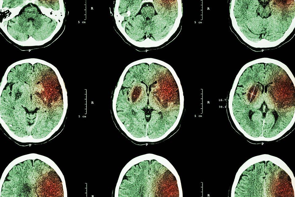
Your brain is the hungriest organ you have. It makes up only about 2 percent of your body weight, yet it demands roughly 20 percent of your oxygen and glucose, and it has almost no way to store fuel of its own. That means brain tissue depends on a constant, second-by-second delivery of oxygen-rich blood through the carotid and vertebral arteries. When that delivery stops, neurons cannot switch to a backup plan the way muscle can. Within minutes they begin to fail, and soon after, they begin to die. A stroke (also called a cerebrovascular accident, or CVA) is exactly this: a sudden interruption of blood flow to part of the brain, causing that region to lose function.
Two ways the flow fails: ischemic vs hemorrhagic
There are two fundamentally different problems that can starve brain tissue, and telling them apart drives everything you do next. Explore the interactive figure below to see them side by side.
An ischemic stroke is a plumbing blockage. A vessel gets clogged and the tissue downstream is cut off. This is by far the most common type, making up about 87 percent of strokes. The clog comes in two flavors: a thrombotic stroke, where a clot forms right there on top of atherosclerotic plaque in a cerebral artery, and an embolic stroke, where a clot (often a fragment thrown from the heart during atrial fibrillation) travels through the bloodstream and lodges in a smaller brain vessel.
A hemorrhagic stroke is the opposite mechanism: a leak. A weakened or overstretched vessel ruptures and bleeds into or around the brain. Now there are two injuries happening at once. The tissue past the rupture is deprived of flow, and the escaping blood forms a mass that raises intracranial pressure and irritates surrounding tissue. Chronic hypertension and ruptured aneurysms are common culprits. Hemorrhagic strokes are less common but often more deadly.
| Feature | Ischemic (~87%) | Hemorrhagic |
|---|---|---|
| Core problem | Blockage (clot cuts off flow) | Bleed (vessel ruptures) |
| Common causes | Atherosclerosis (thrombotic), A-fib emboli (embolic) | Hypertension, ruptured aneurysm |
| Clot-buster tPA | Can help if given in time | Absolutely contraindicated |
| Typical headache | Often mild or absent | Often sudden, severe ("worst of my life") |
The penumbra, and why time is brain
When an artery blocks, the tissue in the very center of the blocked zone dies quickly. But surrounding that dead core is a rim of tissue called the ischemic penumbra. These neurons are starving and silent, so they have stopped working, but they are not yet dead. They are being kept barely alive by trickles of blood from neighboring vessels. This penumbra is the whole reason we rush. If we can restore flow fast, penumbra neurons can wake back up and the deficit can shrink. If we wait, that salvageable rim converts into permanent dead tissue.
Clinicians estimate the brain loses roughly 1.9 million neurons every minute an ischemic stroke goes untreated. That staggering rate is the reason for the mantra time is brain. Every minute of delay is literally brain tissue you cannot get back.
Recognizing it fast: FAST
Because minutes matter, nurses and the public are taught a simple screen called FAST:
- F - Face: Ask the person to smile. Does one side droop?
- A - Arm: Ask them to raise both arms. Does one drift downward?
- S - Speech: Is speech slurred, absent, or strange? Can they repeat a simple sentence?
- T - Time: If any of these are present, note the time symptoms began and call for emergency help immediately.
That last letter is easy to gloss over but it may be the most important. Treatment eligibility depends entirely on knowing when the patient was last known well.
Why the CT scan comes before the clot-buster
The main clot-dissolving drug is tPA (tissue plasminogen activator, also called alteplase). It works beautifully on a clot, but it is a clot-dissolving (thrombolytic) drug. If the stroke were actually a bleed, giving tPA would pour fuel on a fire, dramatically worsening the hemorrhage. Here is the catch: ischemic and hemorrhagic strokes can look identical at the bedside. You cannot tell them apart by symptoms alone.
That is why an emergency non-contrast CT scan of the head is done first, every time. Its main job is to rule out a bleed. Only once blood is excluded can tPA be considered, and only within a strict window, usually within 3 to 4.5 hours of symptom onset for eligible patients. Some patients may also qualify for a mechanical thrombectomy, where the clot is physically pulled out, within a longer window.
Which side, which deficit
Because motor nerve tracts cross over in the lower medulla (the pyramidal decussation), a stroke on one side of the brain produces weakness on the opposite (contralateral) side of the body. A right-brain stroke causes left-sided weakness, and vice versa. Language adds another wrinkle: in most people the language centers live in the left hemisphere. So a left-brain stroke often produces aphasia, difficulty producing or understanding language, along with right-sided weakness. A right-brain stroke more often spares speech but may cause left-sided neglect, where the patient ignores their affected side entirely.
- Ischemic strokes (~87%) are blockages, thrombotic or embolic; hemorrhagic strokes are bleeds. The treatment for one would harm the other.
- The ischemic penumbra is salvageable tissue around the dead core. Restoring flow fast can rescue it, which is why time is brain.
- Screen quickly with FAST: Face droop, Arm drift, Speech trouble, Time to call for help and note onset.
- A non-contrast CT must rule out a bleed before tPA, because the clot-buster would worsen a hemorrhage; tPA is usually given within 3 to 4.5 hours of onset.
- Deficits are contralateral (opposite side); a left-brain stroke often causes aphasia plus right-sided weakness.
Section 2Raised intracranial pressure
Picture the brain as a delicate organ living inside a hard, sealed box: the skull. In a healthy person that box holds three things, and they share the space in a careful balance. There is the brain tissue itself (about 80% of the volume), the blood flowing through its vessels (about 10%), and the clear cerebrospinal fluid (CSF) that cushions and bathes it (about 10%). Because the skull cannot stretch, the total volume inside is fixed. The gentle pressure these three contents create is called the intracranial pressure (ICP), and normally it sits around 5 to 15 mmHg.
The Monro-Kellie doctrine: a box that cannot expand
The rule that governs all of this is the Monro-Kellie doctrine. It says that because the skull is a rigid, fixed container, the three volumes inside must add up to a constant. If one of them grows, another must shrink to make room, or the pressure will climb. At first the brain is clever about this. It buys time through compensation: it shunts CSF down into the spinal canal and squeezes some venous blood out of the skull. This is why a slowly growing problem can stay silent for a while. But this wiggle room is limited. Once those buffers are used up, even a tiny added volume causes the pressure to shoot up steeply. The patient falls off a compensation cliff, and small changes suddenly produce big, dangerous swings in ICP.
What raises the pressure
Anything that adds volume to that sealed box can raise ICP. The common culprits map neatly onto the three normal contents plus new intruders:
- Bleeding (hemorrhage) - an epidural, subdural, subarachnoid, or intracerebral bleed adds a pool of blood that takes up space.
- Tumor (mass lesion) - a growing mass literally crowds out brain, blood, and CSF.
- Cerebral edema - swelling of the brain tissue itself, often after trauma, stroke, or infection, as fluid leaks into or around cells.
- Hydrocephalus - a backup of CSF, either because too much is made, its flow is blocked, or it is not reabsorbed, so fluid accumulates in the ventricles.
The earliest warning: a change in consciousness
Here is the single most important nursing point. The earliest and most sensitive sign of rising ICP is a change in level of consciousness (LOC). The brain's alertness centers are exquisitely sensitive to pressure and to falling oxygen delivery. So before anything dramatic happens, the patient becomes subtly different: restless, confused, harder to rouse, more drowsy, slower to answer, or newly agitated. Families often notice it first ("she's just not herself"). Trust that. A dropping Glasgow Coma Scale (GCS) score is your red flag long before the classic textbook signs appear. Do not wait for pupils to change.
Cushing's triad: the late, ominous sign
When the pressure inside the skull rises high enough to threaten blood flow to the brainstem, the body launches a last-ditch rescue. To keep pushing blood past the crushing pressure, it drives the systolic blood pressure up. Baroreceptors sense that surging pressure and reflexively slow the heart, producing bradycardia. Meanwhile the pressure squeezes the brainstem's respiratory centers, making breathing irregular (for example, Cheyne-Stokes). Together these three make up Cushing's triad. It is a late and grave finding that signals the brainstem is in trouble.
| Feature | Early rising ICP | Cushing's triad (late) |
|---|---|---|
| Consciousness | Subtle confusion, restlessness, drowsiness, falling GCS | Deeply obtunded or comatose |
| Blood pressure | Often normal | Rising systolic with widening pulse pressure |
| Heart rate | Often normal | Bradycardia |
| Breathing | Usually normal | Irregular (e.g., Cheyne-Stokes) |
| Meaning | Act now, still reversible | Brainstem compromise, herniation near |
Herniation: the feared end result
If the pressure is not relieved, the brain is forced across its normal partitions - under the falx, across the tentorial notch, or down through the foramen magnum at the skull's base. This shifting of brain tissue across its normal boundaries is called herniation. As tissue is pushed downward it crushes the brainstem, which controls breathing, heart rate, and consciousness. The results are catastrophic: a fixed, dilated pupil on the affected side (from cranial nerve III compression), loss of protective reflexes, respiratory arrest, and death. Herniation is the dreaded destination of untreated intracranial hypertension, and everything above is about catching the problem long before this point. Explore the figure below to see how a growing volume drives the pressure up and pushes the brain toward herniation.
- The Monro-Kellie doctrine: the skull is a fixed box holding brain, blood, and CSF. When one volume grows, another must shrink or ICP rises. Compensation buys time, then fails sharply.
- Common causes are a bleed, tumor, cerebral edema, or hydrocephalus - anything that adds volume to the sealed skull.
- The earliest sign is a change in level of consciousness (restlessness, confusion, drowsiness, a falling GCS). Act on it early.
- Cushing's triad - rising systolic BP with widening pulse pressure, bradycardia, and irregular breathing - is a late, ominous sign of brainstem compromise.
- Herniation, brain tissue forced through the foramen magnum onto the brainstem, is the feared and often fatal end result.
Section 3Brain & spinal cord injury
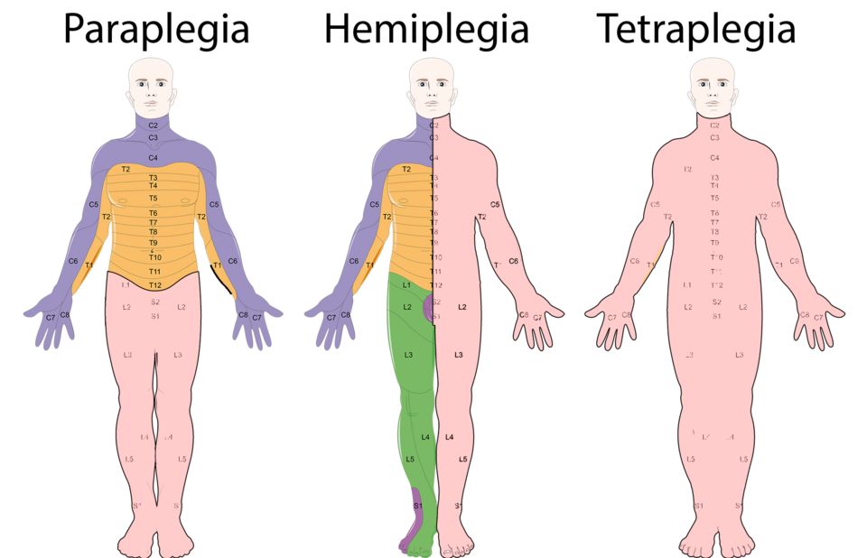
Your brain is soft, delicate tissue floating inside a hard, unforgiving skull, cushioned by a thin layer of cerebrospinal fluid. That fluid normally acts like a shock absorber. But the skull that protects your brain from the outside world becomes a problem during trauma, because the brain can slam against the bone and there is nowhere for swelling to go. This is why head injuries are so dangerous: the box is sealed, and any extra blood or swelling raises pressure on the brain itself.
Concussion vs contusion, and the coup-contrecoup pattern
A concussion is a functional injury. The brain gets shaken hard enough to disrupt how neurons signal, but there is usually no visible bleeding or bruising on imaging. Patients often have a brief loss of consciousness, confusion, headache, and amnesia around the event. A contusion is a step worse: it is an actual bruise of brain tissue, with capillary bleeding and swelling you can often see on CT. Contusions can enlarge over hours, so serial neuro checks matter.
Both are frequently caused by a coup-contrecoup injury. Picture a moving head striking a dashboard. The brain first bruises directly under the point of impact (the coup), then rebounds and slams into the opposite side of the skull (the contrecoup). One blow, two injured areas.
Bleeding between the layers: epidural vs subdural hematoma
Three membranes wrap the brain: the tough outer dura mater, the middle arachnoid, and the delicate pia mater against the brain. Where the bleeding collects, and what vessel is torn, changes how fast the patient crashes.
An epidural hematoma is bleeding above the dura, classically from a torn middle meningeal artery after a temporal skull fracture. Because it is arterial, it fills fast under high pressure. The textbook picture is a lucid interval: the patient is knocked out, wakes up and seems fine, then rapidly deteriorates as the clot expands and compresses the brain. This is a neurosurgical emergency.
A subdural hematoma is bleeding below the dura, from torn bridging veins. Because it is venous, it oozes slowly, so symptoms can take hours, days, or even weeks to appear. It is especially common in older adults and people with chronic alcohol use, because brain atrophy stretches those bridging veins and makes them fragile.
| Feature | Epidural hematoma | Subdural hematoma |
|---|---|---|
| Vessel | Artery (middle meningeal) | Bridging veins |
| Speed | Fast, high pressure | Slow ooze |
| Classic clue | Lucid interval then decline | Gradual, fluctuating symptoms |
| Typical patient | Temporal skull fracture | Older adults, chronic alcohol use |
Spinal cord injury by level
The spinal cord is the main data cable between brain and body. Where it is damaged determines what is lost below that point, because signals can no longer travel past the injury. As a rule, the higher the injury, the more of the body is affected.
- Lumbar/sacral injuries may affect the legs, bladder, and bowel.
- Thoracic injuries can cause paraplegia, loss of function in the legs and lower trunk.
- Cervical injuries can cause tetraplegia (quadriplegia), affecting all four limbs.
The scariest zone is the high cervical cord. The phrase students memorize is "C3, C4, C5 keep the diaphragm alive." The phrenic nerve, which drives the diaphragm, arises from these levels. An injury at or above C3-C5 can paralyze the diaphragm, so the patient cannot breathe on their own and needs immediate ventilatory support. Lower injuries are not off the hook: lesions from about C5 down through the upper thoracic cord knock out the intercostal and abdominal muscles, which shrinks vital capacity and weakens the cough even when the diaphragm still works, so secretions collect and respiratory failure can arrive days later. This is why airway and breathing come first in any suspected cervical injury, and why respiratory status is watched in cord injuries well below the neck.
Two shock states to keep straight
Spinal shock is the temporary loss of all reflexes, movement, and sensation below the injury right after trauma, a kind of neurological stunning that can last days to weeks. Neurogenic shock is a circulatory problem from losing sympathetic tone after a high injury, causing the classic triad of hypotension, bradycardia, and warm dry skin. Later, autonomic dysreflexia is a dangerous, exaggerated reflex in injuries at or above T6, usually triggered by a full bladder or bowel, causing a sudden dangerous spike in blood pressure that is a true emergency.
Explore the interactive figure below to see how these head injuries take shape.
- Concussion is functional (no bleeding); contusion is a true bruise; coup-contrecoup injures both the impact site and the opposite side.
- Epidural = arterial, fast, classic lucid interval; subdural = venous, slower, common in older adults and people with chronic alcohol use.
- Higher spinal injuries affect more of the body; C3-C5 injuries threaten the phrenic nerve and diaphragm, so breathing is the priority - and lower cervical and high thoracic injuries still weaken the intercostals and cough, so watch respiratory status in any injury above about T12.
- Spinal shock = temporary loss of reflexes; neurogenic shock = hypotension with bradycardia; autonomic dysreflexia = dangerous BP spike at or above T6.
Section 4Seizures & epilepsy
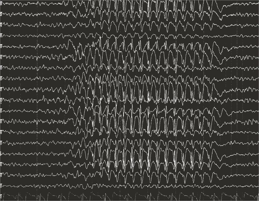
To understand a seizure, start with the normal brain. Your neurons are constantly talking to each other using tiny electrical signals, and in health that chatter is coordinated but not synchronized - different regions fire at different times for different jobs, like a huge orchestra where each section plays its own part. This balance is kept in check by a tug-of-war between excitatory signals (mostly the neurotransmitter glutamate, which says "fire") and inhibitory signals (mostly GABA, which says "settle down"). As long as that balance holds, the brain hums along smoothly.
A seizure is what happens when that balance breaks and a population of neurons suddenly fires all at once - a burst of abnormal, hypersynchronous electrical activity. Instead of the orchestra playing its parts, every instrument blasts the same note together. Depending on where in the brain this happens and how far it spreads, you get very different outward signs. One important distinction: a seizure is a single event, while epilepsy is a chronic condition defined by a tendency toward recurrent, unprovoked seizures (usually diagnosed after two or more, or one seizure plus a high risk of recurrence).
Focal vs. generalized: where the storm begins
Seizures are grouped by where the abnormal firing starts. A focal seizure (older term: partial) begins in one area of one hemisphere. Because only part of the brain is involved, the signs match that region - a twitching hand, an odd smell, a rising stomach sensation, or a period of staring and confusion. The person may stay aware (focal aware) or have altered awareness (focal impaired awareness). A focal seizure can sometimes spread and become generalized.
A generalized seizure begins in networks of both hemispheres at once. Awareness is usually impaired from the outset, though not always - in a myoclonic seizure the person stays fully aware through the sudden jerk, which is exactly why the current classification does not sort generalized seizures by awareness the way it sorts focal ones. The two you will see most often are tonic-clonic and absence.
| Feature | Tonic-clonic (generalized) | Absence |
|---|---|---|
| Classic look | Stiffening then rhythmic jerking | Brief blank staring spell |
| Phases | Tonic (muscles rigid) then clonic (rhythmic jerks) | No motor phases; may have eyelid flutter |
| Duration | Usually 1 to 3 minutes | Often 5 to 10 seconds |
| Awareness | Lost; person falls | Lost briefly; no fall |
| After the event | Postictal confusion, fatigue, sore muscles | Resumes activity, often unaware it happened |
| Typical patient | Any age | Often school-age children |
In a tonic-clonic seizure you may also see cyanosis (breathing pauses during the tonic phase), tongue-biting, and loss of bladder control - all explained by whole-brain firing overriding normal muscle and autonomic control. Absence seizures are so brief they are often mistaken for daydreaming, which is why a child "not paying attention" in class sometimes turns out to be seizing dozens of times a day.
What sets a seizure off: common triggers
Anything that pushes the brain toward excitation - or removes something that was calming it - can lower the seizure threshold:
- Fever in young children - rapid temperature spikes cause febrile seizures, usually between 6 months and 5 years.
- Low sodium (hyponatremia) or low glucose (hypoglycemia) - neurons depend on stable electrolytes and fuel to control firing; when either drops, membranes become unstable.
- Missed antiseizure medication - the most common reason a person with epilepsy breaks through; the calming drug level falls too low.
- Alcohol withdrawal - alcohol suppresses the brain, so stopping abruptly leaves it in a rebound hyperexcitable state.
- Others: sleep deprivation, flashing lights, and infections such as meningitis.
Status epilepticus: the emergency
Status epilepticus is a seizure that lasts 5 minutes or longer, or repeated seizures without full recovery of consciousness in between. This is a true neurologic emergency. Prolonged firing burns through oxygen and glucose faster than the brain can supply them, and the sustained muscle activity drives up temperature and can injure neurons - leading to hypoxia, acidosis, and permanent damage. It requires rapid IV benzodiazepines (such as lorazepam) and airway support.
Nursing priorities during a seizure
Your job is protection, not intervention in the seizure itself - you cannot stop the electrical activity by hand, so focus on preventing injury:
- Protect the airway and head - turn the person to their side (recovery position) to let secretions drain and reduce aspiration risk; cushion the head and remove nearby hazards.
- Do not restrain - holding limbs down can cause fractures or dislocations; let the movements run their course.
- Nothing in the mouth - never insert objects or fingers; it risks broken teeth, bites, and airway blockage.
- Time it - note the start; if it reaches 5 minutes, treat as status epilepticus and get help.
- Stay and observe - loosen tight clothing, provide privacy, and document what you saw (movements, duration, incontinence) for the provider.
- A seizure is a burst of abnormal, hypersynchronous electrical firing; epilepsy is a tendency toward recurrent, unprovoked seizures.
- Focal seizures start in one area (signs match that region); generalized seizures involve both hemispheres - tonic-clonic (stiffen then jerk, postictal confusion) and absence (brief staring, common in children).
- Common triggers lower the seizure threshold: fever in children, low sodium or glucose, missed medication, and alcohol withdrawal - correct the reversible cause.
- Status epilepticus (a seizure 5 minutes or longer, or back-to-back seizures without recovery) is an emergency requiring IV benzodiazepines and airway support.
- During a seizure: turn side-lying to protect the airway, cushion the head, do NOT restrain or put anything in the mouth, and time it.
Section 5Neurodegenerative disease
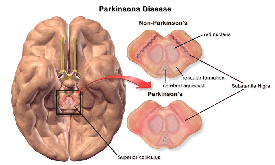
Your brain runs on about 86 billion neurons, and here's the humbling part: for the most part, you were born with nearly all of them, and adult brains make very few new ones. Neurons are not like skin or blood cells that constantly replace themselves. They are meant to last a lifetime. That is exactly why the diseases in this section are so devastating. When these specialized cells die, the body usually cannot grow replacements, so the losses stack up and the damage is progressive and irreversible. Think of it like a city slowly losing its power lines one by one, with no crew coming to repair them.
Normally, healthy neurons keep themselves tidy. They fold their proteins correctly, clear out cellular garbage, and pass chemical messages (neurotransmitters) across tiny gaps called synapses. The neurodegenerative diseases each represent a different way this housekeeping and signaling breaks down, in a different neighborhood of the nervous system.
Alzheimer disease: the memory system clogs first
Alzheimer disease is the most common cause of dementia. The trouble is misfolded protein. Two abnormal proteins build up: beta-amyloid plaques collect in the spaces between neurons, and tau tangles (neurofibrillary tangles) twist up inside them. These deposits are toxic and jam the machinery that neurons use to talk and stay alive, so the cells wither and die. Because the damage often begins in the hippocampus, the brain's memory-forming hub, the first and loudest symptom is usually short-term memory loss, the person forgets recent conversations while old memories stay intact longer. As it spreads across the cortex, it steals language, judgment, and eventually the ability to recognize loved ones and perform basic self-care.
Parkinson disease: a dopamine shortage in the movement circuit
Parkinson disease targets a specific spot deep in the brain called the substantia nigra, a cluster of neurons that make dopamine. Dopamine is the chemical that smooths and starts your movements. As these neurons die off, dopamine drops, and the movement circuit (the basal ganglia) loses its balance. The classic picture is often summarized as TRAP:
- Tremor at rest, often a "pill-rolling" motion of the fingers that eases with purposeful movement
- Rigidity, stiff, cogwheel-like resistance in the limbs
- Akinesia/Bradykinesia, slowed and shrinking movements (small handwriting, a masked face, a shuffling gait)
- Postural instability, poor balance and a tendency to fall, usually a later sign
ALS: the motor neurons fail, but the mind stays sharp
Amyotrophic lateral sclerosis (ALS), also called Lou Gehrig disease, attacks the motor neurons, the wiring that carries "move" commands from the brain and spinal cord to the muscles. Both upper and lower motor neurons degenerate, so patients develop progressive muscle weakness, atrophy, and fasciculations (muscle twitches), often starting in one hand or foot and spreading. The cruel twist is that in most patients cognition, senses, and awareness stay largely intact, the person remains fully alert inside a body that is losing its ability to move, swallow, and eventually breathe. Respiratory muscle failure is typically the life-limiting concern.
| Feature | Alzheimer | Parkinson | ALS |
|---|---|---|---|
| Cells lost | Cortical/hippocampal neurons | Dopamine neurons (substantia nigra) | Upper & lower motor neurons |
| Hallmark pathology | Amyloid plaques + tau tangles | Dopamine loss (Lewy bodies) | Motor neuron degeneration |
| First symptom | Short-term memory loss | Resting tremor, slowness | Focal muscle weakness |
| Cognition | Progressively lost | Often preserved early | Usually preserved |
The common thread tying all three together is the one worth memorizing: a specific population of neurons is slowly and permanently lost, so symptoms only worsen over time. Current treatments slow progression or ease symptoms, they do not regrow the lost cells or cure the disease. That is why nursing care centers on safety, function, dignity, and family support. Explore the figure below to see where each disease strikes in the nervous system.
- Neurodegenerative diseases share one theme: progressive, irreversible loss of specific neurons that the body cannot replace.
- Alzheimer = beta-amyloid plaques + tau tangles, starting in the hippocampus, so short-term memory loss comes first.
- Parkinson = dopamine-neuron loss in the substantia nigra, causing resting tremor, rigidity, bradykinesia, and postural instability (TRAP).
- ALS = upper and lower motor-neuron death causing spreading weakness and atrophy, while cognition and senses usually stay intact.
- Treatments slow or ease symptoms but do not reverse the neuron loss, so care emphasizes safety, function, and support.
Section 6Demyelinating & neuromuscular disorders
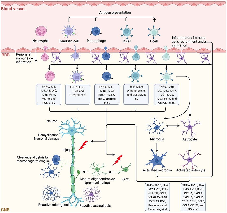
Every time you lift a coffee cup, wiggle your toes, or take a breath, a tiny chemical conversation happens between a nerve and a muscle. That conversation takes place at the neuromuscular junction (NMJ), the specialized synapse where a motor nerve meets a muscle fiber. Understanding this handshake is the key to understanding why the disorders in this section cause weakness in such different patterns.
The neuromuscular junction: how the signal crosses
When an electrical impulse (an action potential) races down a motor neuron and reaches the nerve ending, it triggers the release of a neurotransmitter called acetylcholine (ACh). The ACh floats across the tiny gap (the synaptic cleft) and binds to nicotinic ACh receptors on the muscle membrane. That binding opens ion channels, the muscle membrane depolarizes, and the muscle contracts. An enzyme called acetylcholinesterase then chews up the ACh so the signal switches off cleanly and the muscle can relax. Three ingredients matter: enough ACh released, enough working receptors to catch it, and a healthy nerve to carry the impulse in the first place. Break any one link and you get weakness, but the pattern of that weakness tells you which link failed.
Multiple sclerosis: the insulation frays
Nerve fibers in the central nervous system are wrapped in myelin, a fatty insulation that lets impulses jump quickly from gap to gap. In multiple sclerosis (MS), the body's own immune cells attack and strip that myelin off CNS neurons (the brain, spinal cord, and optic nerves). This is autoimmune demyelination. Where myelin is lost, a plaque or sclerosis forms, and signals slow, scatter, or stop entirely. Because myelin can be damaged almost anywhere in the CNS, symptoms are famously scattered: blurred or double vision from optic neuritis, numbness and tingling, weakness, spasticity, fatigue, and bladder problems. The classic course is relapsing-remitting: flares appear, then partly fade as inflammation settles and some remyelination occurs, though scarring accumulates over time. A helpful teaching phrase is lesions separated in space and time.
Myasthenia gravis: the receptors get blocked
In myasthenia gravis (MG), the nerve and its ACh are fine, but the muscle's receptors are under attack. The immune system makes anti-ACh receptor antibodies that block and destroy nicotinic receptors at the NMJ. With fewer receptors to catch ACh, each signal lands weakly. The hallmark is fatigable weakness: muscles work at first but tire quickly with repeated use and recover with rest. This is why symptoms are typically worse at the end of the day. It classically strikes muscles that work constantly, causing ptosis (drooping eyelids), diplopia (double vision), and trouble chewing, swallowing, or holding the head up. The feared complication is myasthenic crisis, when respiratory muscles weaken and breathing fails.
Guillain-Barre syndrome: the nerves lose their coating from the outside in
In Guillain-Barre syndrome (GBS), the immune system attacks the myelin of peripheral nerves, often a week or two after an infection (frequently a GI or respiratory illness). This is molecular mimicry: antibodies aimed at the bug cross-react with nerve myelin. The signature is ascending paralysis, weakness and diminished reflexes that begin in the feet and climb upward, sometimes reaching the diaphragm and requiring ventilator support. Unlike MG, weakness is usually symmetric and progressive rather than fatigable, and it typically peaks then slowly recovers.
| Feature | Myasthenia gravis | Guillain-Barre |
|---|---|---|
| Target | ACh receptors at NMJ | Peripheral nerve myelin |
| Pattern | Fatigable, often eyes first | Ascending, feet upward |
| Reflexes | Usually preserved | Diminished or absent |
| Trigger | Autoimmune, thymus-linked | Often post-infection |
| Feared crisis | Myasthenic crisis (breathing) | Respiratory failure |
Explore the neuromuscular junction in the figure below to see where ACh crosses and where these diseases strike.
- At the neuromuscular junction, acetylcholine crosses from nerve to muscle and binds receptors to trigger contraction, then acetylcholinesterase clears it.
- Multiple sclerosis is autoimmune demyelination of the CNS, often relapsing-remitting, with scattered symptoms separated in space and time.
- Myasthenia gravis is caused by antibodies against ACh receptors, producing fatigable weakness that worsens with use and improves with rest.
- Guillain-Barre syndrome is peripheral demyelination causing ascending paralysis, often after an infection, with lost reflexes.
- MG and GBS can both weaken the diaphragm, so respiratory monitoring is a nursing priority.
The chapter in one breath
- Ischemic stroke (about 87%) is a clot blocking flow; hemorrhagic stroke is a burst vessel, and the FAST screen (Face droop, Arm drift, Speech slurred, Time to call 911) catches both fast because time is brain.
- Only ischemic strokes get tPA within roughly 3 to 4.5 hours, so a stat CT must rule out bleeding first, since giving a clot-buster into a hemorrhage is deadly.
- Raised intracranial pressure shows late as Cushing's triad (rising BP with widening pulse pressure, bradycardia, irregular breathing) plus a sluggish, blown pupil, all warning the brain is being squeezed toward herniation.
- Traumatic brain injury is tracked with the Glasgow Coma Scale (eyes, verbal, motor); a score of 8 or less usually means secure the airway, and watch for the lucid-interval pattern of an epidural hematoma.
- Spinal cord injury above T6 risks neurogenic shock (warm, dry skin with hypotension and bradycardia) early, and autonomic dysreflexia (a pounding headache with dangerously high BP, often from a full bladder) later.
- Seizures require protecting the patient (side-lying, nothing in the mouth, never restrain) and timing them, because a seizure lasting over 5 minutes is status epilepticus, a true emergency.
- Neurodegenerative diseases differ by mechanism: Parkinson's from dopamine loss (resting tremor, rigidity, bradykinesia), Alzheimer's from plaques and tangles (progressive memory loss), and ALS from upper and lower motor-neuron death (spreading weakness and atrophy with cognition intact). Multiple sclerosis is not degenerative but autoimmune demyelination of the CNS, producing relapsing-remitting deficits.
- Myasthenia gravis causes fatigable weakness that worsens with use (ptosis, diplopia) from antibodies against acetylcholine receptors, while Guillain-Barre syndrome is an ascending paralysis that threatens the diaphragm, so monitor respiratory function closely.
ReferenceWords worth knowing
A quick reference for the terms in this chapter — skim it now, or circle back whenever a word trips you up.
- Bradykinesia
- Slowness in starting and carrying out movement, a core motor sign of Parkinson disease that arises when degeneration of dopamine-producing neurons in the substantia nigra leaves the basal ganglia unable to smoothly initiate voluntary movement.
- Cushing's triad
- A late and ominous set of three vital-sign changes, hypertension with a widening pulse pressure, bradycardia, and irregular (often slow) respirations, signaling dangerously high intracranial pressure and impending brainstem herniation.
- Demyelination
- The loss or breakdown of the fatty myelin sheath that insulates axons, which slows or blocks nerve signal conduction and produces the neurologic deficits seen in diseases like multiple sclerosis.
- Epidural hematoma
- A collection of blood between the skull and the outer dura mater, usually from a torn middle meningeal artery after temporal trauma, classically causing a brief lucid interval before rapid deterioration.
- Hemorrhagic stroke
- A stroke caused by a ruptured blood vessel bleeding into or around the brain, which both deprives tissue of blood and raises intracranial pressure as the expanding blood compresses surrounding neurons.
- Herniation
- The life-threatening displacement of brain tissue across or through rigid intracranial structures (such as under the falx or through the tentorium or foramen magnum) when pressure builds, often compressing the brainstem and its vital centers.
- Intracranial pressure
- The pressure exerted inside the skull by the brain, blood, and cerebrospinal fluid together, normally about 5–15 mmHg, which becomes dangerous when a mass, bleed, or swelling pushes it higher.
- Ischemic stroke
- The most common type of stroke, caused by a clot or narrowing that blocks an artery and cuts off blood flow, starving a region of brain tissue of the oxygen and glucose it needs to survive.
- Monro-Kellie doctrine
- The principle that the skull is a fixed, rigid box, so an increase in any one of its three contents (brain, blood, or cerebrospinal fluid) must be offset by a decrease in another or intracranial pressure will rise.
- Multiple sclerosis
- A chronic autoimmune disease in which the body attacks central nervous system myelin, producing scattered plaques and a relapsing-remitting pattern of symptoms such as vision changes, weakness, and numbness.
- Myasthenia gravis
- An autoimmune disorder in which antibodies block or destroy acetylcholine receptors at the neuromuscular junction, causing fluctuating muscle weakness that worsens with use and improves with rest.
- Penumbra
- The ring of at-risk but still salvageable brain tissue surrounding the dead core of an ischemic stroke, which can be rescued if blood flow is restored quickly, which is why "time is brain."
- Seizure
- A sudden burst of abnormal, excessive electrical activity in the brain's neurons that can cause changes in movement, sensation, behavior, or consciousness.
- Status epilepticus
- A neurologic emergency defined as a seizure lasting 5 minutes or longer, or repeated seizures without full recovery in between, which risks permanent brain injury and requires immediate treatment.
- Subdural hematoma
- A collection of blood between the dura and the arachnoid, usually from torn bridging veins, which often bleeds slowly and may present days to weeks later, especially in older adults and those on anticoagulants.
- tPA
- Tissue plasminogen activator, a clot-dissolving (thrombolytic) drug given within a strict time window for ischemic stroke to break down the blocking clot and restore blood flow, but never used in hemorrhagic stroke.
DrillFlashcards for this unit
These are the Week 5 cards — the same deck as the hub, pulled in here so you can review the moment you finish reading. Tap the card to flip it; hit Shuffle to mix them up. To grade yourself and track mastery across the whole deck, open the full flashcards page.
Check yourself
| Feature | Epidural | Subdural |
|---|---|---|
| Bleed source | Artery (fast) | Vein / bridging veins (slower) |
| Location | Above the dura | Below the dura |
| Onset | Rapid, often with lucid interval | Minutes to weeks; chronic in elderly |
| CT shape | Lens / convex (lemon) | Crescent (banana) |
| Feature | Alzheimer | Parkinson |
|---|---|---|
| Core deficit | Memory / cognition | Movement |
| Pathology | Amyloid plaques & tau tangles | Dopamine loss in substantia nigra |
| Hallmark signs | Short-term memory loss, disorientation | Resting tremor, rigidity, bradykinesia, shuffling gait |
The Endocrine System
The endocrine system is the body’s chemical messaging service — slower than nerves, but with reach that lasts hours to days. When one gland makes too much or too little of a hormone, the ripple effects show up everywhere: blood sugar, heart rate, blood pressure, mood, growth, and water balance. This chapter gives you the one idea that unlocks all of it — feedback — and then walks each major gland from healthy to what goes wrong, mechanism by mechanism.
By the end of this chapter you can
- Trace how the hypothalamus and pituitary command the other glands, and tell a tropic hormone from a direct one.
- Use negative feedback to predict a lab — and use a stimulating-hormone level to place a problem as primary (the gland) vs secondary (the pituitary).
- Contrast the too-much vs too-little state for the adrenal (Cushing’s vs Addison’s), thyroid, and ADH (SIADH vs DI).
- Read the electrolyte shifts that give a disorder away (Addison’s potassium; the sodium in SIADH and DI).
- Explain why Type 1 and Type 2 diabetes are different diseases, name the diagnostic numbers, and tell DKA from HHS.
- Recognize growth-hormone excess by timing (gigantism before the growth plates close, acromegaly after).
- Flag the endocrine emergencies: Addisonian crisis, thyroid storm, myxedema coma, DKA/HHS, and severe hyponatremia.
Section 1The chain of command
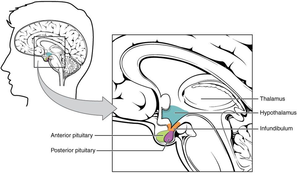
The normal wiring: how orders travel down
Picture the endocrine system as a company. The hypothalamus is the executive on the top floor. It constantly reads the body's status - temperature, blood pressure, stress, hunger, the levels of hormones already circulating - and decides what needs to change. Just beneath it sits its manager, the pituitary gland, tucked into a bony pocket at the base of the brain called the sella turcica. The two are connected by a short stalk (the infundibulum), and the way they talk to each other depends on which half of the pituitary we mean.
For the front half, the hypothalamus does not send nerve signals - it sends chemical memos. It makes tiny releasing hormones and inhibiting hormones (like TRH, CRH, GnRH, GHRH, and the inhibitor somatostatin) and drips them into a private set of blood vessels running down the stalk called the hypophyseal portal system. Think of it as a pneumatic tube between two offices: a dedicated capillary bed that carries the memo straight from the hypothalamus to the anterior pituitary without diluting it in the whole bloodstream first.
The anterior pituitary: a six-product factory
The anterior pituitary is a true factory - it manufactures six hormones. Several are tropic (from a Greek root meaning "to turn toward"): they don't act on body tissue directly, they turn on another gland. Others are direct hormones that act on the body's own tissues. A handy mnemonic is FLAT PiG: FSH, LH, ACTH, TSH (the tropic four) and Prolactin, GH (the two direct ones).
| Hormone | Target | Type |
|---|---|---|
| TSH | Thyroid gland | Tropic |
| ACTH | Adrenal cortex | Tropic |
| FSH & LH | Gonads (ovaries, testes) | Tropic |
| GH | Whole body & liver (via IGF-1) | Direct |
| Prolactin | Breast | Direct |
Notice that GH works partly by proxy: it tells the liver to release IGF-1 (insulin-like growth factor-1), which does much of the actual growth-promoting work in bone and muscle.
The posterior pituitary: a storage locker, not a factory
The back half is different, and students mix this up constantly. The posterior pituitary makes nothing. It is a storage locker. The two hormones it releases - ADH (antidiuretic hormone, also called vasopressin, which tells the kidneys to hold onto water) and oxytocin (uterine contraction in labor and milk let-down) - are actually built in the hypothalamus. Their parent neurons send long axons down the stalk, and the hormone simply travels down the nerve and waits in the terminals until a signal says "release." So the anterior lobe gets chemical memos through blood vessels, while the posterior lobe gets the finished product delivered down a nerve.
Negative feedback: the body's thermostat
The whole system governs itself with negative feedback, exactly like a home thermostat. Set the thermostat to 70 degrees: when the room warms past the setpoint, the furnace shuts off; when it cools, the furnace fires back up. Hormones do the same. Walk the thyroid axis: the hypothalamus releases TRH, which tells the pituitary to release TSH, which tells the thyroid to release T4 (and T3). When circulating T4 climbs high enough, it loops back and switches off both TRH and TSH - the furnace turns off. If T4 falls, the pituitary senses the shortage and cranks TSH up to push the thyroid harder. This is why the single most useful sentence in endocrine nursing is: low thyroid hormone usually means high TSH.
Positive feedback: the rare exception
Occasionally the body wants the opposite - a signal that amplifies itself until an event completes. This is positive feedback, and it is rare precisely because it is explosive. Two classic examples: the oxytocin surge in labor (stretch of the cervix drives more oxytocin, which drives stronger contractions, which stretches the cervix more - until the baby is delivered), and the LH surge at mid-cycle that triggers ovulation. Both shut off once the goal is reached.
The master skill: primary vs secondary vs tertiary
Here is the reasoning trick that separates strong students. When a hormone level is off, ask where the fault lives, and let the tropic hormone point you to it. A primary problem is in the target gland itself (the thyroid, adrenal, or gonad). A secondary problem is in the pituitary. A tertiary problem is up in the hypothalamus.
| Level | Broken part | Thyroid example (low T4) |
|---|---|---|
| Primary | Target gland | Low T4, high TSH (pituitary is shouting) |
| Secondary | Pituitary | Low T4, low TSH (no one is shouting) |
| Tertiary | Hypothalamus | Low T4, low TSH, low TRH |
Once you internalize the chain of command and the direction feedback pushes, endocrine disease stops being memorization and becomes detective work. Explore the wiring below.
- The hypothalamus sends releasing/inhibiting hormones through the hypophyseal portal blood vessels to the anterior pituitary, which manufactures six hormones (TSH, ACTH, FSH, LH, GH, prolactin).
- The posterior pituitary makes nothing - it only stores and releases ADH and oxytocin, which the hypothalamus builds and sends down the nerve.
- Tropic hormones (TSH, ACTH, FSH, LH) act on other glands; direct hormones (GH, prolactin) act on body tissue.
- Negative feedback works like a thermostat: when the final hormone rises it shuts off its own signals, so low thyroid hormone usually drives TSH up. Positive feedback (oxytocin in labor, the LH surge) is the rare, self-amplifying exception.
- Use the tropic hormone level to localize the lesion: primary = target gland, secondary = pituitary, tertiary = hypothalamus.
Section 2Adrenal glands — the cortisol problem
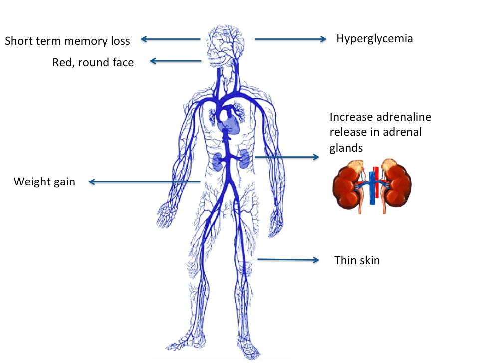
The normal adrenal gland: two organs stacked in one
Perched like little caps on top of each kidney, the adrenal glands are really two organs fused together. The outer cortex makes steroid hormones from cholesterol, and the inner medulla makes the fight-or-flight catecholamines (epinephrine and norepinephrine). The cortex is layered into three zones, and a handy way to remember what each makes is salt, sugar, sex as you travel inward. The outermost zona glomerulosa makes aldosterone (the mineralocorticoid that holds onto salt and water). The middle zona fasciculata makes cortisol (the glucocorticoid that manages sugar and stress). The innermost zona reticularis makes weak androgens (the sex steroids).
What cortisol actually does
Cortisol is your body's built-in stress hormone, released on a daily rhythm and surging whenever you are sick, injured, or frightened. It raises blood glucose by driving gluconeogenesis (the liver building new glucose) so your brain and muscles have fuel in a crisis. It is powerfully anti-inflammatory and immunosuppressive, which is exactly why we bottle it up as drugs like prednisone. And it helps maintain blood pressure by keeping blood vessels responsive to catecholamines. Control runs through the HPA axis: the pituitary releases ACTH, which tells the cortex to make cortisol, and rising cortisol switches ACTH back off (negative feedback). Hold onto that feedback loop, because it is the key to telling these diseases apart.
Cushing: too much cortisol
First, the vocabulary. Cushing syndrome is the umbrella term for any cause of excess cortisol. Cushing disease specifically means a pituitary ACTH-secreting adenoma driving the adrenals. But the single most common cause overall is neither of those: it is exogenous steroid medication taken long-term for asthma, autoimmune disease, or transplants. Adrenal tumors making cortisol directly are a third route.
Every classic sign traces back to too much cortisol. Because cortisol redistributes fat centrally, patients develop a rounded moon face, a buffalo hump between the shoulders, and truncal obesity with thin arms and legs (fat goes to the middle while cortisol breaks down limb muscle). Cortisol thins the skin and weakens collagen, so you see thin, fragile, easily-bruised skin and stretched purple striae across a belly that outgrew its weakened skin. Constant gluconeogenesis produces hyperglycemia (steroid-induced diabetes). Cortisol's mineralocorticoid spillover retains sodium and water, causing hypertension. Bone breakdown leads to osteoporosis, and the immunosuppression brings poor wound healing and infection risk. Mood swings, irritability, and even psychosis round out the picture.
Addison: too little cortisol (primary insufficiency)
Addison disease is primary adrenal insufficiency - the gland itself is destroyed, usually by autoimmune attack. Because the whole cortex fails, the patient loses all three outputs: cortisol, aldosterone, and adrenal androgens. Low aldosterone means sodium and water are lost while potassium is retained, so expect hyponatremia, HYPERkalemia, hypotension, and intense salt craving. Low cortisol brings hypoglycemia, profound weakness and fatigue, and weight loss. The tell-tale finding is bronze hyperpigmentation: with cortisol gone, feedback is lost and the pituitary floods the body with ACTH, and ACTH also stimulates skin melanocytes.
Secondary insufficiency: a pituitary problem
If the pituitary fails instead, ACTH is low. Two clues follow. There is no hyperpigmentation (no excess ACTH to darken skin), and aldosterone is largely spared because aldosterone is driven mainly by the renin-angiotensin system, not ACTH. So these patients get low cortisol but usually keep their sodium and potassium closer to normal.
Pheochromocytoma: the medulla's turn
A pheochromocytoma is a tumor of the adrenal medulla that dumps catecholamines in unpredictable surges. Remember the 5 P's: Pressure (episodic, often severe hypertension), Pain (pounding headache), Palpitations, Perspiration, and Pallor. Between surges the patient may feel completely normal, which is what makes it sneaky.
Putting it together
The fastest way to keep Cushing and Addison straight is a side-by-side look. Explore the interactive figure below, too.
| Feature | Cushing (excess) | Addison (deficiency) |
|---|---|---|
| Cortisol | High | Low |
| Blood glucose | High | Low |
| Blood pressure | High | Low |
| Sodium / Potassium | Na high / K low | Na low / K high |
| Weight & fat | Central gain, moon face | Loss, wasting |
| Skin | Thin, bruises, striae | Bronze hyperpigmentation |
One last feedback check that ties it all together: an adrenal tumor making its own cortisol pushes ACTH down (feedback shuts the pituitary off), while a failed adrenal gland lets ACTH climb up. The direction ACTH moves tells you where the problem lives.
- Cortex = salt (aldosterone), sugar (cortisol), sex (androgens); medulla = catecholamines. Cortisol raises glucose, calms inflammation, and supports blood pressure.
- Cushing = too much cortisol (most often from steroid medication): central obesity, moon face, striae, hyperglycemia, hypertension, thin skin, infection risk.
- Addison (primary) destroys the whole cortex: low cortisol AND aldosterone -> hypotension, hyponatremia, HYPERkalemia, hypoglycemia, salt craving, and bronze skin from high ACTH.
- Secondary insufficiency (pituitary) means low ACTH: no hyperpigmentation and aldosterone is spared. Never stop chronic steroids abruptly - taper to avoid an adrenal crisis (glucocorticoid-only, so potassium usually stays normal).
- Pheochromocytoma = medulla tumor with catecholamine surges (5 P's). Low ACTH points to an adrenal tumor; high ACTH points to a failed adrenal gland.
Section 3Thyroid — the metabolic thermostat
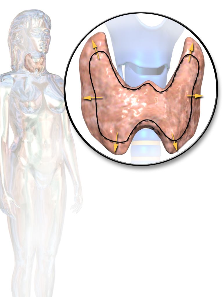
Think of your thyroid as the body's metabolic thermostat - a small butterfly-shaped gland sitting at the front of your neck that decides how fast every cell runs its engine. When it dials the setting up, your heart beats faster, you burn more fuel, and you feel warm. When it dials down, everything slows: heart, gut, brain, and body temperature all ease off. Almost every confusing thyroid symptom makes sense once you remember that one idea - this gland sets the pace of your metabolism.
The normal axis: a three-station relay
Control starts up in the brain. The hypothalamus releases TRH (thyrotropin-releasing hormone), which travels a short distance to the anterior pituitary and tells it to release TSH (thyroid-stimulating hormone). TSH then rides the bloodstream to the thyroid and says, "make hormone." The thyroid responds by producing mostly T4 (thyroxine) - the bulk of its output - and a smaller amount of T3 (triiodothyronine), which is the more active form. Much of the T4 gets converted to T3 out in the tissues, so think of T4 as the storage/delivery version and T3 as the one that actually revs the cell.
Two more facts anchor the whole system. First, the thyroid cannot build these hormones without iodine - it is a required raw ingredient, which is exactly why iodized salt exists. Second, the thyroid has a side job unrelated to metabolism: its C-cells secrete calcitonin, a hormone that gently lowers blood calcium by tucking it back into bone. It is a minor player in humans, but worth knowing it comes from the thyroid.
Hypothyroidism: the thermostat turned down
When the thyroid makes too little hormone, metabolism idles - and every symptom is that slowdown showing up in a different organ. In iodine-sufficient countries the number-one cause is Hashimoto's thyroiditis, an autoimmune attack that gradually destroys thyroid tissue. Worldwide, iodine deficiency is still the leading cause. Other causes include after treatment - post-thyroidectomy or radioactive iodine ablation - and certain medications (such as lithium and amiodarone).
Trace the mechanism and the signs write themselves. A slow metabolism means the body burns less fuel, so patients feel fatigued, gain weight despite a normal appetite, and cannot generate heat well - hence cold intolerance. Slowed cardiac pacing gives bradycardia. Sluggish skin turnover and reduced sweating leave skin dry and hair coarse and thinning. A slow gut means constipation. Reproductive hormones drift, causing menstrual changes (often heavier or irregular periods). Even the nervous system lags, producing the classic slowed, delayed deep-tendon reflexes. Severe, long-standing disease is called myxedema - a puffy, non-pitting swelling from mucin building up in the skin - and its life-threatening extreme is myxedema coma: hypothermia, low blood pressure, and a depressed level of consciousness. That is a true emergency.
Hyperthyroidism: the thermostat cranked up
Too much hormone and metabolism races. The leading cause is Graves' disease, an autoimmune condition in which antibodies against the TSH receptor (TSH-receptor antibodies, TRAb) - specifically the stimulating type, TSI (thyroid-stimulating immunoglobulins) - bind and switch that receptor on, flooring the gas pedal regardless of what the pituitary wants. Other causes include a toxic multinodular goiter (autonomous nodules that make hormone on their own) and thyroiditis (an inflamed gland leaking stored hormone).
The signs mirror hypothyroidism in reverse. Burning fuel too fast causes weight loss despite a big appetite and heat intolerance. The heart speeds up into tachycardia, palpitations, and sometimes atrial fibrillation. The overdriven nervous system brings anxiety, a fine tremor, and brisk hyperreflexia, while a fast gut causes diarrhea. Graves' adds two things unique to its autoantibody attack: bulging eyes, or exophthalmos (from tissue swelling behind the eye), and pretibial myxedema, a thickened patch of skin over the shins. The emergency here is thyroid storm - dangerous fever, extreme tachycardia, and agitation or delirium.
Reading the labs: let TSH tell the story
Because of negative feedback, the pattern of TSH versus T4 tells you both whether there is a problem and where it lives. In a primary problem the thyroid itself is the culprit; in a secondary problem the pituitary is failing to give proper orders, which flips the expected relationship.
| Pattern | TSH | T4 |
|---|---|---|
| Primary hypothyroid | High | Low |
| Primary hyperthyroid | Low | High |
| Secondary (pituitary) hypothyroid | Low or inappropriately normal | Low |
The tell for a secondary problem: a low T4 should drive TSH up, so a low T4 paired with a TSH that is low or "normal" is inappropriate - the pituitary is not doing its job.
Hypo vs hyper at a glance
| Feature | Hypothyroid | Hyperthyroid |
|---|---|---|
| Heart rate | Slow (bradycardia) | Fast (tachycardia, AFib) |
| Weight | Gain | Loss |
| Temperature | Cold intolerance | Heat intolerance |
| GI | Constipation | Diarrhea |
| Mood | Sluggish, low | Anxious, restless |
| Skin | Dry, coarse | Warm, moist |
Keep that one thermostat image in mind and the whole gland makes sense - explore the interactive figure below to see the axis and its feedback in motion.
- The thyroid sets metabolic speed: hypothyroid slows everything (fatigue, weight gain, cold, bradycardia, constipation); hyperthyroid speeds everything (weight loss, heat, tachycardia, anxiety, diarrhea).
- The axis runs TRH (hypothalamus) → TSH (pituitary) → T4 and active T3 (thyroid), and it needs iodine; negative feedback makes TSH the key lab.
- Hashimoto's is the top cause of hypothyroidism (iodine-sufficient areas); Graves' - via TSI/TRAb antibodies - is the top cause of hyperthyroidism and can add exophthalmos.
- Labs: primary hypothyroid = high TSH/low T4; primary hyperthyroid = low TSH/high T4; a low or inappropriately normal TSH with a low T4 points to a secondary (pituitary) cause.
- Emergencies are myxedema coma (hypo) and thyroid storm (hyper); after thyroid surgery, watch for hypocalcemia via Trousseau and Chvostek signs.
Section 4Diabetes mellitus
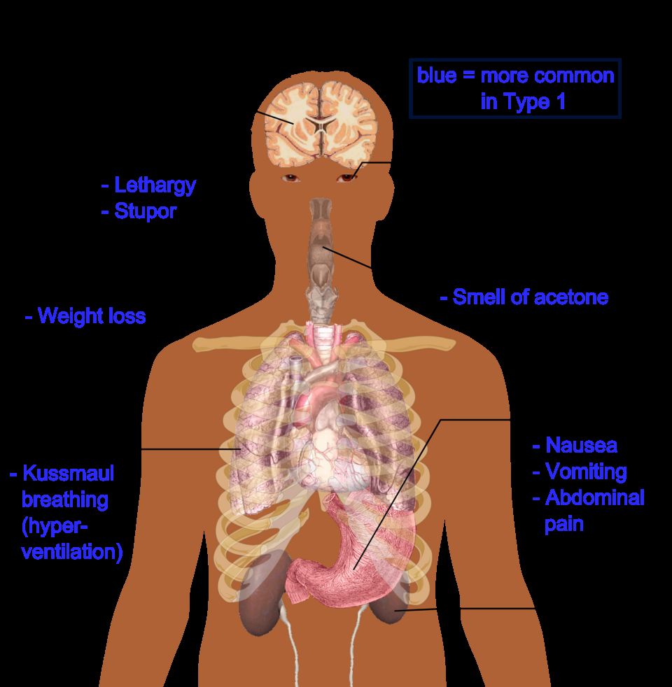
Before we talk about what breaks in diabetes, it helps to know how your body normally manages fuel. Think of your bloodstream as a delivery highway and glucose (blood sugar) as the cargo every cell wants for energy. The traffic controller is your pancreas, and specifically tiny clusters of cells inside it called the islets of Langerhans.
Normal fuel control: the fed state and the fasting state
Inside those islets, beta cells make insulin and alpha cells make glucagon. These two hormones pull in opposite directions, and that tug-of-war keeps blood sugar in a safe range. After a meal (the fed state), glucose rises, beta cells release insulin, and insulin acts like a key: it unlocks cells so glucose can move in to be burned or stored, and it tells the liver to bank extra glucose as glycogen. Between meals or overnight (the fasting state), glucose falls, so alpha cells release glucagon, which tells the liver to break glycogen back down and release glucose so your brain never runs dry. In short, insulin stores fuel and lets glucose into cells; glucagon raises blood sugar. Diabetes is what happens when the insulin side of that system fails.
Type 1: an absolute insulin deficiency
In type 1 diabetes, the immune system mistakenly attacks and destroys the beta cells. This is an autoimmune process, and we can often detect islet autoantibodies in the blood. Because the beta cells are gone, there is essentially no insulin at all, which we call an absolute insulin deficiency. It usually appears in younger, leaner people, and because the body cannot make insulin, these patients always require insulin injections to live. Without insulin, glucose cannot get into cells, so it piles up in the blood while the cells effectively starve.
Type 2: insulin resistance plus a relative shortfall
In type 2 diabetes, insulin is present, but the cells stop responding to it well. This is insulin resistance. The pancreas compensates by making extra insulin, but over time it cannot keep up, leaving a relative insulin deficiency. Type 2 is gradual, often silent for years, and strongly tied to obesity, metabolic syndrome, and older age. One physical clue is acanthosis nigricans, velvety dark patches in skin folds like the neck and armpits that signal chronic high insulin.
Why the three P's happen: osmotic diuresis
Whichever type, once blood glucose climbs high enough, the kidneys cannot reabsorb it all, so glucose spills into the urine. Sugar in the urine acts like a sponge, dragging water out with it. This is osmotic diuresis, and it explains the classic three P's: polyuria (peeing large volumes), which causes dehydration and triggers polydipsia (intense thirst), while starving cells drive polyphagia (hunger). Weight loss and blurry vision often ride along.
How we diagnose it
Diagnosis rests on standard numbers. Any one of these (usually confirmed on a repeat) makes the diagnosis:
- Fasting plasma glucose ≥ 126 mg/dL
- Random glucose ≥ 200 mg/dL with classic symptoms
- A1c ≥ 6.5 percent (a 3-month average of blood sugar)
- 2-hour OGTT ≥ 200 mg/dL (oral glucose tolerance test)
The long game: chronic complications
Years of high glucose damage blood vessels in two patterns. Microvascular (small-vessel) damage causes retinopathy (vision loss), nephropathy (kidney failure), and neuropathy (nerve damage, often numb, tingling feet). Macrovascular (large-vessel) damage speeds up coronary artery disease, stroke, and peripheral arterial disease. Combine numb feet with poor circulation and you get the feared diabetic foot ulcers and slow, poor wound healing.
Gestational diabetes
Pregnancy hormones naturally cause some insulin resistance. When the pancreas cannot keep up, gestational diabetes results. It is often silent, so we routinely screen pregnant patients (usually around 24 to 28 weeks). Extra maternal glucose crosses to the baby, who overproduces insulin and grows too large, called macrosomia. After delivery that sugar supply vanishes, but the baby's high insulin persists, causing neonatal hypoglycemia.
The two emergencies: DKA vs HHS
In type 1, with no insulin at all, the body burns fat for fuel and makes acidic ketones, tipping into diabetic ketoacidosis (DKA). In type 2, there is usually just enough insulin to block ketones, but sugar and dehydration climb to extremes in hyperosmolar hyperglycemic state (HHS).
| Feature | DKA | HHS |
|---|---|---|
| Usual type | Type 1 | Type 2 |
| Glucose | Usually > 250 mg/dL | Often > 600 mg/dL |
| Ketones | Present | Minimal or absent |
| Blood pH | Acidotic (low) | Near normal |
| Breathing | Kussmaul (deep, rapid) | Not Kussmaul |
| Onset | Rapid (hours) | Slow (days) |
| Core treatment | IV fluids, insulin, careful potassium | IV fluids, insulin, careful potassium |
Both are treated with the same core idea: rehydrate with IV fluids, give insulin to lower glucose and shut off ketone production, and replace potassium carefully (insulin drives potassium into cells, so levels can crash dangerously). Explore the comparison figure below.
- Insulin (from beta cells) lets glucose into cells and stores fuel; glucagon (from alpha cells) raises blood sugar between meals.
- Type 1 is autoimmune, an absolute insulin deficiency that always needs insulin and is DKA-prone; type 2 is insulin resistance plus a relative deficiency, tied to obesity and HHS-prone.
- High glucose spills into urine and drags water with it (osmotic diuresis), producing the three P's: polyuria, polydipsia, and polyphagia.
- Diagnose with fasting glucose ≥ 126, random ≥ 200 with symptoms, A1c ≥ 6.5 percent, or 2-hour OGTT ≥ 200 mg/dL; chronic highs cause microvascular and macrovascular damage.
- DKA (type 1) has ketones, acidosis, and Kussmaul breathing; HHS (type 2) has very high glucose with little ketosis; both need IV fluids, insulin, and careful potassium. Cold and clammy means give sugar fast.
Section 5Growth hormone — timing is everything
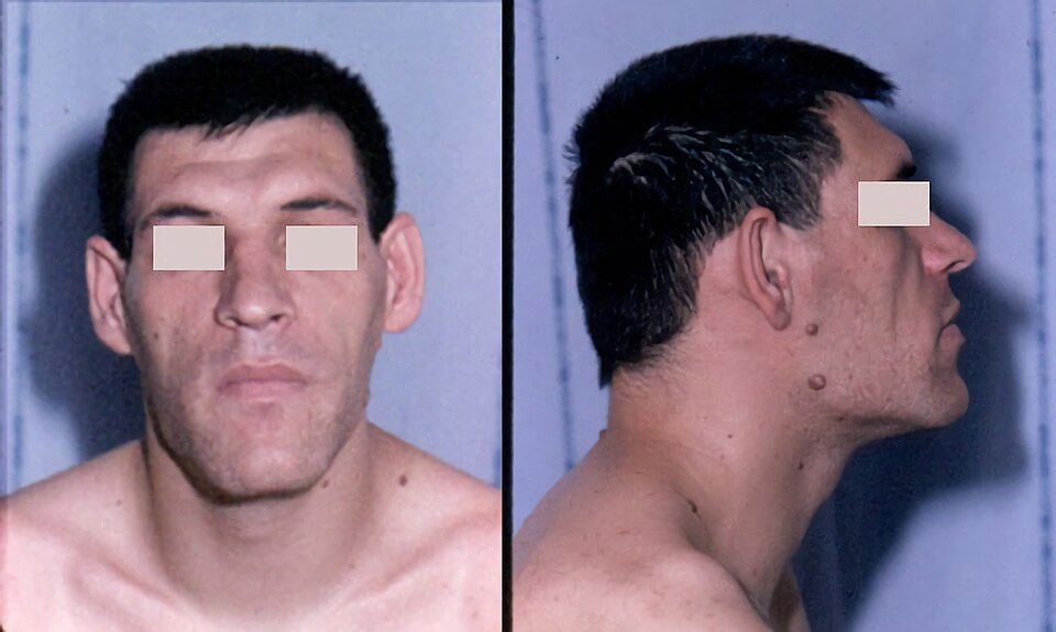
Somewhere near the base of your brain, a pea-sized gland called the anterior pituitary releases growth hormone (GH, somatotropin) in pulses, mostly overnight. GH is your body's build order: it tells bone, muscle, and soft tissue to grow. Like most hormones, it works on a push-pull system. The hypothalamus sends down GHRH (growth hormone-releasing hormone) to press the gas, and somatostatin to press the brake. The balance of those two, plus signals like sleep, stress, exercise, and low blood sugar, decides how much GH gets out.
How GH actually builds you
GH works two ways at once. It acts directly on tissues, and it acts indirectly by traveling to the liver and telling it to make IGF-1 (insulin-like growth factor 1). IGF-1 is the messenger that does much of the real growth work at the bone and muscle, which is why we lean on it for testing later. Together they lengthen and thicken bone at the growth zones, add muscle, and support organ and connective tissue. GH also nudges metabolism: it pushes the body to burn fat and it tends to raise blood glucose by opposing insulin. Hold onto that last fact, because it explains a lot of what goes wrong when there is too much GH.
When there is too little GH
In a child, not enough GH means the skeleton grows slowly but proportionally - a smaller child who is still nicely in proportion, head to limbs to trunk. This is proportional short stature, and it is different from conditions where the limbs and trunk are mismatched. In an adult, growth is already done, so GH deficiency shows up more quietly: fatigue, reduced muscle and strength, more central body fat, and a general sense of low vitality - changes in body composition rather than height.
Too much GH: why timing decides everything
Here is the single most important idea, and it is all about the epiphyseal (growth) plates - the cartilage zones at the ends of long bones where new bone is laid down to make you taller. While those plates are open (childhood and adolescence), bone can still lengthen. Once they fuse after puberty, that lengthening door is closed for good.
So excess GH lands very differently depending on when it strikes. Before the plates close, extra GH lets the whole skeleton keep lengthening - the child grows abnormally tall all over. This is gigantism. After the plates fuse, bone can no longer get longer, so the growth signal has nowhere to go but outward - bone and soft tissue thicken instead. This is acromegaly.
Acromegaly, sign by sign - and why
Because acromegaly builds outward, the clues are about things getting bigger and coarser over years, often so slowly the patient does not notice. As bone and soft tissue in the hands and feet expand, patients report a rising ring and shoe size. The face coarsens: the brow and jaw thicken, producing a protruding jaw called prognathism, and the teeth may spread apart as the jaw enlarges. The tongue enlarges (macroglossia), the voice may deepen, and internal organs enlarge too (organomegaly). Soft-tissue swelling in the wrist compresses the median nerve, giving carpal tunnel syndrome - numbness and tingling in the hand. Because GH opposes insulin, many patients develop glucose intolerance or frank diabetes. And overactive sweat glands cause noticeable sweating (hyperhidrosis).
The usual culprit - and its neighbors
Both conditions are almost always caused by a GH-secreting pituitary adenoma, a benign tumor of the anterior pituitary. Because that tumor sits in a tight bony pocket near important structures, it can cause mass effects as it grows: headache from local pressure, and classically bitemporal hemianopsia - loss of the outer (temporal) half of each visual field - when the tumor presses upward on the optic chiasm, where the nasal retinal fibers cross.
| Feature | Gigantism | Acromegaly |
|---|---|---|
| Timing of GH excess | Before growth plates close (childhood) | After growth plates fuse (adulthood) |
| Growth plates | Still open | Fused |
| What changes | Whole skeleton lengthens | Bone and tissue thicken |
| Main result | Abnormal tall height | Enlarging hands, feet, face (thickness) |
| Usual cause | GH-secreting pituitary adenoma | GH-secreting pituitary adenoma |
Picture a growth plate opening and closing and it all clicks into place - explore it below.
- GH from the anterior pituitary is balanced by GHRH (stimulates) and somatostatin (inhibits); it acts partly through liver-made IGF-1 and tends to raise blood glucose.
- The deciding factor for GH excess is whether the epiphyseal growth plates are open: open plates give gigantism (tall all over), fused plates give acromegaly (bones thicken).
- Acromegaly shows enlarging hands and feet, prognathism with spreading teeth, macroglossia, carpal tunnel syndrome, glucose intolerance, and sweating - all from outward growth.
- The usual cause is a GH-secreting pituitary adenoma, which can also cause headache and bitemporal hemianopsia from pressure on the optic chiasm.
- Diagnose with an elevated IGF-1 plus failure of GH to suppress after an oral glucose load.
Section 6ADH — the body’s water valve
The normal setup: one hormone, one job
Antidiuretic hormone (ADH), also called vasopressin, really does have a single mission: tell the kidneys to hang on to water. To understand its disorders, start with how the body decides when to hold water. Tucked in the hypothalamus are tiny sensors called osmoreceptors. Their whole job is to taste the blood - not for sugar or salt specifically, but for how concentrated it is (its osmolality). When you sweat on a hot day or forget to drink, the blood gets saltier and more concentrated. The osmoreceptors feel that and sound the alarm.
Here is a detail students love once it clicks: the hypothalamus makes ADH, but it does not release it directly. It sends the hormone down to the posterior pituitary, which acts as a storage tank and release valve. When the osmoreceptors fire, the posterior pituitary squirts stored ADH into the bloodstream. ADH then travels to the kidney and lands on the collecting duct - the final stretch of tubing before urine leaves the kidney. There it inserts little water doorways called aquaporins into the tubule wall. Water slips back out of the forming urine and into the blood. Result: the blood is re-diluted, and only a small amount of concentrated urine leaves the body. When you are well hydrated, ADH switches off, the aquaporins pull back, and you make lots of pale, dilute urine. That is the water valve, opening and closing all day.
Now the two disorders. They are near-perfect mirror images, so learn one and you almost get the other for free.
SIADH: too much ADH (the body is soaked)
SIADH stands for syndrome of inappropriate antidiuretic hormone. The key word is inappropriate - ADH is being released when the body does not need it. The valve is jammed open. Common culprits include central nervous system injury (head trauma, stroke, brain surgery, meningitis), small-cell lung cancer (the tumor secretes its own ADH), certain medications (SSRIs, carbamazepine, some chemo agents), and the stress states of surgery, pain, and nausea, which all nudge ADH upward.
With ADH stuck on, the kidneys reabsorb too much water. That extra water dilutes everything in the blood, so the serum sodium falls - a dilutional hyponatremia. Notice this is a water problem, not a salt-loss problem: the total sodium is roughly normal, just swimming in too much fluid. Because the retained water spreads through the whole body rather than pooling as obvious swelling, patients usually look euvolemic (they may gain a little weight but often have no dramatic edema). Meanwhile the urine is the opposite of the blood: small in volume and highly concentrated, because the kidney is clinging to water.
The danger is neurologic. Sodium keeps the fluid balance around brain cells steady; when it drops, water shifts into the neurons and the brain swells. Expect headache, confusion, lethargy, and in severe or fast drops, seizures. For chronic, mild, or asymptomatic SIADH the fix is simple in concept: fluid restriction. Take away the excess water and the sodium drifts back up. A seizing or obtunded patient is a different emergency — fluid restriction is far too slow, so 3% hypertonic saline is given in small boluses to pull the brain out of danger. Either way the correction rate is capped at roughly 6 to 8 mEq/L in 24 hours; raising sodium faster than that can strip myelin from brainstem neurons, an irreversible injury called osmotic demyelination syndrome.
Diabetes insipidus: too little ADH (the body cannot hold water)
Diabetes insipidus (DI) is the flip side. Either the brain does not make or release enough ADH (central DI, often after head injury, pituitary surgery, or a tumor), or the kidney simply cannot respond to the ADH it receives (nephrogenic DI, from kidney disease or drugs like lithium). Either way, the aquaporins never open. Water pours straight through into the urine.
The picture is dramatic: huge volumes of dilute, pale urine (sometimes many liters a day), progressive dehydration, and a rising serum sodium - hypernatremia - because water is leaving while sodium stays behind and gets concentrated. The body fights back with intense, unrelenting thirst. Central DI is treated by replacing the missing hormone with desmopressin (DDAVP), a synthetic ADH.
A quick word on the name
Do not let "diabetes" fool you - DI has nothing to do with blood sugar. The word diabetes just means a large flow of urine. The last name tells you the type: insipidus means tasteless (the old test was literally tasting the dilute urine), while mellitus means sweet, from the sugar spilling in diabetes mellitus. Same big-urine complaint, completely different cause.
SIADH vs DI side by side
| Feature | SIADH (too much ADH) | DI (too little ADH / no response) |
|---|---|---|
| ADH level / action | High - valve stuck open | Low or ineffective - valve stuck shut |
| Serum sodium | Low (hyponatremia) | High (hypernatremia) |
| Urine concentration | Concentrated | Dilute |
| Urine volume | Low | Very high |
| Volume status | Euvolemic / fluid-overloaded | Dehydrated (volume depleted) |
| Core treatment | Fluid restriction | Desmopressin (central DI) |
These two live at opposite ends of the same dial - the ADH water valve. Picture that valve opening and closing on the kidney tubule, and explore it below.
- ADH is made in the hypothalamus, stored and released by the posterior pituitary, and tells the kidney collecting duct to reabsorb water by inserting aquaporins; osmoreceptors trigger it when blood is too concentrated.
- SIADH = too much ADH: water retention, dilutional euvolemic hyponatremia, low-volume concentrated urine, and neuro symptoms - treat chronic or mild cases with fluid restriction, seizures or coma with 3% hypertonic saline, and cap the rise at about 6 to 8 mEq/L per day to avoid osmotic demyelination. Remember: Soaked, Sodium low.
- DI = too little ADH (central) or a kidney that cannot respond (nephrogenic): large volumes of dilute urine, dehydration, hypernatremia, intense thirst - central DI is treated with desmopressin.
- SIADH and DI are exact opposites on sodium, urine, and volume status - learn one and invert it for the other.
- Diabetes insipidus has nothing to do with blood sugar: insipidus means tasteless urine, mellitus means sweet.
The chapter in one breath
- Negative feedback is the master key: a low target hormone drives its pituitary signal up (low T4 means high TSH), while a high one shuts the signal off.
- Cushing's is too much cortisol (moon face, central obesity, hyperglycemia, thin easily-bruised skin, hypertension); Addison's is too little (hypotension, weight loss, hyperkalemia, bronze hyperpigmentation).
- Thyroid logic: hypo runs slow (fatigue, cold, weight gain), hyper runs fast (tachycardia, heat, weight loss), and a goiter can accompany either.
- Type 1 diabetes is absolute insulin deficiency prone to DKA (ketones, acidosis, Kussmaul breathing); Type 2 is insulin resistance prone to HHS (extreme hyperglycemia, profound dehydration, minimal ketones).
- Growth hormone excess before the growth plates fuse causes gigantism; the same excess after fusion causes acromegaly.
- ADH extremes mirror each other: too much is SIADH (water retention, dilutional hyponatremia), too little is diabetes insipidus (massive water loss, hypernatremia).
- Never stop steroids abruptly: taper corticosteroids to prevent an adrenal crisis, and stress-dose during illness or surgery.
- Post-thyroidectomy safety: watch for hypocalcemia from parathyroid injury (Chvostek/Trousseau signs, tingling, tetany) and for airway compromise from neck swelling.
ReferenceWords worth knowing
A quick reference for the terms in this chapter — skim it now, or circle back whenever a word trips you up.
- Aldosterone
- A mineralocorticoid hormone from the adrenal cortex that tells the kidneys to reabsorb sodium and water while excreting potassium, thereby raising blood volume and blood pressure.
- Aquaporin
- A water-channel protein inserted into kidney tubule membranes under the influence of antidiuretic hormone, letting water be reabsorbed and concentrating the urine.
- Catecholamines
- The fight-or-flight hormones epinephrine and norepinephrine released by the adrenal medulla that increase heart rate, blood pressure, and blood glucose.
- Cortisol
- The main glucocorticoid from the adrenal cortex that raises blood glucose, mobilizes fat and protein, and suppresses inflammation as part of the stress response.
- Exophthalmos
- Abnormal forward bulging of the eyeballs, classically seen in Graves disease when immune-driven tissue swelling pushes the eyes outward.
- Goiter
- A visible enlargement of the thyroid gland, which can occur whether thyroid hormone output is high, low, or normal.
- Hyperpigmentation
- Darkening of the skin that occurs in primary adrenal insufficiency because high ACTH also stimulates melanin-producing cells.
- IGF-1
- Insulin-like growth factor 1, a hormone made mainly by the liver in response to growth hormone that carries out most of growth hormone's effects on tissue growth.
- Insulin resistance
- A condition in which body cells respond poorly to insulin, so the pancreas must secrete more of it to keep blood glucose controlled.
- Ketoacidosis
- A dangerous metabolic state, typical of type 1 diabetes, in which severe insulin lack forces fat breakdown into acidic ketones that lower blood pH.
- Myxedema
- The severe, advanced form of hypothyroidism marked by non-pitting facial and tissue swelling from mucopolysaccharide buildup, and in crisis by profound slowing of body functions.
- Negative feedback
- The self-regulating loop in which a rising hormone level signals its own source to shut down further release, keeping the hormone within a stable range.
- Osmotic diuresis
- Excessive urine production that happens when high blood glucose spills into the urine and pulls water along with it, causing dehydration.
- Primary vs secondary endocrine disorder
- A primary disorder arises from a defect in the hormone-producing gland itself, whereas a secondary disorder arises from the pituitary (too little stimulating hormone) and a tertiary disorder from the hypothalamus above it - many nursing texts group both under secondary.
- Thyroid storm
- A life-threatening surge of thyroid hormone activity producing high fever, racing heart, agitation, and possible cardiovascular collapse.
- Tropic hormone
- A hormone, usually from the pituitary, whose target is another endocrine gland that it stimulates to release its own hormone.
DrillFlashcards for this unit
These are the Week 6 cards — the same deck as the hub, pulled in here so you can review the moment you finish reading. Tap the card to flip it; hit Shuffle to mix them up. To grade yourself and track mastery across the whole deck, open the full flashcards page.
Check yourself
| Feature | Cushing | Addison |
|---|---|---|
| Cortisol | High | Low |
| Blood glucose | High | Low |
| Blood pressure | High | Low |
| Serum sodium | High | Low |
| Serum potassium | Low | High |
| Weight | Gain (central) | Loss |
| Feature | Type 1 | Type 2 |
|---|---|---|
| Core defect | Autoimmune beta-cell loss | Insulin resistance |
| Insulin level | Absent | Normal to high early |
| Typical onset | Childhood/young, lean | Adult, often obese |
| Ketosis risk | High (DKA) | Low (HHS instead) |
Growth hormone excess
Gigantism is GH excess before growth-plate closure, producing very tall stature; acromegaly is GH excess after closure, causing enlargement of hands, feet, jaw, and facial features rather than height.
Posterior pituitary / ADH
SIADH is too much ADH: water is retained, urine is concentrated, and serum sodium falls (dilutional hyponatremia). DI (diabetes insipidus) is too little ADH (or renal resistance): huge volumes of dilute urine, thirst, and rising serum sodium.
| Feature | SIADH | DI |
|---|---|---|
| ADH effect | Excess | Deficient |
| Urine output | Low, concentrated | High, dilute |
| Serum sodium | Low | High |
| Volume status | Retained water | Dehydration |
Hematology: The Blood
Blood is a liquid organ — it carries oxygen, stops leaks, and defends the body — so when it goes wrong, the whole body feels it. This chapter covers how blood cells are made, the anemias that starve tissues of oxygen, the clotting and bleeding disorders, the rules of blood typing and transfusion, and the cancers of the blood itself.
By the end of this chapter you can
- Describe hematopoiesis and the role of erythropoietin.
- Classify the common anemias and match each to its cause.
- Explain hemostasis and the major bleeding and clotting disorders.
- Apply ABO/Rh compatibility and respond to a transfusion reaction.
- Distinguish leukemia, lymphoma, and multiple myeloma.
Section 1Blood, marrow & how cells are made
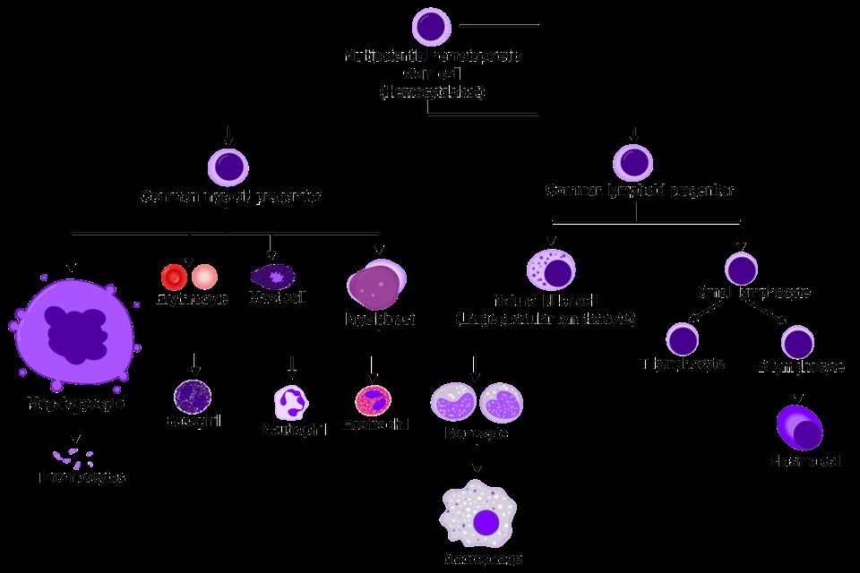
Think of blood as a river that touches every cell you own. It looks like a simple red liquid, but it is actually a connective tissue - living cells suspended in fluid. Blood delivers oxygen and nutrients, hauls away carbon dioxide and waste, carries hormones and immune defenders, helps clot a wound, and even balances your temperature and pH. When something goes wrong with blood, the effects show up everywhere at once, because everything depends on it.
What blood is made of
If you spin a tube of blood in a centrifuge, it separates into layers. The top ~55% is plasma - a straw-colored fluid that is mostly water plus proteins (albumin, which holds fluid inside vessels; clotting factors; and antibodies), electrolytes, glucose, and wastes. The bottom ~45% is packed red cells; that percentage is the hematocrit. A thin middle layer, the buffy coat, holds white cells and platelets.
- Red blood cells (erythrocytes) - carry oxygen. By far the most numerous.
- White blood cells (leukocytes) - the immune army (neutrophils, lymphocytes, monocytes, eosinophils, basophils).
- Platelets (thrombocytes) - cell fragments that plug leaks and start clotting.
| Feature | Red cells | Platelets |
|---|---|---|
| Main job | Carry oxygen | Stop bleeding (hemostasis) |
| Has a nucleus? | No (mature RBCs) | No - they are cell fragments |
| Typical lifespan | About 120 days | About 8 to 10 days |
| Made from | Bone marrow stem cells | Megakaryocytes in marrow |
Hematopoiesis - where blood cells are born
Every blood cell traces back to one ancestor: the hematopoietic stem cell (HSC) living in the red bone marrow (in adults, mainly the pelvis, sternum, vertebrae, and ends of long bones). This process is called hematopoiesis. A stem cell is pluripotent - it can become any blood cell - and it makes copies of itself so the factory never runs out. As daughter cells mature, they commit down one of two big lines: the myeloid line (red cells, platelets, and most white cells) or the lymphoid line (B and T lymphocytes). Because the marrow constantly replaces cells that wear out, anything that damages the marrow - chemotherapy, radiation, leukemia crowding it out, or aplastic anemia - can drop all the cell lines at once, a state called pancytopenia.
Erythropoietin - the kidney's oxygen alarm
How does the body know to make more red cells? The kidneys act as oxygen sensors. When they detect low oxygen reaching their own tissue (hypoxia) - from blood loss, high altitude, or lung disease - they release a hormone called erythropoietin (EPO). EPO travels to the bone marrow and tells it to ramp up red cell production. This is a beautiful negative-feedback loop: more red cells means more oxygen delivery, which quiets the alarm and lowers EPO again.
This explains a classic clinical picture. In chronic kidney disease, the damaged kidneys stop making enough EPO, so the marrow never gets the signal - the patient develops anemia of chronic kidney disease even though the marrow itself is healthy. That is why these patients are often treated with synthetic EPO. It also explains why people living at high altitude naturally run higher hemoglobin and hematocrit: chronic low oxygen keeps EPO switched on. Endurance athletes are the instructive exception - training does raise total red cell mass, but it expands plasma volume even more, so the measured hematocrit often reads normal or slightly low, a dilutional picture called athlete's pseudoanemia.
Hemoglobin - how red cells carry oxygen
A red cell is essentially a bag stuffed with hemoglobin (Hgb), the iron-containing protein that actually binds oxygen. Each hemoglobin molecule has four heme groups, and each heme holds one iron atom that grabs one oxygen molecule - so one hemoglobin carries up to four oxygen molecules. The red cell's flexible biconcave disc shape gives it lots of surface area and lets it squeeze through tiny capillaries.
This is why the pieces have to line up. You need enough iron (to build heme), enough vitamin B12 and folate (to build the cell), and a healthy marrow responding to EPO. Take away any one and oxygen delivery falls. Low hemoglobin is anemia, and its symptoms flow directly from the mechanism: fatigue and pallor because tissues are starved of oxygen, and tachycardia and shortness of breath because the heart speeds up to move the remaining oxygen faster. Nurses usually watch the hemoglobin and hematocrit together as the fastest read on a patient's oxygen-carrying capacity.
- Blood is a connective tissue: ~55% plasma (water plus albumin, clotting factors, antibodies) and ~45% cells - red cells, white cells, and platelets.
- Hematopoiesis makes all blood cells from pluripotent stem cells in the red bone marrow, splitting into myeloid and lymphoid lines; marrow damage can cause pancytopenia.
- The kidneys sense low oxygen and release erythropoietin to drive red cell production - so chronic kidney disease causes anemia from lost EPO.
- Hemoglobin is the iron-based protein that binds oxygen; each molecule carries up to four O2, and building it needs iron, B12, and folate.
- Low hemoglobin (anemia) starves tissues of oxygen, producing fatigue, pallor, and compensatory tachycardia - watch Hgb and hematocrit together.
Section 2Anemias
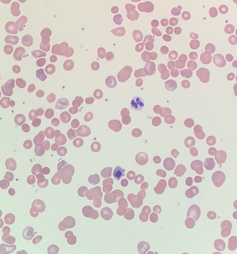
Every one of your cells needs a steady supply of oxygen to make energy, and oxygen does not travel through blood on its own. It rides on hemoglobin, the iron-rich protein packed inside your red blood cells (RBCs). Healthy bone marrow constantly builds new RBCs, stuffs them with hemoglobin, and releases them to circulate for about 120 days. When there is not enough functional hemoglobin to carry the oxygen your tissues need, we call that anemia. Notice the wording: anemia is not simply "low blood." It is low oxygen-carrying capacity, whether from too few RBCs, too little hemoglobin per cell, or hemoglobin that does not work.
Because the problem is tissue oxygen delivery, the classic symptoms make sense once you trace the mechanism. Tissues starved of oxygen produce less energy, so patients feel fatigue and weakness. With fewer red cells reflecting light through the skin and mucous membranes, you see pallor (pale skin, conjunctiva, nail beds). The heart tries to compensate by pumping faster to move what little oxygen there is, causing tachycardia. And because the blood is oxygen-poor, patients breathe faster and feel short of breath, especially with exertion, which is dyspnea. These are the shared symptoms of almost every anemia; the differences lie in why the hemoglobin is missing.
Iron-deficiency anemia (microcytic)
Iron is the core of the heme molecule that binds oxygen. Without enough iron, the marrow makes red cells that are small (microcytic, low MCV) and pale (hypochromic) because they simply cannot fill with hemoglobin. This is the most common anemia worldwide. Causes usually fall into three buckets: blood loss (heavy menstrual periods, or slow GI bleeding from ulcers or colon cancer, which quietly drains iron), increased demand (pregnancy, infancy, rapid growth), and poor intake or absorption (diets low in iron, or gut conditions). Lab clues include low ferritin (the body's iron storage form) and low serum iron.
B12 and folate deficiency (macrocytic)
Vitamin B12 and folate are required to build DNA. When they run short, developing red cells cannot divide properly, so they grow large and fragile instead, producing macrocytic (high MCV) megaloblastic anemia. Folate deficiency comes from poor diet (few leafy greens), alcohol use, or pregnancy demand. B12 deficiency has a special cause worth memorizing: pernicious anemia. Absorbing B12 from food requires intrinsic factor, a protein made by the stomach's parietal cells. In pernicious anemia, an autoimmune attack destroys those cells, intrinsic factor disappears, and ordinary dietary B12 cannot be absorbed no matter how much is eaten. Replacement is therefore given by injection, or as high-dose oral B12 (1,000 to 2,000 mcg daily), which works because about 1 percent of a swallowed dose crosses the gut by passive diffusion without any intrinsic factor at all. One key distinction: because B12 also maintains the nervous system, its deficiency adds neurologic signs (numbness, tingling, balance problems) that folate deficiency does not.
| Feature | Iron deficiency | B12 / folate deficiency |
|---|---|---|
| Cell size (MCV) | Small (microcytic) | Large (macrocytic) |
| Core problem | Too little hemoglobin | Faulty DNA / cell division |
| Typical cause | Blood loss, low intake | Poor diet, pernicious anemia |
| Neuro symptoms | No | Yes (B12 only) |
Hemolytic anemias and sickle cell disease
Hemolytic anemias occur when red cells are destroyed faster than the marrow can replace them (hemolysis means RBC breakdown). Because dying cells spill their contents, you often see jaundice from the released bilirubin. The classic inherited example is sickle cell disease. A single genetic change produces abnormal hemoglobin called HbS. When oxygen levels drop, or with dehydration, infection, or cold, HbS molecules stick together and warp the soft round RBC into a rigid, crescent (sickle) shape. These stiff cells jam small vessels, blocking blood flow in what is called a vaso-occlusive crisis - episodes of severe, sudden pain as tissues downstream are starved of oxygen. The sickled cells are also fragile and are destroyed early, driving chronic anemia.
Finally, aplastic anemia is a marrow-failure state where the bone marrow stops producing blood cells altogether, so RBCs, white cells, and platelets all fall together (pancytopenia). Explore the different anemia types in the figure below.
- Anemia is low functional hemoglobin, reducing oxygen delivery; shared signs (fatigue, pallor, tachycardia, dyspnea) all stem from tissue hypoxia and cardiac compensation.
- Iron-deficiency anemia is microcytic and most common; a new case in an older adult means rule out GI bleeding.
- B12 and folate deficiencies are macrocytic; only B12 deficiency causes neurologic signs, and pernicious anemia is loss of intrinsic factor blocking B12 absorption.
- Sickle cell disease (HbS) sickles under low oxygen, causing painful vaso-occlusive crises; manage with hydration and pain control, adding oxygen when the patient is hypoxic.
- Aplastic anemia is bone marrow failure producing pancytopenia.
Section 3Bleeding & clotting disorders
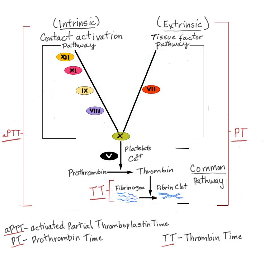
Every time you get a paper cut, a beautifully choreographed emergency response kicks in to stop the bleeding within minutes. This process is called hemostasis (literally "blood standing still"), and it happens in overlapping steps. Understanding the normal sequence is the key to understanding what goes wrong in bleeding and clotting disorders.
Normal hemostasis: how the body plugs a leak
The moment a blood vessel is injured, three things happen in quick succession. First, vasoconstriction: the vessel clamps down to slow blood flow to the area. Second, the platelet plug forms. Platelets are tiny cell fragments that normally glide past smooth vessel walls, but when they touch exposed collagen at an injury, they stick, activate, change shape, and clump together, helped by a bridging protein called von Willebrand factor (vWF). This soft plug is a quick fix. Third comes the coagulation cascade, a chain reaction of clotting proteins (factors) that activate one another in sequence. The cascade has an intrinsic and extrinsic pathway that converge, ultimately turning prothrombin into thrombin. Thrombin then converts fibrinogen into fibrin, tough protein threads that weave through the platelet plug like rebar in concrete, creating a stable clot. Later, the clot is dissolved by fibrinolysis once healing is underway.
Thrombocytopenia and ITP: too few platelets
Thrombocytopenia means a low platelet count (usually below 150,000/mm3, with serious bleeding risk often under 20,000). With too few platelets, the first plug is weak. Patients show petechiae (pinpoint red spots), purpura, easy bruising, nosebleeds, and gum bleeding. In immune thrombocytopenic purpura (ITP), the immune system makes antibodies that tag the patient's own platelets, and the spleen destroys them. It often follows a viral illness in children and is more chronic in adults. Treatment targets the immune attack, using corticosteroids, IVIG, and sometimes splenectomy.
Hemophilia and von Willebrand disease: the factor problems
Hemophilia is an inherited deficiency of a clotting factor, so the platelet plug forms fine but the fibrin mesh never sets properly. Both common types are X-linked recessive, which is why they classically affect males, whose mothers are carriers. Hemophilia A is a deficiency of factor VIII; hemophilia B (Christmas disease) is a deficiency of factor IX. Clinically they look identical: hemarthrosis (deep bleeding into joints), muscle bleeds, and prolonged bleeding after surgery or trauma. Lab work shows a prolonged aPTT with a normal platelet count. Treatment is replacing the missing factor.
Von Willebrand disease is the most common inherited bleeding disorder and is usually autosomal (affecting both sexes equally). Missing or defective vWF means platelets cannot stick well, and because vWF also carries factor VIII, that factor drops too. So it blends both worlds: mucosal, platelet-type bleeding (heavy menstrual periods, nosebleeds) plus a mild factor picture.
| Feature | ITP | Hemophilia A/B | von Willebrand |
|---|---|---|---|
| Core defect | Platelets destroyed | Factor VIII or IX missing | vWF missing/defective |
| Inheritance | Acquired (autoimmune) | X-linked recessive | Usually autosomal |
| Bleeding type | Petechiae, mucosal | Deep joints, muscles | Mucosal, menstrual |
| Key lab | Low platelet count | Prolonged aPTT | Low vWF, sometimes low VIII |
DIC and DVT: when clotting itself becomes the disease
Disseminated intravascular coagulation (DIC) is a dangerous paradox. Triggered by sepsis, trauma, obstetric complications, or cancer, the cascade fires everywhere at once, throwing tiny clots throughout the microcirculation. This consumes platelets and clotting factors faster than the body can replace them. The result is a patient who is both clotting and bleeding: microclots damage organs while the depleted supply leaves them oozing from IV sites, gums, and incisions. Labs show low platelets, prolonged PT/aPTT, low fibrinogen, and elevated D-dimer. DIC is always secondary, so treatment means fixing the underlying cause.
Clotting can also go wrong in a single vessel. A deep vein thrombosis (DVT) is a clot in a deep vein, usually of the leg. Virchow's triad names the three ingredients that promote it: venous stasis (immobility), endothelial injury (vessel-wall damage), and hypercoagulability (blood that clots too readily). The feared complication is a piece breaking off and traveling to the lungs as a pulmonary embolism.
You can explore hemostasis, bleeding, and clotting in the interactive figure below.
- Hemostasis runs in order: vasoconstriction, platelet plug (needs vWF), then the coagulation cascade making thrombin and fibrin.
- Platelet/vWF problems cause immediate superficial bleeding (petechiae, mucosal); factor problems cause delayed deep bleeding (joints, muscles).
- ITP is autoimmune platelet destruction; hemophilia A (factor VIII) and B (factor IX) are X-linked; von Willebrand disease is the most common inherited bleeding disorder.
- DIC consumes platelets and factors, so the patient clots AND bleeds at once, with a high D-dimer; it is always secondary to another problem.
- DVT forms via Virchow's triad (stasis, endothelial injury, hypercoagulability) and can embolize to the lungs.
Section 4Blood types & transfusion
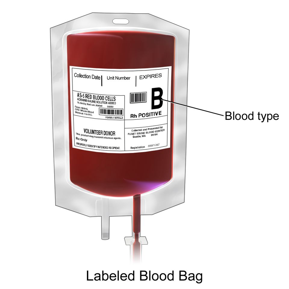
Every drop of blood carries a kind of ID badge. On the surface of your red blood cells sit tiny protein and sugar markers called antigens, and floating in your plasma are antibodies, immune proteins that attack any antigen your body does not recognize as its own. Your immune system learns early in life to leave your own red cells alone while staying armed against foreign ones. This self-versus-nonself recognition is the whole reason transfusions must be matched. Give someone blood carrying an antigen their plasma has antibodies against, and those antibodies clamp onto the donor cells and destroy them, sometimes with life-threatening speed.
The ABO system
The ABO system sorts blood into four types based on which antigens sit on the red cells. Type A cells carry the A antigen, type B carry B, type AB carry both, and type O carry neither. Here is the elegant, slightly counterintuitive part: your plasma automatically makes antibodies against whatever antigen you lack. So type A blood has anti-B antibodies, type B has anti-A, type O has both anti-A and anti-B, and type AB has neither.
Because type O cells carry no A or B antigen, there is nothing on them for a recipient's antibodies to attack, which is why O-negative is called the universal donor, the go-to in a trauma emergency before typing is done. Because type AB plasma carries no anti-A or anti-B antibodies, an AB person can receive any ABO type, making AB-positive the universal recipient.
| Blood type | Antigen on red cell | Antibody in plasma |
|---|---|---|
| A | A | anti-B |
| B | B | anti-A |
| AB | A and B | none |
| O | none | anti-A and anti-B |
The Rh factor and hemolytic disease of the newborn
On top of ABO sits a second marker, the Rh factor (also called the D antigen). If your red cells carry it, you are Rh-positive; if not, Rh-negative. Unlike ABO, Rh-negative people do not come with ready-made anti-Rh antibodies. They only build them after being exposed to Rh-positive blood, a process called sensitization.
This matters most in pregnancy. If an Rh-negative mother carries an Rh-positive baby, a small leak of fetal blood, often during delivery, can sensitize her. The first baby is usually fine, but in a later Rh-positive pregnancy her anti-Rh antibodies can cross the placenta and destroy the fetal red cells. This is hemolytic disease of the newborn (HDN), also called erythroblastosis fetalis, causing fetal anemia, jaundice, and heart failure.
The fix is RhoGAM (Rh immune globulin), given around 28 weeks and again within 72 hours of delivery. RhoGAM works by mopping up any stray fetal Rh-positive cells before the mother's immune system can notice them and start making its own permanent antibodies, so sensitization never happens.
Type, screen, and crossmatch
Before any transfusion, the lab types the blood (identifies ABO and Rh), screens the plasma for unexpected antibodies, and crossmatches by mixing donor cells with recipient plasma to confirm they do not clump. Clumping, called agglutination, is the visible warning that antibodies are attacking, and it means that unit is unsafe. This careful matching is why transfusion is normally so safe.
When a transfusion goes wrong
Reactions still happen, and telling them apart guides your response.
- Acute hemolytic reaction is the feared one, usually from an ABO mismatch (often a clerical or ID error). Recipient antibodies destroy donor cells, releasing hemoglobin that can trigger fever, chills, flank or back pain, dark urine, hypotension, and kidney failure.
- Febrile non-hemolytic reaction is the most common: fever and chills from the recipient reacting to donor white blood cells, uncomfortable but usually not dangerous.
- Allergic reaction is a response to donor plasma proteins, ranging from hives and itching up to anaphylaxis.
For any suspected reaction, the priority is the same: stop the transfusion immediately. Keep the IV line open with normal saline (using fresh tubing so no more blood enters), then check vital signs, notify the provider and blood bank, and send the blood bag and tubing back for analysis.
Explore the ABO and Rh types in the figure below.
- ABO type is set by antigens on red cells; plasma carries antibodies against the antigens you lack, so O-negative is the universal donor and AB-positive the universal recipient.
- Rh-negative people make anti-Rh antibodies only after exposure; RhoGAM prevents maternal sensitization and hemolytic disease of the newborn.
- Blood is typed, screened, and crossmatched to catch agglutination before it reaches the patient.
- Acute hemolytic reactions (ABO mismatch) are the most dangerous; febrile and allergic reactions are more common but usually milder.
- For any suspected transfusion reaction, stop the transfusion first, then keep the line open with saline and notify the provider and blood bank.
Section 5Blood cancers
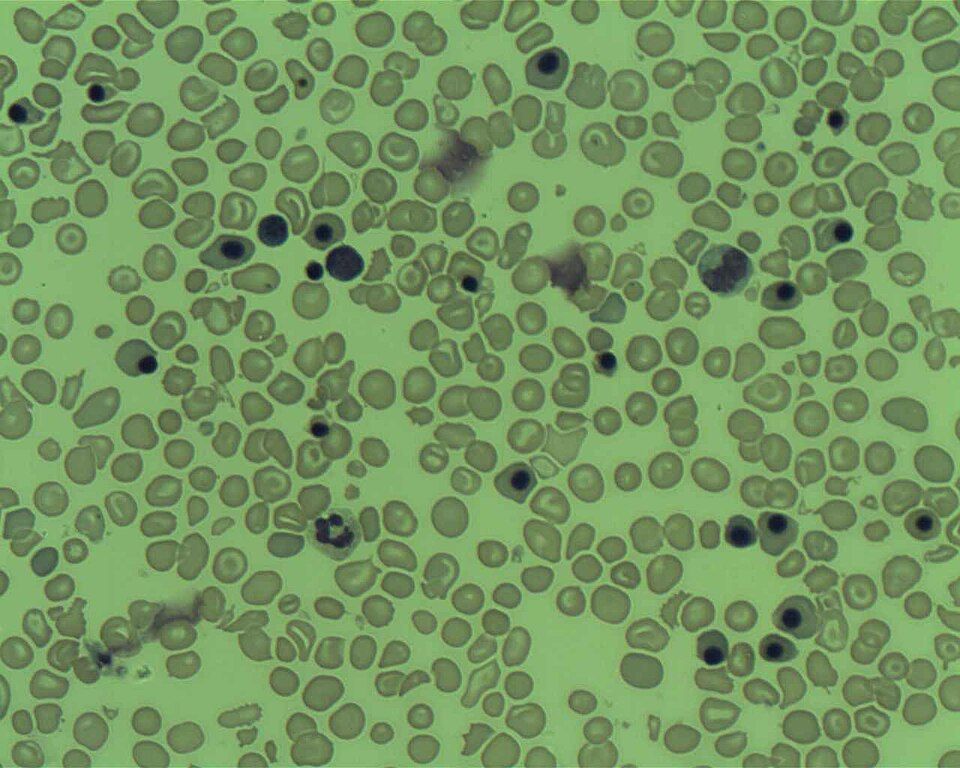
Your bone marrow is a busy blood factory. Deep inside your big bones, hematopoietic stem cells divide and mature into three great families of blood cells: red cells that carry oxygen, platelets that plug leaks, and white cells that fight infection. Normally this factory is tightly controlled. Every cell knows when to grow, when to stop, and when to die. Blood cancers happen when one line of blood cells loses that control and multiplies without a brake. The trouble is not just the abnormal cells themselves, but the fact that they take up space, resources, and function that healthy cells need. Understanding which cell went rogue and where it piles up explains almost every symptom your patient will have.
Leukemia: the marrow gets crowded out
Leukemia is cancer of the white-cell-forming lines in the bone marrow. Immature, useless white cells (often called blasts) reproduce so fast that they physically crowd out normal production. Picture a factory floor jammed with defective units so nothing else can be built. That single mechanism explains the classic triad: too few red cells means anemia (fatigue, pallor, shortness of breath), too few platelets means thrombocytopenia (easy bruising, petechiae, bleeding gums), and too few functional white cells means dangerous infection risk, even though the total white count on the lab report may look high.
Leukemias are sorted two ways. Acute vs chronic describes tempo: acute leukemias explode with immature blasts and make patients sick in days to weeks, while chronic leukemias smolder for months to years with more mature-looking cells. Myeloid vs lymphocytic describes the parent line: myeloid cells give rise to red cells, platelets, and granulocytes, while lymphocytic cells give rise to lymphocytes. Combine them and you get the four big names.
| Type | Who it usually affects | Key feature |
|---|---|---|
| ALL (acute lymphocytic) | Children most often | Most common childhood cancer; responds well to treatment |
| AML (acute myeloid) | Older adults | Blasts may show Auer rods |
| CLL (chronic lymphocytic) | Older adults | Often found by accident on routine bloodwork |
| CML (chronic myeloid) | Middle-aged adults | Linked to the Philadelphia chromosome |
Lymphoma: cancer that builds tumors in lymph tissue
Where leukemia floods the marrow and blood, lymphoma is a solid cancer of lymphocytes that grows into masses inside lymph nodes and lymphoid organs like the spleen. The hallmark sign is painless lymphadenopathy: a firm, swollen, non-tender node in the neck, armpit, or groin. Patients often also have B symptoms, which is the textbook name for the trio of drenching night sweats, unexplained fever, and weight loss.
Lymphomas split into two branches. Hodgkin lymphoma is defined by a specific giant abnormal cell, the Reed-Sternberg cell, on biopsy. It usually starts in one node region and spreads in an orderly, predictable pattern, which helps make it one of the more curable cancers. Non-Hodgkin lymphoma is a large, diverse group without Reed-Sternberg cells; it tends to be more scattered and unpredictable at diagnosis and is far more common overall.
Multiple myeloma: rogue plasma cells attack bone and kidney
Plasma cells are the mature antibody-factory version of B lymphocytes. In multiple myeloma, one plasma cell line turns malignant and churns out a single useless antibody, called a monoclonal (M) protein, while crowding the marrow. Its damage spells out neatly with the mnemonic CRAB: Calcium elevated, Renal (kidney) injury, Anemia, and Bone lesions.
- Bone lesions: myeloma cells activate osteoclasts that eat away bone, creating punched-out lytic lesions, deep bone pain, and pathologic fractures.
- Hypercalcemia: the dissolving bone dumps calcium into the blood, causing confusion, constipation, and thirst.
- Kidney injury: the flood of M protein and calcium clogs and damages the renal tubules.
- Anemia and infection: marrow crowding plus antibodies that do not actually work.
Explore the interactive figure below to see how leukemia fills the marrow, how lymphoma builds masses in nodes, and how myeloma eats into bone.
- Leukemia crowds the bone marrow with abnormal white cells, causing anemia, bleeding (thrombocytopenia), and infection; classified acute vs chronic and myeloid vs lymphocytic (ALL, AML, CLL, CML).
- Lymphoma forms solid masses in lymph nodes: Hodgkin shows Reed-Sternberg cells and spreads predictably; non-Hodgkin is more common and more scattered. Watch for painless lymphadenopathy plus B symptoms.
- Multiple myeloma is malignant plasma cells; remember CRAB: hyperCalcemia, Renal injury, Anemia, Bone lytic lesions, driven by a monoclonal M protein.
- The shared red flags across blood cancers are fatigue, frequent infections, and easy bruising; a high WBC in leukemia still means the patient is immunocompromised.
The chapter in one breath
- Hematopoiesis from a single bone-marrow stem cell makes all blood cells; erythropoietin from the kidney drives red-cell production, so kidney failure often causes anemia.
- Blood splits into plasma (fluid, proteins, clotting factors) and formed elements: RBCs carry oxygen via hemoglobin, WBCs fight infection, and platelets plug leaks.
- Iron-deficiency anemia makes RBCs small and pale (microcytic, hypochromic) with low ferritin, causing fatigue, pallor, and pica.
- Vitamin B12/folate deficiency causes large immature cells (macrocytic, megaloblastic anemia); only B12 loss adds neuro signs, and B12 needs stomach intrinsic factor to absorb.
- Sickle cell anemia is inherited hemoglobin S that warps RBCs when deoxygenated, triggering painful vaso-occlusive crises, while hemolytic anemias destroy cells early and raise bilirubin.
- Clotting fails from too few platelets (thrombocytopenia), missing factors (hemophilia), or widespread clotting-then-bleeding (DIC); watch for petechiae, bruising, and prolonged bleeding.
- Transfusion safety hinges on ABO and Rh typing: type O is the universal RBC donor, AB the universal recipient, and a mismatch triggers a life-threatening hemolytic reaction.
- Leukemias flood the marrow with abnormal white cells (crowding out normal lines), lymphomas grow in lymph nodes (Hodgkin vs non-Hodgkin), and multiple myeloma is malignant plasma cells eating bone.
ReferenceWords worth knowing
A quick reference for the terms in this chapter — skim it now, or circle back whenever a word trips you up.
- ABO system
- The main red-cell blood-group classification (A, B, AB, O) based on which sugar antigens sit on the red-cell surface, so a patient's plasma carries antibodies against the antigens they lack and a mismatched transfusion triggers a dangerous hemolytic reaction.
- Anemia
- A reduced number of red blood cells or amount of hemoglobin, which lowers the blood's oxygen-carrying capacity and usually causes fatigue, pallor, and shortness of breath.
- DIC (disseminated intravascular coagulation)
- A life-threatening emergency in which widespread clotting is switched on throughout the small vessels, using up clotting factors and platelets so fast that the patient paradoxically both clots and bleeds at the same time.
- Erythropoietin
- A hormone released mainly by the kidneys in response to low oxygen that signals the bone marrow to make more red blood cells.
- Hematopoiesis
- The ongoing process, occurring mostly in red bone marrow, by which stem cells mature into all the formed elements of blood: red cells, white cells, and platelets.
- Hemoglobin
- The iron-containing protein inside red blood cells that binds oxygen in the lungs and releases it to the tissues, and also gives blood its red color.
- Hemophilia
- An inherited (usually X-linked) bleeding disorder caused by a missing or deficient clotting factor (factor VIII in type A, factor IX in type B), leading to prolonged bleeding, often into joints and muscles.
- Hemostasis
- The body's coordinated process of stopping bleeding, moving from blood-vessel spasm to a platelet plug and finally a stable fibrin clot.
- Intrinsic factor
- A protein made by the stomach's parietal cells that binds vitamin B12 so it can be absorbed in the small intestine, without which B12 deficiency develops.
- Leukemia
- A cancer of the blood-forming tissue in which the marrow overproduces abnormal, immature white blood cells that crowd out healthy cells, leaving the patient prone to infection, bleeding, and anemia.
- Macrocytic
- Describing red blood cells that are abnormally large, a pattern often seen in vitamin B12 or folate deficiency.
- Microcytic
- Describing red blood cells that are abnormally small, a pattern classically seen in iron-deficiency anemia.
- Pernicious anemia
- A type of macrocytic anemia caused by loss of intrinsic factor (often from autoimmune damage to the stomach lining), which prevents vitamin B12 absorption and impairs red-cell maturation.
- Sickle cell disease
- An inherited disorder in which abnormal hemoglobin S makes red cells stiffen into a crescent shape under low-oxygen stress, so the rigid cells block small vessels and cause painful crises, hemolysis, and organ damage.
- Thrombocytopenia
- An abnormally low platelet count, which raises the risk of easy bruising and prolonged or spontaneous bleeding.
- Virchow's triad
- The three conditions that together promote abnormal clot formation: sluggish blood flow (stasis), vessel-wall injury, and increased blood clottability (hypercoagulability).
DrillFlashcards for this unit
These are the Week 8 cards — the same deck as the hub, pulled in here so you can review the moment you finish reading. Tap the card to flip it; hit Shuffle to mix them up. To grade yourself and track mastery across the whole deck, open the full flashcards page.
Check yourself
Cardiovascular
The cardiovascular system is a pump and a plumbing network that never rests, and small failures ripple everywhere. This chapter runs from the pressure in the pipes to the muscle of the pump itself: hypertension, coronary disease and heart attack, heart failure, valve problems, the rhythms on the EKG, and the diseases of the peripheral vessels.
By the end of this chapter you can
- Explain what sets blood pressure and how hypertension damages organs.
- Distinguish stable angina from a myocardial infarction and name its markers.
- Contrast left- and right-sided, systolic and diastolic heart failure.
- Tell valve stenosis from regurgitation.
- Read the basic EKG waves and recognize dangerous arrhythmias and peripheral vascular disease.
Section 1Blood pressure & hypertension
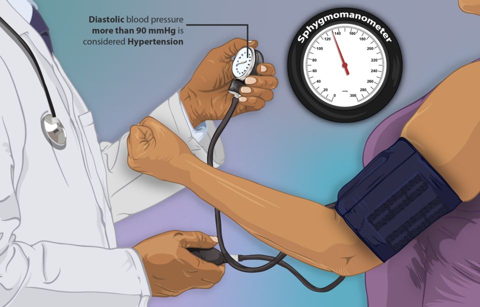
Every time your heart beats, it pushes blood against the walls of your arteries. Blood pressure is simply the force of that push. We write it as two numbers, like 120/80 mmHg: the top (systolic) is the pressure when the heart squeezes, and the bottom (diastolic) is the pressure when the heart relaxes between beats. Healthy pressure keeps blood moving to every organ without hammering the vessels so hard they get damaged.
What actually sets the pressure
Two things multiplied together decide your blood pressure: how much blood the heart pumps per minute and how tight the pipes are. In textbook terms, BP = cardiac output (CO) times systemic vascular resistance (SVR). Cardiac output is heart rate times stroke volume (how much blood leaves the heart with each beat). Systemic vascular resistance is how narrow or constricted the arterioles are. Think of a garden hose: turn up the water flow (more cardiac output) or squeeze the nozzle tighter (more resistance), and pressure rises. This is why a racing heart, extra fluid volume, or clamped-down vessels all push the number up.
How the body keeps it steady
Your body corrects blood pressure minute-to-minute and day-to-day using two main systems.
Baroreceptors are stretch sensors in the carotid arteries and aortic arch. When pressure drops, they sense less stretch and tell the brainstem to speed the heart and constrict vessels; when pressure spikes, they do the opposite. This is the fast, second-by-second reflex, and it is why you may feel dizzy standing up too quickly before the reflex catches up.
The RAAS (renin-angiotensin-aldosterone system) is the slower, longer-term controller. When the kidneys sense low pressure or low blood flow, they release renin. Renin kicks off a chain that makes angiotensin II, a powerful vasoconstrictor that tightens vessels (raising SVR). Angiotensin II also triggers the adrenal glands to release aldosterone, which tells the kidneys to hold onto sodium and water, raising blood volume and therefore cardiac output. Both actions raise pressure. Many blood-pressure medications (ACE inhibitors, ARBs) work by interrupting this exact pathway.
When pressure stays too high: hypertension
Hypertension means blood pressure that stays elevated over time, usually defined as 130/80 mmHg or higher on repeated readings. It shows up in two flavors.
| Feature | Primary (essential) | Secondary |
|---|---|---|
| How common | About 90-95% of cases | Roughly 5-10% of cases |
| Cause | No single identifiable cause; genetics, aging, salt, weight, stress | An identifiable underlying condition |
| Typical triggers | Family history, obesity, high-sodium diet, inactivity | Kidney disease, renal artery narrowing, pheochromocytoma, sleep apnea, certain drugs |
| Onset clue | Gradual, often midlife | Sudden, very high, or resistant to treatment; sometimes young patients |
| Treatment focus | Lifestyle plus medication | Fix the underlying cause |
Primary hypertension has no single fixable cause; it emerges from a mix of genetics, aging vessels, high sodium intake, and often extra weight. Secondary hypertension is driven by a specific, identifiable problem, so treating that problem can actually cure the high pressure.
The silent killer and its targets
Hypertension earns the nickname the silent killer because it usually causes no symptoms for years. There is no headache, no warning; the person feels fine while the elevated pressure quietly grinds away at organs. By the time damage shows, it can be advanced.
That damage is called target-organ damage, and it strikes vessel-rich organs:
- Heart: the left ventricle must pump against higher resistance, so it thickens (left ventricular hypertrophy), and over time can fail; coronary arteries also stiffen, raising heart-attack risk.
- Brain: weakened, pressure-battered vessels can rupture or clot, causing stroke; chronic damage contributes to vascular dementia.
- Kidneys: the tiny filtering vessels scar and narrow, leading to chronic kidney disease and eventually failure.
- Eyes: the retinal vessels are visible windows onto this damage; hypertensive retinopathy can blur or steal vision.
- Blood pressure equals cardiac output times systemic vascular resistance, so more fluid, a faster heart, or tighter vessels all raise it.
- Baroreceptors give fast, moment-to-moment correction; the RAAS gives slower control by constricting vessels (angiotensin II) and retaining salt and water (aldosterone).
- Primary (essential) hypertension is 90-95% of cases with no single cause; secondary hypertension has an identifiable, often treatable cause.
- Hypertension is the silent killer because it causes no symptoms while quietly harming the heart, brain, kidneys, and eyes (target-organ damage).
- Because patients feel fine, adherence teaching is critical; drugs like ACE inhibitors and ARBs work by blocking the RAAS pathway.
Section 2Coronary artery disease, angina & MI
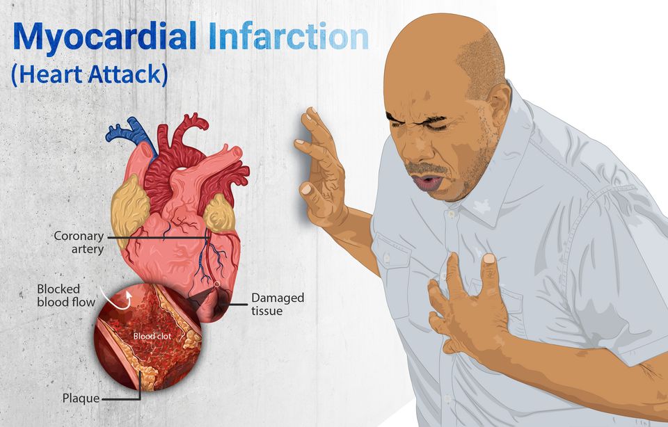
Your heart is a muscle, and like any muscle it needs a steady supply of oxygen-rich blood to keep working. But here is the twist: even though gallons of blood rush through its chambers every hour, the heart muscle itself cannot use that blood directly. Instead, it feeds itself through its own dedicated plumbing, the coronary arteries, which branch off the aorta and wrap around the outside of the heart. When those vessels are open and healthy, supply easily keeps up with demand, and the muscle never notices. Trouble begins when the pipes get narrow.
How the coronary arteries get clogged
Over years, cholesterol, inflammatory cells, and cellular debris build up inside the artery wall, forming a fatty deposit called a plaque. This slow, silent process is atherosclerosis. As the plaque grows, it stiffens the vessel and squeezes the channel (the lumen) smaller, so less blood can get through. This is coronary artery disease (CAD). For a long time the narrowing causes no symptoms because a resting heart does not demand much. The problem shows up when the heart is asked to work harder.
Angina: the heart's cry for oxygen
Angina is chest pain or pressure caused by myocardial ischemia - the muscle is not getting enough oxygen for the work it is doing. It is a demand-versus-supply problem. In stable angina, a fixed plaque leaves just enough room for resting blood flow. But when someone climbs stairs, shovels snow, or gets upset, the heart rate and force climb, oxygen demand outstrips the narrowed supply, and the muscle aches. The classic tip-off is that it is predictable: it comes on with exertion and fades within minutes of rest or a nitroglycerin tablet, which dilates vessels and eases the heart's workload. Importantly, in stable angina the muscle is starved but not yet dying.
Unstable angina is a different, more dangerous animal. Here a plaque has become fragile and partly disrupted, and pain occurs at rest, lasts longer, or is suddenly worse than the patient's usual pattern. It is one of the acute coronary syndromes (ACS) and signals that a heart attack may be brewing.
| Feature | Stable angina | Unstable angina |
|---|---|---|
| Trigger | Predictable exertion or stress | Often at rest, no clear trigger |
| Plaque | Fixed, stable narrowing | Disrupted, unstable plaque |
| Relief | Rest or nitroglycerin, minutes | Poorly relieved, may persist |
| Muscle status | Ischemic, not dying | At high risk, emergency |
When the pipe closes: myocardial infarction
A myocardial infarction (MI) - a heart attack - happens when an unstable plaque ruptures. The body treats the exposed plaque like a wound and rushes platelets and clotting factors to it, forming a blood clot, or thrombus. If that clot completely blocks the artery, blood flow stops cold. The muscle downstream is suddenly cut off from oxygen, and the starved heart muscle begins to die within about 20 to 30 minutes, and the damage spreads over the next few hours. This is infarction - irreversible tissue death (necrosis), not just the reversible starvation of angina. This is why nurses live by the phrase time is muscle: every minute the artery stays blocked, more heart is lost for good.
Recognizing an MI
The hallmark is chest pain - often crushing, heavy, or squeezing pressure, sometimes radiating to the left arm, jaw, or back, and frequently paired with sweating (diaphoresis), nausea, and shortness of breath. But nurses must stay alert for atypical presentations: women, older adults, and people with diabetes may have only fatigue, indigestion, or breathlessness with no dramatic chest pain, because neuropathy and different symptom patterns blunt the classic warning. Three tools confirm the diagnosis:
- ECG: a STEMI (ST-elevation MI) shows a raised ST segment, meaning the full thickness of muscle is being injured by a completely blocked artery - a true emergency needing immediate reopening of the vessel.
- Troponin: when heart cells die, they leak the protein troponin into the blood. A rising troponin level over serial draws is the most specific sign that muscle has actually been damaged.
- The story: the pattern and timing of the pain ties it together.
- Atherosclerosis narrows the coronary arteries over years, reducing blood flow to the heart muscle (coronary artery disease).
- Stable angina is reversible ischemia from a demand-supply mismatch, relieved by rest or nitroglycerin; unstable angina occurs at rest and warns of an impending MI.
- A myocardial infarction results from plaque rupture and a thrombus that blocks the artery, causing irreversible muscle death.
- Recognize an MI by chest pain (watch for atypical presentations), ST-elevation on ECG, and rising troponin - and remember that time is muscle.
Section 3Heart failure
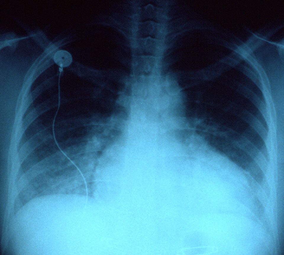
Picture the heart as a two-sided pump working every second of your life. The right side collects oxygen-poor blood returning from the body and pushes it to the lungs; the left side receives freshly oxygenated blood from the lungs and drives it out to every organ. When the heart is healthy, the amount of blood coming in matches the amount going out, and tissues get exactly the oxygen they need. Heart failure is what happens when the pump can no longer keep up with the body's demand. It does not mean the heart has stopped. It means the heart is failing to move blood forward efficiently, so blood backs up behind the weak side and organs downstream go under-supplied.
Left-sided failure: the backup floods the lungs
The left ventricle is the strongest chamber because it has to push blood to the whole body. When it weakens, blood cannot move forward fast enough, so it dams up behind the left side, back into the lungs' capillaries. Rising pressure forces fluid out of those capillaries and into the air sacs, a state called pulmonary congestion or pulmonary edema. This is why left-sided failure is a lung story. Patients feel short of breath (dyspnea), especially on exertion. Lying flat lets fluid redistribute toward the lungs, so they wake up gasping or need to prop up on pillows, which is orthopnea and paroxysmal nocturnal dyspnea. On auscultation you hear crackles (rales), the sound of air bubbling through fluid-filled alveoli. A frothy or pink-tinged cough can appear as fluid leaks into airways.
Right-sided failure: the backup floods the body
When the right ventricle fails, blood dams up behind it, into the systemic veins that were trying to return blood to the heart. Now the congestion shows up in the body, not the lungs. You see jugular venous distention (JVD) as neck veins bulge under the backed-up pressure, peripheral edema in the ankles and legs where gravity pulls the leaked fluid, and hepatomegaly as blood engorges the liver. The single most common cause of right-sided failure is left-sided failure itself: the lung congestion eventually strains the right ventricle, which is why untreated left failure often becomes biventricular.
| Feature | Left-sided failure | Right-sided failure |
|---|---|---|
| Blood backs up into | Lungs (pulmonary circulation) | Body (systemic veins) |
| Hallmark signs | Dyspnea, orthopnea, crackles | JVD, peripheral edema, hepatomegaly |
| Classic complaint | Cannot breathe lying flat | Swollen legs, weight gain |
| Common cause | Hypertension, prior MI | Left-sided failure, lung disease |
Systolic vs diastolic: a squeeze problem or a filling problem
Failure can come from two different mechanical faults. In systolic failure, the ventricle is weak and cannot contract forcefully, so it ejects a smaller fraction of the blood it holds. This is measured as a reduced ejection fraction (EF), often under 40% (normal is usually 55 to 70%), and is called HFrEF (heart failure with reduced ejection fraction). In diastolic failure, the ventricle contracts fine but is stiff and cannot relax and fill properly, so less blood is available to pump even though the squeeze percentage looks normal. This is HFpEF (preserved ejection fraction). The key teaching point: a normal EF does not rule out heart failure.
Compensation that turns against you
When cardiac output drops, the body reacts as if it were bleeding. The sympathetic nervous system fires, raising heart rate and squeezing vessels to prop up blood pressure. The kidneys, sensing low flow, activate the renin-angiotensin-aldosterone system (RAAS), which retains sodium and water and constricts arteries. In the short term these buy time. Over weeks, though, they backfire: the extra fluid worsens the congestion, the higher pressure makes the failing heart work harder, and chronic overdrive drives ventricular remodeling, where the heart enlarges and stiffens. This vicious cycle is why the mainstay drugs (ACE inhibitors, ARBs, beta-blockers) work by blocking these compensations.
The stretched, overworked ventricles release B-type natriuretic peptide (BNP), a hormone that tries to promote fluid loss. Because BNP rises with wall stretch, a high level is a useful marker that shortness of breath is coming from heart failure rather than the lungs. Explore the left- versus right-sided comparison below.
- Heart failure is a pump that cannot meet the body's demand; blood backs up behind the failing side.
- Left-sided failure floods the lungs (dyspnea, orthopnea, crackles); right-sided failure floods the body (JVD, peripheral edema, hepatomegaly).
- Systolic failure (HFrEF) is a weak squeeze with low EF; diastolic failure (HFpEF) is a stiff, poorly filling ventricle with normal EF.
- RAAS and sympathetic activation compensate at first but eventually worsen congestion and drive remodeling.
- BNP rises with ventricular stretch and helps confirm that dyspnea is cardiac in origin.
Section 4Valve disorders
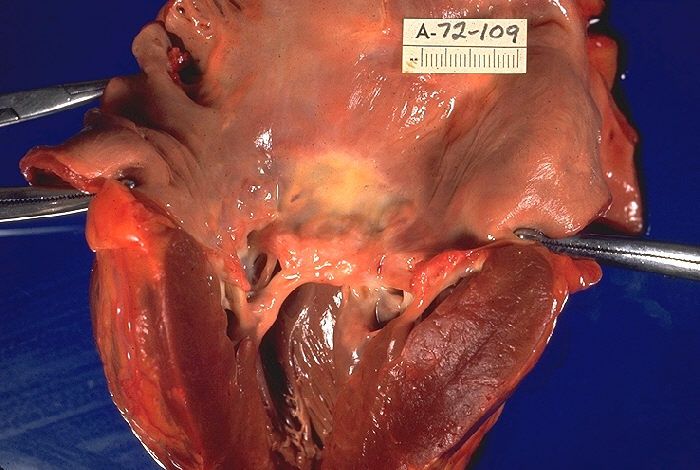
Picture your heart as a house with four rooms and four one-way doors between them. Those doors are your valves, and their whole job is simple: let blood move forward and never let it slip backward. When the heart squeezes, the doors ahead swing open easily; when it relaxes, they snap shut tight so nothing leaks back. Two of these doors do the heavy lifting on the left side of the heart, the side that pushes blood out to the whole body: the mitral valve (between the left atrium and left ventricle) and the aortic valve (between the left ventricle and the aorta). When a valve stops opening well or stops sealing well, the heart has to work harder for every single beat, and over time that extra work reshapes the chamber behind it.
The two ways a valve fails: stenosis vs. regurgitation
There are really only two things that can go wrong with a door. Either it is hard to push open, or it does not shut all the way. That is the entire concept.
Stenosis means the valve has become stiff, thick, or narrowed, so the opening is too small. The chamber behind it must generate much higher pressure to force blood through the tight gap, like trying to breathe through a pinched straw. Regurgitation (also called insufficiency or a leaky/incompetent valve) means the valve does not seal, so some blood sloshes backward with each beat. Now the chamber has to pump the same blood twice, handling both the normal load and the leaked-back volume. Stenosis is mainly a pressure problem; regurgitation is mainly a volume problem, and that distinction predicts how the heart changes.
| Feature | Stenosis | Regurgitation |
|---|---|---|
| Basic defect | Valve too narrow, hard to open | Valve leaks, does not close |
| Main burden | Pressure overload | Volume overload |
| Chamber response | Concentric hypertrophy (thick walls) | Eccentric hypertrophy/dilation (stretched) |
| Classic murmur timing | Systolic (aortic) or diastolic (mitral) | Depends on valve; e.g., holosystolic in MR |
The two lesions you will see most
Aortic stenosis (AS) is the classic pressure problem. The aortic door stiffens, often from age-related calcification, so the left ventricle must slam against a tight opening to eject blood. To make that pressure, the ventricle wall thickens (concentric hypertrophy). The classic triad you learn for symptomatic AS is angina, syncope, and exertional dyspnea, often signaling a narrowed valve area. Patients frequently have a crescendo-decrescendo systolic murmur.
Mitral regurgitation (MR) is the classic volume problem. The mitral door does not seal, so with each squeeze, blood jets backward into the left atrium instead of only moving forward into the aorta. The left atrium and left ventricle stretch and enlarge (eccentric hypertrophy) to hold the extra volume. A holosystolic (pansystolic) murmur best heard at the apex is typical.
Rheumatic heart disease: a delayed autoimmune injury
One cause deserves its own spotlight because it is so testable and so preventable. Rheumatic heart disease follows acute rheumatic fever, which itself follows an untreated group A streptococcal throat infection (think untreated strep throat). The immune system makes antibodies against the bacteria, but through molecular mimicry those antibodies cross-react with the body's own tissues, including heart valves. Years of smoldering inflammation scar and fuse the valve leaflets. The mitral valve is the most commonly damaged, classically producing mitral stenosis. This is why treating strep throat with antibiotics is genuinely heart-protective.
Why chronic valve disease drifts toward heart failure
Here is the through-line that ties it all together. Whether the chamber is straining against high pressure (stenosis) or drowning in extra volume (regurgitation), it copes for a while by remodeling, thickening or stretching to keep output up. This compensation can last years, and patients often feel fine. But muscle that is chronically overworked eventually cannot keep up. Thick, stiff walls relax poorly and fill poorly; stretched, dilated walls squeeze weakly. Either way, the ventricle's ability to move blood forward drops, pressure backs up into the lungs, and you get the picture of heart failure: dyspnea, fatigue, and fluid congestion. Understanding which chamber is overloaded and how tells you exactly where the backup and symptoms will appear. Explore the figure below to see stenosis and regurgitation side by side.
- Two failure modes: stenosis is a stiff, narrowed valve (pressure overload) while regurgitation is a leaky valve that allows backflow (volume overload).
- Signature lesions: aortic stenosis drives left-ventricular concentric hypertrophy; mitral regurgitation dilates the left atrium and ventricle (eccentric hypertrophy).
- Rheumatic heart disease stems from untreated group A strep via molecular mimicry and classically scars the mitral valve, so treating strep throat protects the heart.
- The common endpoint: chronic pressure or volume overload compensates for years, then the overworked chamber fails, backing blood up into the lungs as heart failure.
- Watch symptom onset: a compensated patient can feel well for years; new symptoms (especially in aortic stenosis) signal decompensation and need prompt evaluation.
Section 5The EKG & arrhythmias
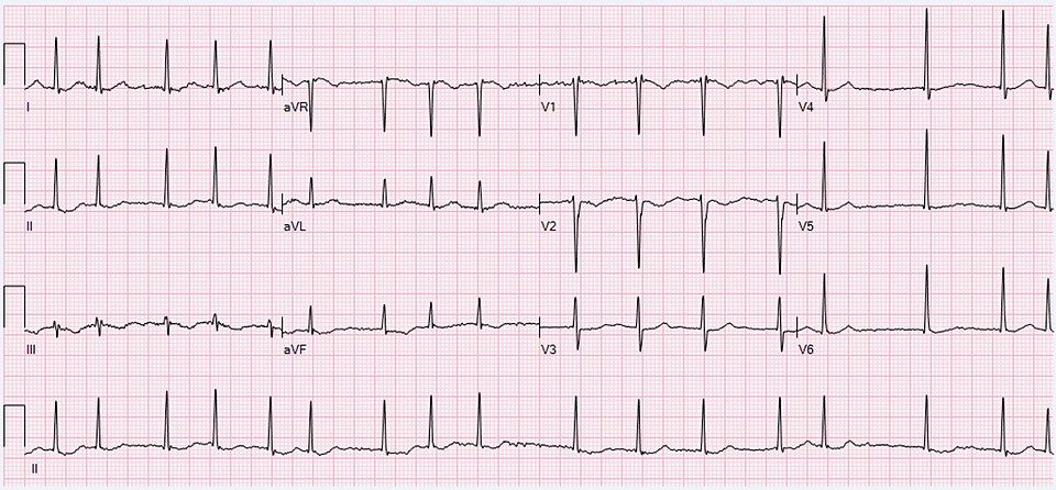
Your heart has its own built-in electrical wiring, and every heartbeat starts as a tiny spark. Before we talk about what goes wrong, let us walk the normal path that spark travels, because every arrhythmia is really just a detour, a delay, or a short-circuit somewhere along that road.
The normal conduction system: the heart's spark and wire
The signal begins in the sinoatrial (SA) node, a patch of cells high in the right atrium. The SA node is the heart's natural pacemaker because it fires fastest, usually 60 to 100 times a minute in an adult at rest. Its spark spreads across both atria and squeezes them, then funnels down to the atrioventricular (AV) node. The AV node is the gatekeeper: it deliberately delays the signal a fraction of a second so the atria can finish emptying blood into the ventricles before the ventricles contract. From there the current races down the bundle of His, splits into the right and left bundle branches, and fans out through the Purkinje fibers, which fire both ventricles almost at once for a strong, coordinated pump.
Reading the P-QRS-T
An EKG (ECG) is simply a voltage recording of that electrical trip. Each normal beat draws three shapes:
- P wave = atrial depolarization (the atria firing and contracting).
- QRS complex = ventricular depolarization (the ventricles firing). It is tall and narrow because the Purkinje system spreads current so fast.
- T wave = ventricular repolarization (the ventricles resetting for the next beat).
To read a strip, check rate and rhythm. For rate, count the QRS complexes in a 6-second strip and multiply by 10, or use the interval between beats. For rhythm, ask three questions: Is there a P wave before every QRS? Is every QRS narrow? Are the intervals regular? A normal sinus rhythm answers yes to all three. When one of those answers turns to no, you have an arrhythmia (dysrhythmia).
Atrial fibrillation and its stroke risk
In atrial fibrillation (AFib) the atria stop firing from the SA node and instead quiver chaotically from many misfiring spots. On the strip the P waves disappear, replaced by a wavy baseline, and the QRS complexes march by irregularly irregular (no pattern at all). The danger is not the fast pulse itself but what happens to the blood: because the atria only quiver instead of squeezing, blood pools and stagnates, especially in the left atrial appendage. Stagnant blood clots (recall Virchow's triad, stasis being one leg), and a clot that breaks loose travels to the brain, causing an embolic stroke. That is why AFib patients are usually placed on anticoagulants.
Heart blocks: a traffic jam at the AV node
A heart block is a delay or failure of the signal passing from atria to ventricles. In first-degree block every beat gets through, just slowly (a long PR interval). In second-degree block some atrial signals reach the ventricles and some are dropped. In third-degree (complete) block nothing gets through: the atria and ventricles beat on their own, ignoring each other, and the ventricles rely on a slow backup pacemaker, often too slow to sustain blood pressure. Complete block frequently needs a pacemaker.
The lethal rhythms
Some arrhythmias are true emergencies because the ventricles, the pumping chambers, fail to move blood.
| Rhythm | What the ventricles do | Response |
|---|---|---|
| Ventricular tachycardia (VT) | Fire far too fast from a ventricular focus; wide QRS, little filling time | Emergency; if no pulse, defibrillate + CPR |
| Ventricular fibrillation (VF) | Only quiver; no organized squeeze, no cardiac output | Immediate defibrillation + CPR |
| Asystole | No electrical activity at all; flatline | CPR + epinephrine; not shockable |
VT and VF are shockable because a defibrillator can reset chaotic ventricular activity back to a coordinated rhythm. Asystole is not shockable, there is no electrical activity to reorganize, so the answer is high-quality CPR and drugs, not a shock.
When an arrhythmia is an emergency
Rhythm names matter less at the bedside than how the patient looks. Watch for signs of poor perfusion: chest pain, shortness of breath, low blood pressure, cool clammy skin, confusion, or fainting (syncope). A rhythm producing no pulse, VT without a pulse, VF, or asystole, means starting CPR and calling a code immediately. An unstable but conscious patient still needs urgent intervention. Explore the interactive EKG below to practice spotting P waves, QRS complexes, and the difference between an organized and a chaotic strip.
- The impulse travels SA node to AV node (which delays it) to bundle of His to Purkinje fibers; the EKG records this as the P wave (atria), QRS (ventricles firing), and T wave (ventricles resetting).
- Read a strip by checking rate and rhythm: a P before every narrow QRS at a regular interval is normal sinus rhythm.
- Atrial fibrillation is irregularly irregular with no P waves; quivering atria let blood pool and clot, raising stroke risk, so patients are usually anticoagulated.
- Heart blocks are AV conduction delays; complete (third-degree) block often needs a pacemaker.
- VT, VF, and asystole are lethal; VF and pulseless VT are shockable with defibrillation, but asystole is not, treat it with CPR and epinephrine.
Section 6Peripheral vascular disease
Your legs are fed by a two-way plumbing system. Arteries carry bright, oxygen-rich blood down from the heart under high pressure to feed the muscles, skin, and nerves. Veins carry the used, oxygen-poor blood back up to the heart against gravity, helped by one-way valves and the squeeze of your calf muscles. When either half of this system fails, the tissue downstream suffers, but it suffers in very different ways depending on whether the problem is inflow (arterial) or outflow (venous). Peripheral vascular disease is the umbrella term for both.
Arterial disease (PAD): not enough blood getting in
Peripheral arterial disease (PAD) is usually caused by atherosclerosis - the same cholesterol plaque that clogs coronary arteries, now narrowing the arteries of the legs. Less blood can squeeze past the plaque, so the muscles are starved of oxygen, especially when they demand more.
The classic early sign is intermittent claudication: a cramping, aching pain in the calf, thigh, or buttock that predictably comes on with walking and reliably eases with rest. The mechanism is simple supply-and-demand - exercising muscle needs more oxygen than the narrowed artery can deliver, so it switches to anaerobic metabolism and lactic acid builds up, causing pain. Rest lowers the demand, the ache resolves, and the cycle repeats at the same walking distance.
As disease worsens, the pain-free walking distance shortens until pain arrives at rest - a burning ache in the forefoot and toes, worst at night when the leg is flat in bed. Chronic ischemia leaves the skin shiny, hairless, and thin, with thickened nails and punched-out ulcers over the toes and pressure points, because starved tissue cannot maintain normal growth. Patients often report that dangling the leg over the side of the bed relieves rest pain, because gravity helps pull the meager blood flow downhill. If a plaque suddenly thromboses or a clot embolizes from the heart, the limb tips from chronic to acute limb ischemia - a surgical emergency whose hallmark is the six P's: pain, pallor, pulselessness, poikilothermia (the limb drifts to room temperature and feels cool), paresthesia, and paralysis. Paresthesia and paralysis are the late nerve-and-muscle findings; when they appear, the limb is close to being lost.
Venous disease: not enough blood getting out
Chronic venous insufficiency (CVI) is the mirror image. Here the arteries deliver blood fine, but the veins cannot return it. Damaged or incompetent valves - often after years of standing, obesity, pregnancy, or a prior clot - let blood flow backward and pool in the legs. This raises pressure in the veins and capillaries, a state called venous hypertension.
That backed-up pressure pushes fluid out into the tissues, producing chronic edema that is usually worst by the end of the day and improves overnight. Red blood cells also leak out and break down, depositing iron (hemosiderin), which stains the skin a permanent brownish discoloration around the lower leg and ankle - a change called hemosiderin staining. The same congested skin often turns red, itchy, scaly, and weepy, an eczema-like inflammation named stasis dermatitis. Over time the skin becomes leathery and tight (lipodermatosclerosis). Because the tissue is congested and poorly nourished, it breaks down into venous ulcers, classically over the inner ankle (the medial malleolus), where venous pressure is highest.
Telling an arterial ulcer from a venous ulcer
Ulcers are where students most often mix these up, so match the wound to its mechanism. An arterial ulcer forms because tissue is starved, so it appears where flow is poorest and looks dead. A venous ulcer forms because tissue is congested and weepy, so it appears where pooling is worst and looks wet.
| Feature | Arterial ulcer | Venous ulcer |
|---|---|---|
| Usual location | Toes, heels, bony points, lateral foot | Around the medial (inner) ankle |
| Wound edges | Well-defined, punched-out, deep | Irregular, shallow, sloping |
| Wound bed | Pale, gray, or dry; little drainage | Ruddy/beefy red; heavy drainage |
| Surrounding skin | Cool, pale, shiny, hairless | Warm, brown-stained, edematous |
| Pain | Severe, worse with elevation | Mild-moderate, better with elevation |
| Pulses | Diminished or absent | Usually present |
DVT, PE, and one word on aneurysm
A deep vein thrombosis (DVT) is a clot in a deep leg vein, favored by Virchow's triad: venous stasis, vessel-wall injury, and hypercoagulability (immobility, surgery, and estrogen are common real-world triggers). The leg is often warm, red, swollen, and tender. The danger is not the leg itself - it is that a piece can break loose, travel to the lungs, and lodge as a pulmonary embolism (PE), causing sudden shortness of breath, pleuritic chest pain, and a drop in oxygen. That is why we never vigorously massage a suspected DVT. Separately, an abdominal aortic aneurysm (AAA) is a balloon-like weakening of the aortic wall that may present as a pulsatile abdominal mass and can be fatal if it ruptures. Explore the arterial-versus-venous figure below to see these patterns side by side.
- PAD starves tissue of inflow: intermittent claudication (pain with walking, relieved by rest) plus a limb that is cool, pale, pulseless, and painful - worse with elevation.
- Chronic venous insufficiency is an outflow/pooling problem: edema, brownish hemosiderin staining, and ulcers around the medial ankle - better with elevation.
- Arterial ulcers are punched-out, dry, and on toes/pressure points; venous ulcers are shallow, wet, and around the inner ankle.
- DVT (Virchow's triad) can throw a clot to the lungs as a life-threatening pulmonary embolism - do not massage the leg.
- An abdominal aortic aneurysm is a weakened, ballooning aorta that can be fatal if it ruptures.
The chapter in one breath
- Blood pressure is cardiac output times systemic vascular resistance; hypertension (usually ≥130/80) is the silent, mostly asymptomatic disease that thickens vessels and overworks the heart until it damages the brain, kidneys, and eyes.
- Coronary artery disease is atherosclerotic plaque starving the myocardium; when a plaque ruptures and a clot fully blocks flow, the muscle downstream infarcts, giving the crushing chest pain, elevated troponin, and ST changes of an MI.
- Left-sided heart failure backs blood up into the lungs, so the classic signs are pulmonary: crackles, dyspnea, orthopnea, and frothy sputum from pulmonary edema.
- Right-sided heart failure backs blood up into the body, so you see peripheral congestion: dependent edema, jugular venous distention, ascites, and a congested, tender liver.
- Valve disorders come in two flavors: stenosis (a stiff, narrow valve the heart must strain to push through) and regurgitation (a leaky valve that lets blood flow backward), both forcing the chamber behind them to hypertrophy or dilate over time.
- The EKG tracks the electrical wave through the heart, where the P wave is atrial depolarization, the QRS is ventricular depolarization, and the T wave is ventricular repolarization.
- Atrial fibrillation is the most common serious arrhythmia, an irregularly irregular quiver that pools blood in the atria and throws clots, which is why these patients need anticoagulation to prevent stroke.
- Lethal rhythms demand instant action: ventricular fibrillation and pulseless ventricular tachycardia are shockable (defibrillate plus CPR), while true asystole is not shocked but treated with CPR and epinephrine.
- Peripheral vascular disease splits by side: arterial insufficiency gives pain with walking (claudication), cool pale pulseless feet, and painful distal wounds, while venous insufficiency gives warm swollen legs, brownish skin, and weepy medial-ankle ulcers.
ReferenceWords worth knowing
A quick reference for the terms in this chapter — skim it now, or circle back whenever a word trips you up.
- Angina
- Angina is chest pain or pressure that happens when a section of heart muscle temporarily gets less oxygen-rich blood than it needs, usually because a coronary artery is narrowed by plaque.
- Atherosclerosis
- Atherosclerosis is the slow buildup of fatty, cholesterol-rich plaque inside artery walls that stiffens and narrows the vessel and can eventually block blood flow.
- Atrial fibrillation
- Atrial fibrillation is a fast, disorganized quivering of the atria instead of a coordinated squeeze, which pools blood in the atria and raises the risk of clots and stroke.
- BNP (B-type natriuretic peptide)
- BNP is a hormone released by stretched, overworked heart muscle, so a high blood level often signals that the heart is failing to keep up with its fluid load.
- Cardiac output
- Cardiac output is the amount of blood the heart pumps out each minute, calculated as heart rate multiplied by stroke volume (usually about 4 to 8 L/min in adults).
- Claudication
- Claudication is cramping leg pain that comes on with walking and eases with rest, caused by narrowed arteries that can't deliver enough blood to exercising muscles.
- Ejection fraction
- Ejection fraction is the percentage of blood in a filled ventricle that gets pumped out with each beat, normally around 55 to 70 percent.
- Embolus
- An embolus is a clot, plaque fragment, air bubble, or other mass that travels through the bloodstream until it lodges in and blocks a smaller vessel downstream.
- Heart failure
- Heart failure is the condition in which the heart can no longer pump strongly enough or fill well enough to meet the body's needs, causing fluid backup and fatigue.
- Hypertension
- Hypertension is persistently high blood pressure (usually 130/80 mmHg or above) that forces the heart to work harder and gradually damages vessel walls and organs.
- Myocardial infarction
- Myocardial infarction, or heart attack, is the death of heart muscle caused when a coronary artery is fully blocked and that region of tissue is starved of oxygen.
- RAAS (renin-angiotensin-aldosterone system)
- RAAS is a hormone cascade that raises blood pressure by tightening blood vessels and telling the kidneys to hold onto salt and water when blood flow drops.
- Regurgitation
- Regurgitation is the backward leaking of blood through a heart valve that doesn't close tightly, forcing the heart to pump extra to make up for what flows the wrong way.
- Stenosis
- Stenosis is the abnormal narrowing or stiffening of a heart valve or vessel that restricts forward blood flow and makes the heart push harder to move blood through.
- Systemic vascular resistance
- Systemic vascular resistance is the total opposition to blood flow created by the tone (constriction or dilation) of the body's arteries, a major driver of blood pressure.
- Troponin
- Troponin is a protein released into the blood when heart muscle cells are injured, so a rising level is a key marker used to confirm a myocardial infarction.
DrillFlashcards for this unit
These are the Week 9 cards — the same deck as the hub, pulled in here so you can review the moment you finish reading. Tap the card to flip it; hit Shuffle to mix them up. To grade yourself and track mastery across the whole deck, open the full flashcards page.
Check yourself
Pulmonary
Every cell in the body runs on oxygen, and the lungs are where it gets in and carbon dioxide gets out. This chapter follows a breath from the air to the bloodstream and back — how ventilation and perfusion must match, what happens when airways narrow or a clot lodges, and how to read the story in a patient’s lung sounds.
By the end of this chapter you can
- Explain ventilation, perfusion, and V/Q mismatch (dead space vs shunt).
- Contrast chronic bronchitis with emphysema in COPD.
- Describe the three problems in asthma and rescue vs controller inhalers.
- Recognize a pulmonary embolism and its setup (Virchow’s triad).
- Match lung sounds (crackles, wheezes, stridor) to their causes.
Section 1How breathing works: ventilation, perfusion & V/Q
Every breath you take is really two jobs happening at once, and they have to be perfectly coordinated. Ventilation is the movement of air in and out of the lungs, delivering fresh oxygen to the tiny air sacs called alveoli and carrying carbon dioxide away. Perfusion is the flow of blood through the pulmonary capillaries that wrap around those alveoli. Think of it like a delivery system: ventilation brings the packages (oxygen) to the loading dock, and perfusion is the truck fleet that picks them up. If the packages arrive but no trucks show up, or the trucks show up but there are no packages, the whole system stalls. For gas exchange to work, air and blood must meet at the same place, at the same time.
Matching air to blood: the V/Q ratio
Nurses describe how well ventilation and perfusion line up using the V/Q ratio, where V is ventilation and Q is perfusion (blood flow). In an ideal, healthy lung the overall ratio is about 0.8 usually, because we perfuse a touch more than we ventilate. That number is not the same everywhere in the lung, though. Because of gravity, the bases (bottoms) of the lungs get more blood flow, while the apices (tops) get relatively more air, so the ratio naturally varies from top to bottom. What matters clinically is that when air and blood are reasonably balanced, oxygen crosses into the blood and carbon dioxide is blown off efficiently. When they fall out of balance, we call it a V/Q mismatch, and it comes in two flavors.
Two ways it goes wrong: dead space and shunt
A high V/Q means ventilation without matching perfusion. The air arrives, but no blood is there to pick up the oxygen. This wasted ventilation is called dead space. The classic cause is a pulmonary embolism, where a clot blocks a pulmonary artery. The alveoli fill with air, but the blood truck never arrives.
A low V/Q means perfusion without matching ventilation. Blood keeps flowing past alveoli that are not getting fresh air, so it returns to the heart still low on oxygen. This is called a shunt. Think pneumonia filling alveoli with pus, pulmonary edema flooding them with fluid, or atelectasis (collapsed alveoli). One clinical clue: pure shunt often responds poorly to supplemental oxygen, because the extra oxygen cannot reach blood flowing past blocked alveoli.
| Feature | Dead space (high V/Q) | Shunt (low V/Q) |
|---|---|---|
| What is missing | Perfusion (blood flow) | Ventilation (air) |
| Plain-English picture | Air arrives, no blood to load it | Blood arrives, no fresh air to load |
| Classic cause | Pulmonary embolism | Pneumonia, pulmonary edema, atelectasis |
| Response to oxygen | Good (the still-perfused lung takes up the extra oxygen) | Poor and refractory (blood bypasses any ventilated alveolus) |
Gas exchange across the alveolar membrane
Where air and blood actually meet is the alveolar-capillary membrane, an incredibly thin barrier just a few cells thick. Gases cross it by diffusion, always moving from high pressure to low pressure. Oxygen is more concentrated in the alveolus than in the incoming blood, so it diffuses into the blood; carbon dioxide is more concentrated in the blood, so it diffuses out into the alveolus to be exhaled. Anything that thickens this membrane (fluid, scar tissue, inflammation) slows diffusion and worsens gas exchange, even when ventilation and perfusion are otherwise present.
Hypoxemia vs hypoxia, and how oxygen travels
These two words sound alike but mean different things, and nursing exams love to test the difference. Hypoxemia is low oxygen level in the blood (measured as a low PaO2 on an arterial blood gas or a low SpO2 on the pulse oximeter). Hypoxia is low oxygen reaching the tissues themselves. They often travel together, but not always. Severe anemia, for example, can cause tissue hypoxia even when the blood oxygen level (PaO2) looks normal, because there simply are not enough carriers to deliver it.
And those carriers are the key to the whole system: about 97 percent of oxygen in the blood rides bound to hemoglobin inside red blood cells, with only a tiny fraction dissolved in plasma. Explore the figure below to see ventilation, perfusion, and V/Q matching working together.
- Ventilation (air in and out) and perfusion (blood flow past alveoli) must match at the same place and time for gas exchange to work; the normal V/Q ratio is about 0.8.
- Dead space is ventilation without perfusion (high V/Q, classic cause pulmonary embolism); shunt is perfusion without ventilation (low V/Q, classic causes pneumonia, edema, atelectasis).
- Gases cross the thin alveolar-capillary membrane by diffusion from high to low pressure; a thickened membrane slows oxygen uptake.
- Hypoxemia is low oxygen in the blood; hypoxia is low oxygen in the tissues, and they do not always occur together.
- About 97 percent of oxygen is carried bound to hemoglobin, so oxygen delivery depends on carriers, not just lung function.
Section 2Obstructive disease: COPD
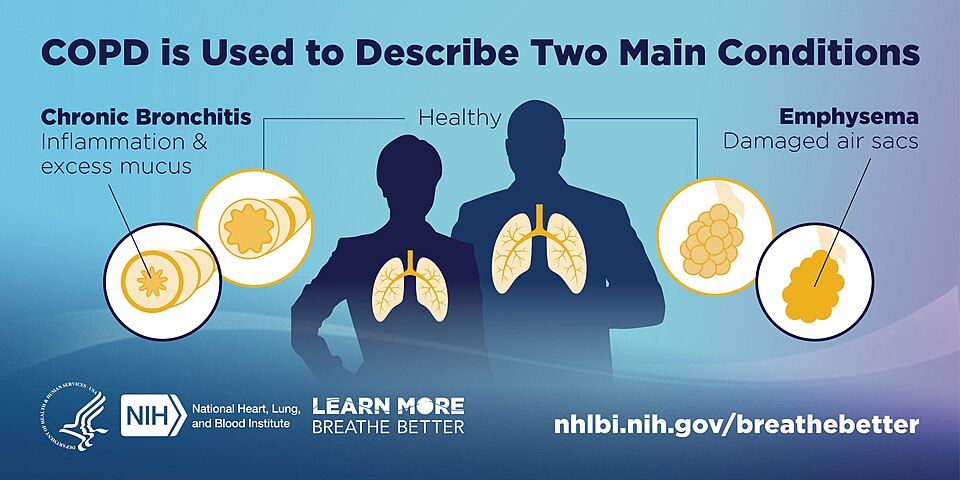
Every time you breathe, air travels down your windpipe, splits into smaller and smaller tubes (the bronchi and bronchioles), and finally reaches millions of tiny balloon-like sacs called alveoli. This is where oxygen slips into the blood and carbon dioxide slips out. Two things make this work: the airways must stay open, and the lung tissue must be springy. That springiness, called elastic recoil, is what quietly squeezes air back out when you exhale. Normal exhalation is passive and effortless.
In chronic obstructive pulmonary disease (COPD), that easy exhale breaks down. The airways narrow and the springiness is lost, so air flows in but gets stuck on the way out. Think of COPD as a disease of getting air OUT, not in. Stale air piles up behind the blockage, a problem called air trapping, and the lungs stay over-inflated. This is why patients often push air out through pursed lips and why their exhale takes so long.
Why air gets trapped
COPD is really two overlapping problems, and most patients have some of each.
Chronic bronchitis is an airway problem. Long-term irritation inflames the bronchial lining, so it swells and the mucus glands grow larger and pump out thick secretions. The airway is now narrowed by both swelling and mucus, and the tiny sweeping hairs (cilia) that clear that mucus are damaged. Mucus sits and grows bacteria, so infections keep coming. It is defined clinically by a a chronic productive cough for at least 3 months in each of 2 consecutive years.
Emphysema is a tissue problem. The walls between alveoli are destroyed and merge into fewer, larger, floppy sacs. That destroys surface area for gas exchange and destroys the elastic recoil that normally props the small airways open and drives exhalation. Without that support, airways collapse on exhale and trap air even more.
| Feature | Chronic bronchitis (blue bloater) | Emphysema (pink puffer) |
|---|---|---|
| Core problem | Inflamed, mucus-clogged airways | Destroyed alveolar walls, lost recoil |
| Classic look | Cyanotic, often overweight, edematous | Thin, barrel chest, leaning forward |
| Cough & mucus | Heavy productive cough | Minimal cough, little sputum |
| Oxygen level | Lower early (hypoxemia, blue tint) | Kept closer to normal by fast breathing |
| Breathing style | Wet, congested | Pursed-lip, working hard to exhale |
The nicknames are memory hooks, not real patient types. The blue bloater is cyanotic and puffy from fluid backup; the pink puffer stays pinker by breathing fast and hard. The over-inflated, air-trapped chest that becomes rounded and rigid is called a barrel chest (an increased anteroposterior diameter).
The leading cause
Cigarette smoking is by far the number-one cause. Smoke keeps the airways inflamed and floods the lung with enzymes that digest alveolar walls, while overwhelming the body's natural defense against those enzymes. Over years, this drives both the bronchitis and the emphysema. (A smaller group inherits alpha-1 antitrypsin deficiency, which removes that natural defense and causes emphysema even in nonsmokers.) Quitting smoking is the single most powerful thing that slows the disease.
When the strain reaches the heart
Chronically low oxygen in the lungs makes lung blood vessels clamp down (hypoxic pulmonary vasoconstriction). That raises pressure in the pulmonary arteries (pulmonary hypertension), and the right side of the heart must pump against it. Over time the right ventricle enlarges and fails, a condition called cor pulmonale. Nurses see this as jugular vein distension, liver enlargement, and peripheral edema, which is part of why the blue bloater looks swollen.
You can explore how the clogged airway and the destroyed alveoli differ in the interactive figure below.
- COPD is trouble getting air OUT: narrowed airways and lost elastic recoil cause air trapping and a prolonged, labored exhale.
- Chronic bronchitis is inflamed, mucus-filled airways (productive cough, blue bloater); emphysema is destroyed alveolar walls with lost recoil (barrel chest, pink puffer).
- Smoking is the leading cause and quitting is the most effective way to slow progression.
- Chronic hypoxia and pulmonary hypertension can strain the right heart into cor pulmonale (edema, JVD).
- Give oxygen carefully, usually targeting SpO2 88 to 92 percent, since very high-flow O2 can worsen CO2 retention.
Section 3Asthma
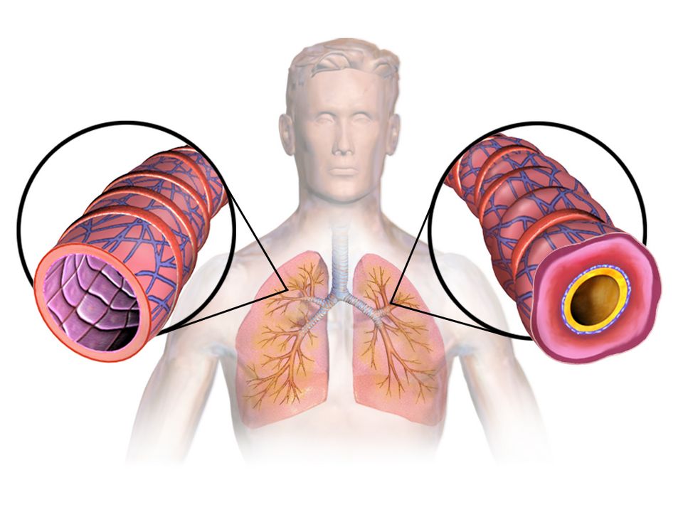
Take a slow breath in. Air rushes down your windpipe, splits into two bronchi, then branches again and again into ever-smaller bronchioles before reaching the tiny air sacs where gas exchange happens. Those small airways are wrapped in a thin layer of smooth muscle, lined with a moist membrane, and are supposed to stay open and springy so air moves in and out with almost no effort. In a healthy lung, breathing out is passive and quiet: the airways stay wide, and the air leaves easily.
In asthma, those small airways become hyperreactive. They overreact to things that should be harmless, and the airway narrows. The key word for asthma is reversible obstruction: unlike the fixed damage of emphysema, an asthma attack can open back up with treatment or even on its own. But while it is happening, air gets trapped, and the person has to work hard to move it.
Three problems at once
An asthma attack is not one thing going wrong; it is three processes hitting the airway together, which is why it can escalate fast.
- Bronchospasm. The ring of smooth muscle around the bronchioles squeezes and tightens, physically narrowing the tube. This is the fast part, and it is what rescue inhalers target.
- Airway inflammation. The lining swells (edema) as immune cells release histamine, leukotrienes, and other mediators. A swollen wall makes an already-narrow tube even tighter.
- Mucus. Irritated airways pump out thick mucus that clogs the passage from the inside.
Muscle squeeze plus swollen walls plus mucus plugs equals a much smaller opening for the same amount of air. Explore the interactive figure below to see how the bronchospasm and inflammation stack up.
What sets it off
Asthma airways are twitchy, and different people have different triggers. Common ones include allergens (pollen, dust mites, pet dander, mold), exercise (especially in dry air), cold air, respiratory infections like the common cold, cigarette smoke, and strong odors or air pollution. Part of good asthma care is helping the patient identify and avoid their personal triggers.
Why it sounds the way it does
When air is forced through those narrowed airways, it makes a high-pitched musical whistle called a wheeze, classically heard on expiration. Here is the mechanism that trips up new nurses: getting air in is easier than getting it out. Breathing in naturally widens the airways; breathing out lets them collapse a little. So air gets trapped, and you see prolonged expiration (a long, labored breath out) as the patient struggles to empty their lungs.
The two inhalers
Patients often carry two inhalers that do very different jobs, and confusing them is dangerous.
| Feature | Rescue (reliever) | Controller (preventer) |
|---|---|---|
| Drug class | Short-acting beta-agonist (SABA), e.g. albuterol | Inhaled corticosteroid (ICS) |
| What it does | Relaxes bronchial smooth muscle fast (bronchodilation) | Reduces underlying airway inflammation over time |
| When to use | During an attack, for quick relief | Every day, even when feeling well |
| Onset | Minutes | Days to weeks of regular use |
The rescue inhaler opens the muscle spasm in minutes but does nothing for the inflammation. The controller quiets the inflammation but is useless in a sudden attack. A patient reaching for their rescue inhaler more and more often is a red flag that their disease is poorly controlled.
When it will not break: status asthmaticus
Status asthmaticus is a severe, prolonged attack that does not respond to the usual rescue treatment. It is a medical emergency. The patient tires out, gas exchange fails, and carbon dioxide climbs. Watch for the silent chest, use of accessory muscles, a rising respiratory rate that then slows as they fatigue, cyanosis, and a rising PaCO2 on blood gas (a dangerous sign, because a tiring asthmatic should normally be blowing off CO2, not retaining it). These patients need aggressive care and often intensive monitoring.
- Asthma is reversible airway obstruction driven by three things at once: bronchospasm, inflammation/edema, and mucus.
- Common triggers include allergens, exercise, cold air, and respiratory infections.
- Expect wheezing and prolonged expiration; a silent chest means almost no air is moving and is an ominous emergency sign.
- Rescue SABA inhalers (albuterol) relieve spasm fast; daily controller inhaled corticosteroids treat the inflammation but do not help an acute attack.
- Status asthmaticus is a severe attack unresponsive to standard treatment; a rising PaCO2 signals impending respiratory failure.
Section 4Pulmonary embolism
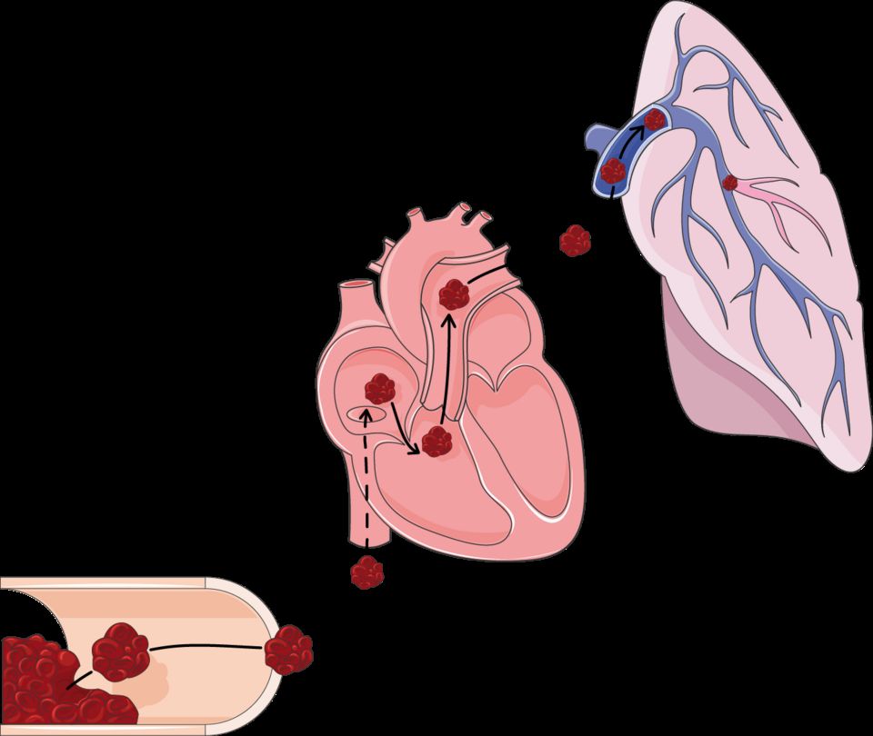
Every breath you take is only half the job. Your lungs pull in oxygen, but that oxygen is useless unless blood flows past the air sacs to pick it up and carry it to the rest of your body. Normally, the right side of your heart pumps blood through the pulmonary arteries into a lacy network of capillaries wrapped around millions of tiny air sacs called alveoli. There, oxygen loads onto red blood cells and carbon dioxide unloads. Air (ventilation) and blood (perfusion) meet in perfect partnership. A pulmonary embolism (PE) is what happens when that partnership is suddenly broken by a plug in the pipe.
What actually goes wrong
A PE is a clot that lodges in a pulmonary artery and blocks blood flow to part of the lung. Most of the time the clot did not form in the lung at all. It usually starts as a deep vein thrombosis (DVT) in a leg or pelvic vein, then a piece breaks loose. Once free, that traveling clot is called an embolus. It rides the venous blood up to the right heart, gets pumped into the pulmonary circulation, and wedges wherever the vessel becomes too narrow to pass. The lung tissue downstream still gets air, but no blood arrives to collect the oxygen.
Why the clot forms: Virchow's triad
Clots do not appear at random. Back in the 1800s, Rudolf Virchow described three conditions that set the stage, and nurses still use his triad today to spot at-risk patients:
- Venous stasis (blood pooling and sitting still). Long flights, bed rest, surgery, immobility, and heart failure all slow the flow so clotting factors accumulate instead of washing away.
- Hypercoagulability (blood that clots too easily). Pregnancy, estrogen-containing contraceptives, cancer, dehydration, and inherited clotting disorders tip the balance toward clotting.
- Endothelial injury (damage to the vessel's inner lining). Trauma, surgery, central lines, and inflammation expose the surfaces that trigger clot formation.
When two or three of these overlap, a DVT becomes far more likely, and a DVT is the seed of most PEs.
Dead space: ventilation without perfusion
Here is the core problem in one phrase: ventilation without perfusion. The blocked region of lung is still being filled with air, but no blood flows past it, so that air is wasted. Physiologists call this wasted, air-but-no-blood region alveolar dead space. Gas exchange simply cannot happen there. Meanwhile, blood that should have gone to that region is shoved into the remaining open vessels, and the mismatch drops the oxygen level in the blood. The result is hypoxemia (low blood oxygen), which is why a PE is a true medical emergency: a large clot can crash oxygen levels and strain the right heart within minutes.
Why the signs happen
PE symptoms come on suddenly, and each one traces straight back to the mechanism:
- Sudden dyspnea (breathlessness) and hypoxia because a chunk of lung can no longer oxygenate blood, so the body is starved of oxygen and screams for more air.
- Pleuritic chest pain (sharp pain that worsens with a deep breath) because the oxygen-starved lung tissue and its lining, the pleura, become irritated and inflamed.
- Tachycardia (fast heart rate) because the heart speeds up to push blood past the blockage and compensate for falling oxygen. You may also see tachypnea, anxiety, and a cough.
| Feature | Normal lung unit | Pulmonary embolism |
|---|---|---|
| Air in alveoli | Yes | Yes (still ventilated) |
| Blood flow past alveoli | Yes | No (clot blocks perfusion) |
| Gas exchange | Efficient | Fails in the blocked zone |
| Result | Normal oxygen | Dead space, hypoxemia |
Explore the figure below to see a clot lodged in a pulmonary artery and how it cuts off blood flow to the lung beyond it.
- A PE is usually a leg DVT that broke loose, traveled as an embolus, and lodged in a pulmonary artery, blocking blood flow to part of the lung.
- Virchow's triad (stasis, hypercoagulability, endothelial injury) sets up the clot in the first place, so prevention targets these three.
- The blocked lung is ventilated but not perfused, creating alveolar dead space (ventilation without perfusion) and dropping blood oxygen.
- Classic signs are sudden dyspnea, pleuritic chest pain, tachycardia, and hypoxia, each explained by lost gas exchange and the body's compensation.
- PE is a medical emergency; a swollen, tender calf is an early clue that a DVT could embolize.
Section 5Pneumonia & respiratory infections
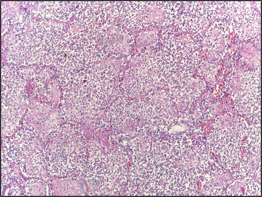
Every time you take a breath, air travels down to about 300 million tiny air sacs called alveoli. These sacs are wrapped in a fine mesh of capillaries, and their walls are astonishingly thin, roughly one cell thick, so oxygen can slip across into the blood while carbon dioxide slips out. This trade is called gas exchange, and it depends on those air sacs staying open, dry, and full of air. Pneumonia is what happens when that clean, air-filled space becomes a battleground.
What goes wrong: the alveoli flood
Pneumonia is an infection of the lung tissue itself, most often caused by bacteria (classically Streptococcus pneumoniae), but also viruses and fungi. When a pathogen reaches the alveoli, the immune system rushes in. White blood cells, fluid, and cellular debris pour into the air sacs, creating pus and inflammatory exudate. The problem is that a sac full of fluid and pus can no longer hold air. Oxygen cannot cross a wall of liquid nearly as well as it crosses air, so gas exchange breaks down. This is why the hallmark of pneumonia is hypoxemia (low blood oxygen) and shortness of breath, or dyspnea. When enough neighboring alveoli fill and clump into a solid, airless region, we call it consolidation, and it shows up as a white patch on a chest X-ray.
The classic signs, and the mechanism behind each
Pneumonia tends to announce itself with a recognizable cluster:
- Fever and chills are the body's response to infection. Chemical signals called pyrogens reset the brain's thermostat upward to make the environment hostile to microbes.
- Productive cough brings up thick, often yellow, green, or rust-colored sputum, which is the pus and debris being cleared from flooded airways.
- Tachypnea and tachycardia (fast breathing and heart rate) are compensations. The body speeds up to move more air and pump oxygen-poor blood faster.
- Pleuritic chest pain, a sharp pain that worsens with a deep breath, appears when inflammation reaches the lining of the lung.
On imaging, that patch of consolidation confirms the flooded region. In older or immunocompromised patients, though, the picture is often quieter, and confusion or simply "not acting right" may be the only early sign.
Where you caught it matters
Nursing care changes depending on where the infection was acquired, because the likely organisms and their drug resistance differ.
| Feature | Community-acquired (CAP) | Hospital-acquired (HAP / VAP) |
|---|---|---|
| Where acquired | Out in daily life; onset before or within 48 hrs of admission | 48+ hrs after admission; VAP is a subtype tied to a ventilator |
| Common organisms | S. pneumoniae, respiratory viruses, atypicals | Often Pseudomonas, Staph aureus including MRSA |
| Drug resistance | Usually lower | Often higher, harder to treat |
| Typical severity | Ranges mild to severe | Frequently sicker, higher mortality |
Ventilator-associated pneumonia (VAP) deserves special note: a breathing tube bypasses the natural filtering and cough defenses of the upper airway, giving bacteria a direct highway into the lungs. This is why nurses follow a "VAP bundle," including keeping the head of the bed elevated and providing regular oral care.
Who is most at risk
Anyone whose defenses are weakened is more vulnerable. This includes the very young and adults over 65, smokers, and people with chronic lung disease such as COPD. It also includes patients who are immunocompromised, those with difficulty swallowing (who may aspirate food or secretions into the lungs), and anyone whose cough reflex is blunted by sedation or a decreased level of consciousness.
A word on tuberculosis
Tuberculosis (TB) is a distinct, airborne infection from Mycobacterium tuberculosis in which the immune system walls the bacteria inside a granuloma; it can stay dormant for years as latent TB (no symptoms, not contagious) before reactivating into active TB with cough, weight loss, and night sweats. Explore the interactive figure below to see how infection fills the alveoli.
- Pneumonia fills the alveoli with fluid and pus, so oxygen cannot cross into the blood, producing hypoxemia, dyspnea, and consolidation on imaging.
- Classic signs are fever, productive cough, crackles from fluid-filled sacs re-opening, and pleuritic chest pain; older adults may only show confusion.
- Community-acquired pneumonia (CAP) is usually less resistant; hospital-acquired (HAP) and ventilator-associated (VAP) pneumonia involve tougher organisms like Pseudomonas and MRSA.
- Highest risk: the very young, adults over 65, smokers, COPD, immunocompromised, and those who aspirate; vaccines and deep breathing help prevent it.
- TB is a separate airborne infection walled into granulomas, existing as latent (dormant, non-contagious) or active disease.
Section 6Lung sounds & assessment
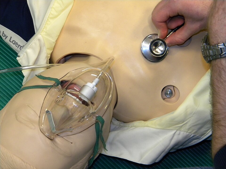
Every time you breathe, air rushes down your windpipe, splits into smaller and smaller tubes, and finally reaches millions of tiny air sacs called alveoli. When those airways are open and the air sacs are dry and elastic, air moves smoothly and quietly. That smooth flow is exactly what a stethoscope is listening for. When you place your stethoscope on someone's chest, you are eavesdropping on how easily air is getting in and out, and whether anything, fluid, mucus, swelling, or scarring, is getting in the way.
Normal breath sounds: the quiet baseline
Over most of the lung fields you should hear vesicular breath sounds, a soft, low, rustling whoosh that is louder on inspiration and fades quickly on expiration. Over the large central airways (the trachea and around the sternum) sounds are naturally louder and more tubular (bronchial and bronchovesicular). The key idea for students: normal air movement is clear and quiet. Anything extra you hear layered on top is called an adventitious (added) sound, and each type tells a different mechanical story about what is happening inside the airways.
Crackles: air popping open through fluid
Crackles (older term: rales) are brief, discontinuous popping or bubbling sounds, often compared to Velcro pulling apart or hair rubbed between your fingers. Mechanistically, they happen when air suddenly pushes open small airways and alveoli that were stuck shut or filled with fluid. You hear them most on inspiration because that is when the collapsed spaces snap open. Crackles usually point toward fluid or collapse in the air sacs, classically pneumonia (inflammatory exudate) or heart failure (fluid backing up into the lungs, called pulmonary edema). In heart failure they often start at the lung bases, where gravity pools fluid.
Wheezes: air squeezing through narrowed tubes
Wheezes are high-pitched, musical, whistling sounds, usually loudest on expiration. Picture blowing through a nearly pinched straw. They happen when airways are narrowed by bronchospasm, swelling (edema), or mucus, so air is forced through a tight channel and the walls vibrate. Wheezes are the signature of asthma and COPD. One important safety note: a wheeze that suddenly goes silent in someone struggling to breathe is ominous, it can mean airflow has dropped so low that no sound is being made (a "silent chest"), which is an emergency, not an improvement.
Stridor: the upper-airway alarm bell
Stridor is a harsh, high-pitched crowing sound heard without a stethoscope, typically on inspiration. It comes from obstruction high up in the upper airway, the larynx or trachea, from swelling (croup, epiglottitis, anaphylaxis) or a foreign body. Treat stridor as an airway emergency: the tube that keeps a person alive is narrowing, and it can close.
Pleural friction rub: surfaces that should glide, grating instead
The lungs are wrapped in two thin membranes (the pleura) that normally slide silently past each other. When they become inflamed (pleuritis, often from infection), they grate together, producing a pleural friction rub, a low-pitched, grating, leathery sound heard on both inspiration and expiration, often with pain on breathing.
| Feature | Crackles | Wheezes |
|---|---|---|
| Sound | Popping / bubbling | Musical / whistling |
| Cause | Fluid or collapsed air sacs | Narrowed airways |
| Timing | Mainly inspiration | Mainly expiration |
| Points toward | Pneumonia, heart failure | Asthma, COPD |
Pulse oximetry: seeing the result of all this airflow
Listening tells you how air is moving; the pulse oximeter (SpO2) tells you the result, how well oxygen is actually reaching the blood. It clips on a finger and estimates the percent of hemoglobin carrying oxygen, usually 95 to 100% in healthy adults. When crackles, wheezes, or obstruction block oxygen from reaching the alveoli, the SpO2 often falls (hypoxemia).
Tie this back to the arterial blood gas (ABG) from Week 1. The pulse ox is a quick, noninvasive estimate; the ABG is the precise lab measurement, giving true PaO2, PaCO2, and pH. Remember the oxyhemoglobin dissociation curve: because of its S-shape, an SpO2 that drops below roughly 90% often signals a much larger fall in PaO2 than the number suggests. Trust the ABG for the full picture, especially for carbon dioxide, which the pulse ox cannot see at all.
Explore the interactive figure below to hear how normal breath sounds differ from crackles, wheezes, stridor, and a friction rub.
- Normal breath sounds are clear and quiet; any added (adventitious) sound signals something interfering with airflow.
- Crackles = fluid or reopening air sacs (pneumonia, heart failure), mostly on inspiration; wheezes = narrowed airways (asthma, COPD), mostly on expiration.
- Stridor is a harsh inspiratory sound from upper-airway obstruction, treat it as an emergency; a pleural friction rub is grating and painful from inflamed pleura.
- Pulse oximetry (SpO2, usually 95 to 100%) estimates oxygen saturation noninvasively but cannot measure CO2, reads unreliably or falsely low in a cold or poorly perfused finger, and reads falsely high in carbon monoxide poisoning.
- The ABG confirms true oxygenation, ventilation, and pH; always weigh the number against how the patient actually looks.
The chapter in one breath
- Ventilation is air moving in and out of alveoli; when it drops (hypoventilation), CO2 backs up and the pH falls, giving respiratory acidosis.
- Perfusion is blood flow through the pulmonary capillaries, and gas exchange only works where ventilated alveoli meet perfused capillaries.
- A normal V/Q ratio is about 0.8; a blocked airway (pneumonia, mucus) creates a low-V/Q shunt, while a blocked vessel (PE) creates a high-V/Q dead space.
- COPD is chronic airflow limitation from emphysema plus chronic bronchitis, where air-trapping and a barrel chest cause a prolonged expiratory phase and chronically retained CO2.
- Asthma is reversible bronchospasm with airway inflammation and mucus, producing expiratory wheezing; a silent chest with a rising CO2 signals dangerous fatigue, not improvement.
- Pulmonary embolism is a clot (often from a DVT) lodging in the pulmonary artery, causing sudden dyspnea, pleuritic chest pain, tachycardia, and hypoxemia with a low CO2 from hyperventilating.
- Pneumonia fills alveoli with inflammatory exudate, so you see fever, productive cough, crackles, and consolidation that impairs oxygenation.
- Adventitious lung sounds localize the problem: crackles mean fluid or reopening alveoli, wheezes mean narrowed airways, and stridor means a dangerous upper-airway obstruction.
ReferenceWords worth knowing
A quick reference for the terms in this chapter — skim it now, or circle back whenever a word trips you up.
- Bronchospasm
- Sudden tightening of the smooth muscle wrapping the bronchi and bronchioles that narrows the airways, making it harder to move air (especially out) and often causing wheezing.
- Chronic bronchitis
- A form of COPD defined clinically as a productive cough lasting at least 3 months in each of 2 consecutive years, caused by chronic airway inflammation, mucus-gland hypertrophy, and excess sputum.
- Consolidation
- Lung tissue that has filled with fluid, pus, blood, or inflammatory cells instead of air (as in pneumonia), making that region solid and impairing gas exchange.
- COPD (chronic obstructive pulmonary disease)
- A progressive, largely irreversible airflow-limiting disease—usually from smoking—that blends emphysema and chronic bronchitis and traps air on exhalation.
- Crackles
- Brief, discontinuous popping or bubbling sounds heard (usually on inspiration) when collapsed or fluid-filled alveoli and small airways suddenly snap open, often signaling fluid or infection.
- Dead space
- Air that is inhaled but does not take part in gas exchange—the conducting airways (anatomic) plus alveoli that are ventilated but not perfused (alveolar); added together the two make up physiologic, or total, dead space.
- Emphysema
- A form of COPD in which the alveolar walls are destroyed and enlarged, reducing surface area for gas exchange and the elastic recoil that normally pushes air out.
- Hypoxemia
- A lower-than-normal level of oxygen in the arterial blood (usually a PaO2 below about 80 mmHg or SaO2 under about 95%), which starves tissues of oxygen if uncorrected.
- Perfusion
- The flow of blood through the pulmonary capillaries that surround the alveoli, releasing carbon dioxide to be exhaled and picking up oxygen from the alveolar air.
- Pulmonary embolism
- A blockage of a pulmonary artery—most often by a clot that traveled from a deep vein—that suddenly cuts off blood flow to part of the lung, creating dead space and often causing sharp dyspnea and hypoxemia.
- Shunt
- Blood that passes through the lungs without being oxygenated because it flows past alveoli that are perfused but not ventilated (as in consolidation or collapse), a cause of hypoxemia that oxygen alone often cannot fix.
- Status asthmaticus
- A severe, prolonged asthma attack that does not respond to usual bronchodilators, a medical emergency that can progress to respiratory failure.
- Stridor
- A harsh, high-pitched sound heard mainly on inspiration that signals narrowing or obstruction of the upper airway (larynx or trachea) and warrants urgent attention.
- Ventilation
- The mechanical movement of air into and out of the lungs, bringing oxygen to the alveoli and carrying carbon dioxide away.
- V/Q ratio
- The balance between ventilation (V, airflow) and perfusion (Q, blood flow) in a lung region, normally about 0.8; mismatches in either direction impair gas exchange.
- Wheeze
- A continuous, musical, usually high-pitched whistling sound—often loudest on expiration—produced when air is forced through narrowed lower airways, as in asthma or COPD.
DrillFlashcards for this unit
These are the Week 10 cards — the same deck as the hub, pulled in here so you can review the moment you finish reading. Tap the card to flip it; hit Shuffle to mix them up. To grade yourself and track mastery across the whole deck, open the full flashcards page.
Check yourself
Renal & Urinary
The kidneys are the body’s quiet housekeepers — filtering waste, balancing water and electrolytes, steadying blood pressure, and even making blood. This chapter starts at the nephron and works outward through the ways kidneys fail suddenly or slowly, the stones and infections of the urinary tract, and what the labs are telling you.
By the end of this chapter you can
- Describe the nephron and what GFR, creatinine, and BUN mean.
- Classify acute kidney injury as prerenal, intrarenal, or postrenal.
- Explain chronic kidney disease, uremia, and its complications.
- Distinguish nephrotic from nephritic syndrome.
- Tell cystitis from pyelonephritis and recognize renal colic.
Section 1The nephron: how the kidney cleans blood
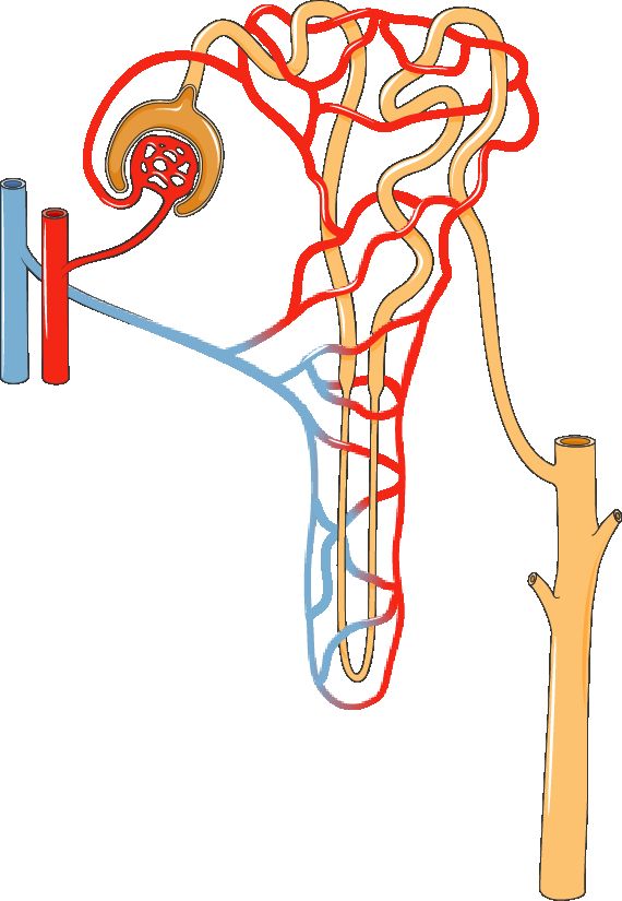
Every minute, more than a liter of blood pours into your kidneys, and by the time it leaves it has been cleaned, balanced, and fine-tuned. The kidney pulls off this trick using millions of tiny units called nephrons - roughly one million per kidney. The nephron is the kidney's functional unit, meaning it is the smallest structure that can do the whole job of making urine. When we talk about kidney disease, we are really talking about nephrons being damaged, overworked, or dying.
The nephron in three steps: filter, reabsorb, secrete
Think of the nephron as a factory with three stations. First comes filtration. Blood enters a tuft of leaky capillaries called the glomerulus, which sits inside a cup called Bowman's capsule. Blood pressure pushes water and small molecules - sodium, glucose, urea, potassium - through the filter into the tubule. This early fluid is called filtrate. Big things like blood cells and proteins are too large to pass, so normally they stay in the blood. That is why finding protein or blood in the urine is a red flag: it usually means the filter is leaking.
Next comes reabsorption along the tubule (the proximal tubule, loop of Henle, and distal tubule). The body cannot afford to dump everything it just filtered, so about 99% of the water and nearly all the glucose and needed sodium are pulled back into the blood. Finally comes secretion, where the tubule actively adds unwanted substances - certain drugs, extra hydrogen ions, extra potassium - into the filtrate to be peed out. What is left after all three steps is urine.
GFR: the number that tells you how well the kidney works
The single best measure of kidney function is the glomerular filtration rate (GFR) - how many milliliters of filtrate all the glomeruli produce per minute. A normal GFR is usually around 90-120 mL/min. When GFR falls, the kidney is filtering less, and wastes start backing up in the blood. GFR is the basis for staging chronic kidney disease (CKD); a GFR under 60 for three or more months defines CKD, and under 15 signals kidney failure. We usually estimate it (eGFR) from a blood test rather than measuring it directly.
The kidney's other jobs (it is not just a filter)
Losing kidney function causes trouble far beyond waste buildup, because the kidney moonlights in several roles:
- Blood pressure - through the renin-angiotensin-aldosterone system (RAAS). When blood flow or pressure drops, the kidney releases renin, which sets off a cascade that constricts vessels and retains sodium and water to raise pressure.
- Red blood cells - the kidney makes erythropoietin (EPO), the hormone that tells bone marrow to build red cells. This is why patients with failing kidneys often develop anemia.
- Activating vitamin D - the kidney converts vitamin D to its active form, which lets the gut absorb calcium. Kidney disease therefore weakens bones and disturbs calcium balance.
- Acid-base balance - the kidney excretes hydrogen ions and reclaims bicarbonate, keeping blood pH steady. Failing kidneys tend toward metabolic acidosis.
Reading the labs: creatinine and BUN
Two blood tests reveal how the nephrons are doing. Creatinine is a waste product from normal muscle breakdown. The body makes it at a steady rate and the kidney clears it, so when creatinine rises, it usually means filtration is falling - it is our everyday marker of GFR. BUN (blood urea nitrogen) measures urea, a waste from protein breakdown. BUN also rises when the kidney struggles, but it is more easily thrown off by dehydration, bleeding, and high-protein intake, so nurses read it alongside creatinine rather than alone. Explore the nephron below to see filtration in action.
| Feature | Creatinine | BUN |
|---|---|---|
| Comes from | Muscle breakdown | Protein breakdown (urea) |
| Usual range | ~0.6-1.2 mg/dL | ~7-20 mg/dL |
| Best for | Estimating GFR / true kidney function | Hydration and overall status |
| Thrown off by | Muscle mass, less so by diet | Dehydration, bleeding, high protein |
- The nephron is the kidney's functional unit; it cleans blood in three steps - filtration at the glomerulus, then reabsorption and secretion along the tubule.
- GFR (usually ~90-120 mL/min) is the key measure of kidney function; a falling GFR defines and stages chronic kidney disease.
- The kidney also controls blood pressure (RAAS), makes erythropoietin for red cells, activates vitamin D for calcium and bone, and manages acid-base balance.
- Creatinine is the everyday marker of filtration; a rising level usually means falling GFR.
- BUN tracks kidney function too but is easily skewed by dehydration, bleeding, and diet, so it is read alongside creatinine.
Section 2Acute kidney injury
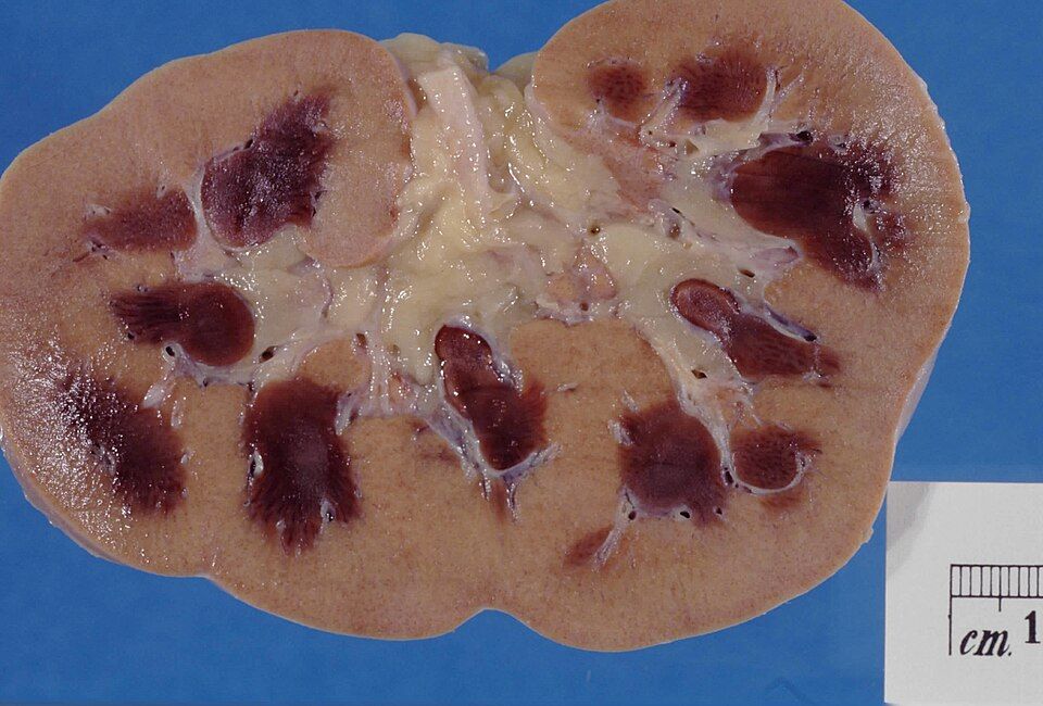
Your kidneys are the body's quiet, constant filters. Every minute, about a fifth of the blood your heart pumps flows through them. Inside each kidney sit roughly a million tiny filtering units called nephrons, and each nephron starts with a knot of capillaries called the glomerulus. Blood pressure pushes fluid and waste across that filter to form urine, while the tubules that follow reclaim the water, sodium, and other things your body wants to keep. Working normally, the kidneys clear metabolic garbage, balance fluid and electrolytes, and help control blood pressure and red blood cell production. When they suddenly stumble, waste backs up fast.
Acute kidney injury (AKI) is a sudden drop in kidney function, unfolding over hours to days. The key word is sudden (unlike chronic kidney disease, which creeps along for months to years). The good news: because it is abrupt, AKI is often reversible if the underlying cause is caught and fixed before the nephrons are permanently damaged.
How we know the kidneys are failing
Two blood tests tell the story. Creatinine is a waste product from muscle that the kidneys normally clear at a steady rate; when filtering drops, creatinine rises. BUN (blood urea nitrogen) is a nitrogen waste from protein breakdown, and it climbs too. A rising creatinine means the glomerular filtration rate (GFR), the volume of filtrate the glomeruli form each minute, is falling. At the same time, the kidneys usually make less urine. Oliguria means putting out less than about 400 mL of urine in 24 hours (often described as under roughly 0.5 mL/kg/hr). Put simply: labs go up, urine goes down.
The three categories: where the problem lives
AKI is sorted by where the trouble sits relative to the kidney itself. This matters because the fix is completely different for each.
| Feature | Prerenal | Intrarenal | Postrenal |
|---|---|---|---|
| Core problem | Poor blood flow to the kidney | Direct damage inside the kidney | Obstruction of outflow after the kidney |
| Typical causes | Dehydration, hemorrhage, heart failure, shock | Acute tubular necrosis, nephrotoxins, prolonged ischemia | Kidney stones, enlarged prostate, tumors, blocked catheter |
| Is tissue damaged yet? | Not yet, usually reversible if flow is restored fast | Yes, tubule cells are injured | Not at first, but backed-up pressure damages tissue over time |
Prerenal AKI is the most common. Anything that drops blood flow (low volume from vomiting or bleeding, a weak heart, or widespread vasodilation in sepsis) starves the glomerulus of the pressure it needs to filter. The kidney tissue is still healthy, so if you restore perfusion quickly (fluids, treat the heart, stop the bleeding), function bounces back. Ignored, though, prolonged low flow becomes ischemic damage and tips into the intrarenal category.
Intrarenal (intrinsic) AKI means the nephron itself is injured. The classic form is acute tubular necrosis (ATN), where the delicate tubule cells die, either from prolonged ischemia or from toxins like certain antibiotics (aminoglycosides), IV contrast dye, or myoglobin released in rhabdomyolysis. Dead tubule cells slough off, clog the tubules, and can't reabsorb properly.
Postrenal AKI is a plumbing problem downstream: urine can't get out. A stone, an enlarged prostate, a tumor, or even a kinked catheter blocks flow. Pressure backs up into the kidney and stops filtration. This type is often the most quickly reversible, relieve the obstruction and urine pours out, but only if caught before back-pressure scars the tissue.
The phases of recovery
When AKI does progress, recovery classically moves through phases. During the oliguric phase (often lasting days to a couple of weeks), urine output is low, and waste, potassium, and fluid pile up. This is the dangerous stretch, watch closely for hyperkalemia (high potassium, which can trigger deadly heart arrhythmias), fluid overload, and metabolic acidosis.
As the tubules heal, the kidney enters the diuretic phase. The nephrons can filter again but haven't regained their concentrating ability, so urine output surges, sometimes several liters a day. Now the risk flips: patients can become dehydrated and lose too much sodium and potassium. Careful fluid and electrolyte replacement is essential. A slower recovery phase follows as function normalizes, though some patients never fully return to baseline.
Curious how the different causes stack up? Explore them in the figure below.
- AKI is a sudden, often reversible drop in kidney function, marked by rising creatinine and BUN with falling GFR and urine output (oliguria).
- Prerenal AKI is poor blood flow to the kidney, intrarenal is direct damage (classically acute tubular necrosis), and postrenal is obstruction of urine outflow.
- Prerenal causes are the most common and are reversible if perfusion is restored before ischemia damages tissue.
- Recovery moves through an oliguric phase (watch for hyperkalemia and fluid overload) then a diuretic phase (watch for dehydration and electrolyte loss).
- Finding the correct category is critical because each is treated differently: restore flow, remove the insult, or relieve the blockage.
Section 3Chronic kidney disease
Your kidneys are quiet workhorses. Every day, about a million tiny filtering units called nephrons in each kidney strain your blood, pulling out waste and extra fluid to make urine while keeping the good stuff (protein, blood cells, most electrolytes) in circulation. They also do jobs that have nothing to do with peeing: they make erythropoietin (EPO) to tell your bone marrow to build red blood cells, activate vitamin D so your intestines can absorb calcium (which then strengthens bone), and balance the blood's acid level. In chronic kidney disease (CKD), nephrons are slowly and permanently destroyed, so all of those jobs start to fail at once.
What CKD is and how we stage it
CKD is a gradual, irreversible loss of nephrons over months to years. Because kidneys have huge reserve, a person can lose a lot of function before feeling sick, so we measure the damage with the glomerular filtration rate (GFR) - how many milliliters of filtrate all the glomeruli squeeze out of the plasma each minute. A normal GFR is usually around 90-120 mL/min, which is only a small slice of the roughly 1.2 L of blood the kidneys receive every minute. CKD is defined as kidney damage or a GFR below 60 for at least 3 months, and it is staged from 1 (mild) to 5 (kidney failure).
| Stage | GFR (mL/min) | What it means |
|---|---|---|
| 1-2 | ≥60 (with damage) | Often silent; caught on labs or urine protein |
| 3 | 30-59 | Complications begin (anemia, bone changes) |
| 4 | 15-29 | Severe; prepare for dialysis or transplant |
| 5 | <15 | Kidney failure; dialysis usually needed |
Why it happens: diabetes and hypertension
The two leading causes are the same two conditions nurses see everywhere. In diabetes, chronically high blood glucose damages the delicate glomerular capillaries (diabetic nephropathy), and one of the earliest warning signs is small amounts of protein leaking into the urine (albuminuria). In hypertension, the constant high pressure batters those same tiny vessels, thickening and scarring them. Cruelly, failing kidneys also raise blood pressure by retaining salt and water and firing the renin-angiotensin system - so hypertension both causes CKD and gets worse because of it, a vicious cycle.
Uremia: when wastes build up
As filtering fails, nitrogen wastes like urea and creatinine climb in the blood. This toxic buildup is called uremia, and because the blood reaches every organ, the symptoms are body-wide. Patients often feel exhausted, nauseated, and itchy (pruritus from waste deposited in skin); they may lose appetite, taste metal, and think slowly. In severe uremia, wastes irritate the heart lining (pericarditis) and the brain (uremic encephalopathy, causing confusion and even seizures). Uremia is why "the kidneys" become a whole-body problem.
The complications and why each one happens
Understanding CKD means tracing each problem back to a lost kidney job:
- Anemia - fewer working cells make less EPO, so the marrow builds fewer red cells. Patients are pale, tired, and short of breath.
- Bone disease (renal osteodystrophy) - kidneys can no longer activate vitamin D or excrete phosphate. Low calcium and high phosphate drive the parathyroids to pump out PTH, which pulls calcium out of bones, leaving them weak.
- Hyperkalemia - the kidneys normally dump excess potassium. When they can't, potassium rises, threatening dangerous cardiac arrhythmias. This is often the most urgent lab.
- Fluid overload - unexcreted salt and water back up, causing edema, hypertension, and pulmonary congestion.
- Metabolic acidosis - kidneys normally excrete acid and regenerate bicarbonate. As that fails, acid accumulates, contributing to fatigue, bone loss, and rapid breathing.
When the kidneys fail
At stage 5, often called end-stage renal disease (ESRD), the kidneys can no longer keep the body's chemistry safe. The two options are dialysis - a machine (hemodialysis) or the abdominal lining (peritoneal dialysis) that filters the blood artificially - or a kidney transplant. Dialysis manages the disease but does not cure it; it replaces only a fraction of true kidney function, which is why prevention and slowing progression matter so much. Explore the stages in the interactive figure below.
- CKD is a slow, irreversible loss of nephrons, staged 1-5 by GFR; a GFR below 60 for over 3 months defines it.
- Diabetes and hypertension are the top two causes and often worsen each other.
- Uremia is the buildup of urea and other wastes, producing whole-body symptoms like fatigue, nausea, itching, and confusion.
- Key complications trace to lost kidney jobs: anemia (low EPO), bone disease (low active vitamin D, high PTH), hyperkalemia, fluid overload, and metabolic acidosis.
- At stage 5 (ESRD), dialysis or transplant replaces failing kidney function; dialysis manages but does not cure.
Section 4Glomerular disease: nephrotic vs nephritic
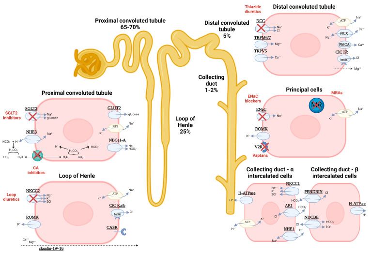
Every minute, your kidneys pull a huge amount of blood through roughly a million tiny filters called glomeruli. Picture each glomerulus as a wadded-up tuft of capillaries wrapped in a cup. Its filtering wall has three layers: the capillary lining (endothelium), a middle mesh called the glomerular basement membrane (GBM), and specialized cells with interlocking foot processes called podocytes. This wall is a smart sieve. It lets water, salts, and small wastes pass into the urine, but it holds back big things the body wants to keep, especially albumin (the main blood protein) and red blood cells. Two features make that possible: the pores are size-selective, and the wall carries a negative charge that repels negatively charged albumin. When this filter breaks, it tends to fail in one of two patterns, and telling them apart is one of the most testable ideas in renal nursing.
Nephrotic syndrome: a very leaky filter
In nephrotic syndrome, the filter becomes too permeable without much inflammation. The podocytes and the charge barrier are damaged, so albumin pours out. The hallmark is massive proteinuria, usually defined as more than 3.5 grams of protein in the urine per day. Losing that much protein drops the blood albumin, producing hypoalbuminemia. Because albumin normally holds fluid inside vessels by oncotic (pulling) pressure, losing it lets fluid leak into tissues, causing the heavy, generalized edema classic for this syndrome, often puffy eyes in the morning and swollen legs and belly by evening. The liver, sensing low protein, ramps up production of many proteins including lipoproteins, so cholesterol and triglycerides climb (hyperlipidemia), and fatty casts can appear in urine. Patients also lose clotting-regulator proteins and immunoglobulins, raising risks of thrombosis and infection. A classic cause in children is minimal change disease; in adults, membranous nephropathy and diabetic or lupus-related disease are common.
Nephritic syndrome: an inflamed filter
In nephritic syndrome, the problem is inflammation, usually immune complexes lodging in the glomerulus and triggering an immune attack. That inflammation physically breaks the capillary wall, so red blood cells escape into the urine. The hallmark is hematuria, often tea- or cola-colored, sometimes with red blood cell casts (tiny cell-molds shaped in the tubules) that are essentially diagnostic of glomerular bleeding. Protein leaks too, but usually in smaller amounts (typically less than 3.5 g/day). Inflammation also swells and clogs the filter, so filtration drops; the kidney retains salt and water, driving hypertension and a milder, often facial edema, plus a rising creatinine (azotemia) and reduced urine output. The teaching classic is post-streptococcal glomerulonephritis (PSGN): a child develops cola-colored urine, puffy eyes, and high blood pressure roughly 1 to 3 weeks after a strep throat or skin infection, as immune complexes deposit in the glomeruli.
How to keep the two straight
A simple anchor: nephrOtic = prOtein (huge protein loss), nephrItic = Inflammation and hematurIa (blood). Nephrotic is a quiet, leaky sieve; nephritic is an angry, bleeding, high-pressure one. Real patients can overlap, but the dominant pattern usually declares itself.
| Feature | Nephrotic | Nephritic |
|---|---|---|
| Core problem | Very leaky filter (podocyte/charge injury) | Inflamed filter (immune-mediated) |
| Proteinuria | Massive (usually >3.5 g/day) | Mild to moderate (usually <3.5 g/day) |
| Hematuria | Absent or minimal | Present; often RBC casts |
| Albumin & lipids | Low albumin, high lipids | Usually near normal |
| Edema | Heavy, generalized (oncotic) | Mild, often facial/periorbital |
| Blood pressure | Often normal | Elevated (salt/water retention) |
| Classic example | Minimal change disease | Post-streptococcal GN |
Explore the interactive figure below to compare the leaky nephrotic filter with the inflamed nephritic one side by side.
- Nephrotic = protein. A leaky filter causes massive proteinuria (usually >3.5 g/day), low albumin, heavy generalized edema, and high lipids.
- Nephritic = inflammation and blood. An inflamed filter causes hematuria (often with RBC casts), milder proteinuria, hypertension, and mild edema.
- Mechanism drives the signs: lost albumin lowers oncotic pressure (nephrotic edema); inflammation clogs filtration and retains salt/water (nephritic hypertension).
- Classic examples: minimal change disease (nephrotic) and post-streptococcal glomerulonephritis, appearing 1 to 3 weeks after strep (nephritic).
- Memory hook: nephrOtic = prOtein, nephrItic = Inflammation/hematurIa.
Section 5Kidney stones
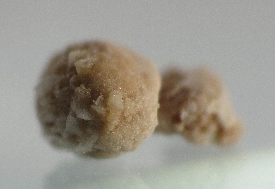
Your kidneys are constantly filtering your blood and dissolving the leftover minerals and salts into your urine. Think of urine as a river carrying dissolved cargo. As long as there is enough water flowing, the minerals stay dissolved and wash out harmlessly. Kidney stones (nephrolithiasis) form when that balance tips: the urine becomes supersaturated, meaning it holds more mineral than it can keep dissolved. The excess starts to crystallize, and those tiny crystals clump together into a hard stone. It is the same physics as making rock candy, except it happens inside the collecting system of the kidney, and nobody wants it.
Why the urine becomes supersaturated
Two things push urine past its saturation point. First, too little water (dehydration) concentrates the minerals, so the same amount of solute is packed into less fluid. Second, too much mineral, from diet, genetics, or metabolic conditions, overloads the urine. When calcium, oxalate, uric acid, or other salts climb high enough, crystals precipitate out. Sluggish, concentrated, standing urine is the perfect environment for a stone to grow, which is why chronic low fluid intake is the single biggest modifiable risk factor.
The pain: renal colic
A stone sitting quietly in the kidney may cause nothing at all. The trouble starts when it drops into the ureter, the narrow tube draining urine to the bladder. The stone lodges and blocks flow, urine backs up, and the ureter stretches and spasms trying to squeeze the stone along. That produces renal colic: sudden, excruciating, wave-like flank pain that classically radiates to the groin as the stone travels downward. Patients are famously restless, unable to find any position that helps, which contrasts with the still, guarded posture of peritonitis. The stone also scrapes the ureteral lining, so you often see hematuria (blood in the urine, sometimes visible, sometimes only on dipstick). Nausea, vomiting, and urinary urgency are common companions because the kidney and gut share nerve pathways.
Types of stones
Calcium stones, usually calcium oxalate, are by far the most common, making up roughly 75 to 80 percent of cases. Knowing the type matters because prevention differs.
| Feature | Calcium stones | Uric acid stones |
|---|---|---|
| Frequency | Most common (~75-80%) | Less common |
| Urine condition | Often normal to alkaline | Favored by acidic urine |
| Key contributors | High calcium/oxalate, dehydration | High purine intake, gout |
| X-ray visibility | Usually radiopaque (show up) | Often radiolucent (hard to see) |
A common point of confusion: cutting dietary calcium usually backfires. Normal calcium intake binds oxalate in the gut so it leaves in the stool instead of the urine, which actually lowers calcium-oxalate stone risk. The real fix is fluid.
Nursing priorities
Management rests on three pillars while the stone passes or is treated:
- Pain control. Renal colic is genuinely one of the worst pains people report. NSAIDs like ketorolac are often first-line because they reduce ureteral spasm and inflammation; opioids may be added. Treat pain aggressively and early.
- Hydration. Encourage generous oral or IV fluids to keep urine dilute and flowing, which helps flush the stone along and prevents new crystals from forming. This is prevention and treatment in one.
- Strain all urine. Have the patient void through a fine mesh strainer to catch the stone. This confirms passage and, crucially, lets the lab analyze the stone's composition so prevention can be targeted.
Watch for warning signs that need escalation: fever (a blocked, infected kidney is an emergency), signs of complete obstruction, or intractable pain and vomiting. Larger stones that will not pass may need lithotripsy (shock waves that fragment the stone) or surgical removal. The interactive figure below lets you explore how a stone lodges in the ureter and produces colic; explore it once you have the mechanism in mind.
- Stones form when urine becomes supersaturated with minerals, driven most often by dehydration or excess mineral load.
- Renal colic is sudden, severe, wave-like flank pain that radiates to the groin, typically with hematuria and a restless patient.
- Calcium (oxalate) stones are the most common type; normal dietary calcium is usually protective, not harmful.
- The three priorities are pain control, aggressive hydration, and straining the urine to catch and analyze the stone.
- Escalate immediately for fever, complete obstruction, or uncontrolled pain and vomiting.
Section 6UTIs & incontinence
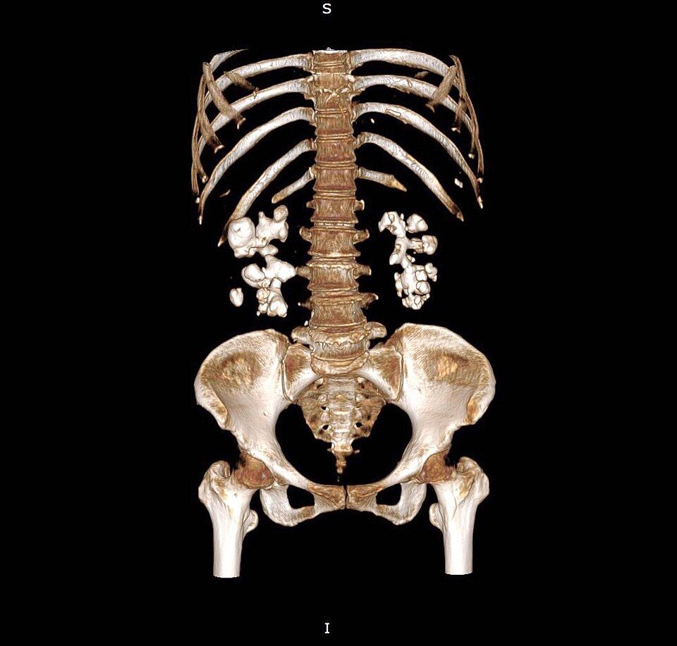
Your urinary tract is basically a plumbing system with a one-way flow and a built-in cleaning stream. The kidneys make urine, the ureters carry it down, the bladder stores it, and the urethra lets it out. Normally that flow is sterile from the bladder up, and every time you empty your bladder you flush out any stray bacteria trying to swim upstream. The lining also fights back with a slightly acidic pH and mucus that makes it hard for microbes to stick. A urinary tract infection (UTI) happens when bacteria, most commonly Escherichia coli from the bowel, colonize the urethra and climb upward faster than the body can rinse them away.
Lower UTI (cystitis) vs upper UTI (pyelonephritis)
Where the bacteria land decides how sick the patient looks. In a lower UTI (cystitis), the infection stays in the bladder and urethra. The inflamed bladder wall becomes irritated and hypersensitive, so patients feel dysuria (burning with urination), frequency, and urgency, often with cloudy or foul-smelling urine and low suprapubic discomfort. Because the infection is localized, patients usually stay afebrile and don't feel systemically ill.
An upper UTI (pyelonephritis) means bacteria have ascended the ureters into one or both kidneys. Now the infection reaches the bloodstream-rich renal tissue, so the body mounts a full systemic response: fever, chills, nausea, and the classic flank pain with costovertebral angle (CVA) tenderness when you percuss over the kidney. Pyelonephritis is the more dangerous picture because it can progress to urosepsis and can scar the kidney over time.
| Feature | Cystitis (lower) | Pyelonephritis (upper) |
|---|---|---|
| Location | Bladder / urethra | Kidney(s) |
| Hallmark symptoms | Dysuria, frequency, urgency | Fever, chills, flank pain |
| Exam finding | Suprapubic tenderness | CVA tenderness |
| Systemic illness | Usually none | Often, can lead to sepsis |
Why women get more UTIs, and the catheter problem
Anatomy is the main reason women get far more UTIs than men. The female urethra is much shorter (roughly 4 cm versus about 20 cm in men), so bacteria have a shorter climb to the bladder. The urethral opening also sits close to the vagina and anus, giving perineal E. coli an easy route. Sexual activity mechanically pushes bacteria toward the urethra, and after menopause, lower estrogen thins the tissue and shifts the protective vaginal flora, raising risk again.
An indwelling (Foley) catheter is a major risk because it bypasses every one of those natural defenses at once. The tube is a direct highway into the bladder, bacteria form a biofilm along its surface where antibiotics and immune cells can't reach, and continuous drainage removes the flushing effect of a normal void. This is why catheter-associated UTIs (CAUTIs) are among the most common hospital-acquired infections, and why the single best prevention is removing the catheter as early as possible.
The older adult who just gets confused
Older adults often skip the textbook burning-and-fever story entirely. Instead, a UTI may show up as new-onset confusion, delirium, or a sudden functional decline such as new incontinence, falls, or refusing to eat. This happens partly because a blunted immune response mutes fever and partly because an aging brain is very sensitive to the systemic inflammation and dehydration that infection brings. The teaching point for nurses: an acute change in mental status in an elder always deserves a workup that includes a urinalysis.
Urinary incontinence at a glance
Incontinence is the involuntary leakage of urine, and it is a symptom, not a normal part of aging. The main types:
- Stress incontinence — leakage with coughing, laughing, or lifting because weak pelvic floor support lets pressure overcome the sphincter.
- Urge incontinence — a sudden, uncontrollable need to void from an overactive detrusor muscle contracting on its own.
- Overflow incontinence — constant dribbling from a bladder that never fully empties, often from obstruction (enlarged prostate) or a weak, underactive detrusor.
- Functional incontinence — a normal urinary tract, but a physical or cognitive barrier (mobility, dementia) keeps the person from reaching the toilet in time.
You can explore the layered urinary tract in the interactive figure below to see exactly where cystitis and pyelonephritis take hold.
- Cystitis (lower UTI) causes dysuria, frequency, and urgency without fever; pyelonephritis (upper UTI) adds fever, chills, and CVA/flank tenderness and can progress to sepsis.
- Women get more UTIs because of a short urethra near the perineum; E. coli is the usual culprit.
- Indwelling catheters bypass natural defenses and cause CAUTIs, so remove them as early as possible.
- In older adults, new confusion or delirium may be the only sign of a UTI, even without fever.
- Incontinence types: stress (leak with pressure), urge (overactive detrusor), overflow (incomplete emptying), and functional (a barrier to reaching the toilet).
The chapter in one breath
- The nephron filters ~180 L/day at the glomerulus; GFR (usually ~90-120 mL/min) is the single best marker of kidney function, and it falls before symptoms appear.
- Acute kidney injury (AKI) is a rapid, often reversible rise in creatinine and drop in urine output, sorted as prerenal (low perfusion, e.g. dehydration/shock), intrarenal (tubule damage like ATN), or postrenal (obstruction).
- Chronic kidney disease (CKD) is irreversible nephron loss over months to years (GFR <60 for 3+ months), driving anemia (low erythropoietin), bone disease (low active vitamin D), and metabolic acidosis.
- In CKD the kidney stops excreting potassium and phosphate, so watch for hyperkalemia (peaked T waves, lethal arrhythmias) as the most urgent electrolyte threat.
- Nephrotic syndrome is a leaky filter: massive proteinuria (>3.5 g/day), low albumin, generalized edema, and high cholesterol - think holes letting protein out.
- Nephritic syndrome is an inflamed filter: hematuria (often tea-colored urine with RBC casts), hypertension, and mild-to-moderate proteinuria - think inflammation letting blood out.
- Kidney stones (nephrolithiasis), most often calcium oxalate, cause sudden severe flank pain radiating to the groin (renal colic) with hematuria; the patient writhes and cannot get comfortable.
- UTIs usually start as cystitis (dysuria, frequency, urgency, suprapubic pain) and can ascend to pyelonephritis (flank pain, fever, CVA tenderness); confusion may be the only sign in older adults.
- Urinary incontinence has distinct types - stress (leak with cough/sneeze from weak pelvic floor) versus urge (sudden strong need from an overactive detrusor) - and each is managed differently.
ReferenceWords worth knowing
A quick reference for the terms in this chapter — skim it now, or circle back whenever a word trips you up.
- Acute kidney injury (AKI)
- A sudden, often reversible drop in kidney function (over hours to days) that lets waste products like creatinine and BUN build up in the blood, usually with reduced urine output.
- Acute tubular necrosis (ATN)
- The most common intrarenal cause of AKI, in which the tubule cells are injured and die, most often from prolonged ischemia (low blood flow) or nephrotoxins such as certain antibiotics or contrast dye.
- BUN (blood urea nitrogen)
- A blood test measuring urea, a nitrogen waste from protein breakdown that the kidneys normally clear, so a rising BUN often signals reduced kidney function or dehydration.
- Chronic kidney disease (CKD)
- A gradual, permanent loss of kidney function lasting three months or longer, staged by the GFR and usually caused over time by diabetes or hypertension.
- Creatinine
- A waste product from normal muscle metabolism that the kidneys filter and excrete steadily, so a rising serum creatinine is one of the most reliable markers that kidney function is declining.
- Dialysis
- A treatment that artificially filters wastes, excess fluid, and electrolytes from the blood (by hemodialysis or peritoneal dialysis) when the kidneys can no longer do the job adequately.
- Erythropoietin (EPO)
- A hormone made mostly by the kidneys that tells the bone marrow to produce red blood cells, which is why failing kidneys often cause anemia.
- GFR (glomerular filtration rate)
- The volume of plasma the glomeruli filter — that is, the volume of filtrate they form — each minute (normally about 90–120 mL/min), used as the best overall measure of how well the kidneys are working.
- Glomerulus
- The tiny tuft of capillaries at the start of each nephron where blood is first filtered, allowing water and small wastes through while normally holding back blood cells and large proteins.
- Nephron
- The functional filtering unit of the kidney (about one million per kidney) that filters blood and fine-tunes the urine through its glomerulus and connected tubules.
- Nephritic syndrome
- A pattern of glomerular inflammation marked by blood in the urine (hematuria), mild-to-moderate protein loss, hypertension, and often reduced GFR.
- Nephrotic syndrome
- A pattern of glomerular damage marked by heavy protein loss in the urine (usually more than 3.5 g/day), low blood albumin, widespread edema, and high blood lipids.
- Oliguria
- Abnormally low urine output, usually defined as less than about 400 mL in 24 hours, which is a key warning sign of acute kidney injury.
- Pyelonephritis
- A bacterial infection of the kidney and its collecting system (often from a bladder infection that ascended upward), typically causing fever, chills, and flank pain.
- Renal colic
- Severe, cramping, wave-like flank pain that radiates toward the groin, classically caused by a kidney stone obstructing and stretching the ureter.
- Uremia
- The toxic clinical syndrome of advanced kidney failure in which retained nitrogen wastes cause symptoms such as nausea, fatigue, itching, and confusion.
DrillFlashcards for this unit
These are the Week 11 cards — the same deck as the hub, pulled in here so you can review the moment you finish reading. Tap the card to flip it; hit Shuffle to mix them up. To grade yourself and track mastery across the whole deck, open the full flashcards page.
Check yourself
| Feature | Prerenal | Intrarenal |
|---|---|---|
| Problem site | Before the kidney (blood flow in) | Inside kidney tissue (nephron) |
| Classic cause | Hypovolemia, shock, heart failure | Acute tubular necrosis, nephrotoxins, contrast, glomerulonephritis |
| Reversible? | Yes, if perfusion restored quickly | Slower; depends on tissue recovery |
| Feature | Nephrotic | Nephritic |
|---|---|---|
| Core problem | Leaky filter loses protein | Inflamed, bleeding glomerulus |
| Proteinuria | Massive (usually >3.5 g/day) | Mild to moderate |
| Urine/blood | Foamy urine, low albumin, edema, high lipids | Hematuria (tea-colored), RBC casts, hypertension |
| Feature | Cystitis (bladder) | Pyelonephritis (kidney) |
|---|---|---|
| Location | Lower urinary tract | Upper tract, kidney |
| Symptoms | Dysuria, frequency, urgency, suprapubic pain | All of those PLUS high fever, chills, flank/CVA tenderness, nausea |
| Severity | Usually localized, low-grade | Systemic; can lead to sepsis |
- Vitamin D: the kidney performs the final activation step, converting vitamin D to its active form (calcitriol) so the gut can absorb calcium, this is why CKD causes bone disease.
- Blood pressure: the kidney releases renin, triggering the renin-angiotensin-aldosterone system (RAAS) to retain sodium and water and raise BP.
- Acid-base: the kidney excretes hydrogen ions and reabsorbs/regenerates bicarbonate, so kidney failure often causes a metabolic acidosis.
Reproductive
The reproductive systems bring their own set of disorders — hormonal, structural, infectious, and malignant. This chapter covers the common female and male conditions students meet on the floor, the cancers to screen for, and the sexually transmitted infections that quietly cause the most long-term harm.
By the end of this chapter you can
- Recognize common female disorders (PCOS, endometriosis, fibroids, PID).
- Name the female cancers and how cervical and breast cancer are screened.
- Explain BPH and tell it from prostate cancer.
- Identify testicular torsion as an emergency.
- Distinguish the major bacterial and viral STIs.
Section 1Female reproductive disorders
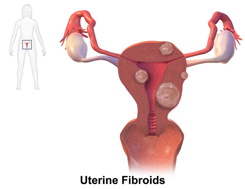
Every month, a healthy uterus rehearses for a pregnancy that usually does not come. This rehearsal is the menstrual cycle, and understanding its normal rhythm makes almost every female reproductive disorder easier to reason through. In the first half (the follicular phase), the pituitary releases FSH, an ovarian follicle matures, and rising estrogen thickens the uterine lining (the endometrium). A surge of LH triggers ovulation around day 14. The emptied follicle becomes the corpus luteum, which pumps out progesterone to hold the lining stable (the luteal phase). If no pregnancy implants, the corpus luteum dies, hormones fall, and the lining sheds as menstruation. Roughly a 28-day loop, though 21 to 35 days is often normal.
When the cycle misbehaves: menstrual disorders
Dysmenorrhea is painful periods. The primary type comes from excess prostaglandins, which cause the uterine muscle to cramp hard and briefly cut off its own blood supply (ischemia) - the same mechanism behind the crampy, labor-like pain. This is why NSAIDs, which block prostaglandin synthesis, work so well. Secondary dysmenorrhea points to an underlying problem like endometriosis or fibroids.
Amenorrhea means absent periods. Primary is never having started by around age 15; secondary is a loss of previously regular cycles for 3 or more months. Pregnancy is the most common cause of secondary amenorrhea and must be ruled out first, but stress, extreme exercise, low body weight, and thyroid or prolactin problems also silence the hypothalamic-pituitary-ovarian axis.
Abnormal uterine bleeding (AUB) covers bleeding that is too heavy (menorrhagia), too frequent, or between periods (metrorrhagia). Often it reflects anovulation: without ovulation there is no progesterone, so estrogen builds the lining unopposed until it outgrows its blood supply and sheds erratically. Chronic heavy loss commonly causes iron-deficiency anemia.
PCOS: the hormone-and-metabolism tangle
Polycystic ovary syndrome is the most common endocrine disorder in reproductive-age women. Think of three linked problems: irregular or absent cycles (from chronic anovulation), androgen excess (causing hirsutism, acne, and scalp thinning), and insulin resistance. The insulin piece is the engine that drives much of the rest: high insulin nudges the ovaries to make more androgens and lowers SHBG, freeing even more testosterone to circulate. The ovaries fill with many small immature follicles - the "polycystic" appearance on ultrasound. Because cycles do not complete, the lining sits under unopposed estrogen, raising the long-term risk of endometrial hyperplasia. PCOS also carries higher risk for type 2 diabetes and metabolic syndrome, which is why lifestyle change and sometimes metformin are frontline.
Endometriosis: the right tissue in the wrong place
In endometriosis, tissue that behaves like endometrium grows outside the uterus - on the ovaries, ligaments, and pelvic peritoneum. It still answers to the monthly hormone cues, so it thickens and bleeds with each cycle. But that blood has nowhere to drain. The result is chronic inflammation, scarring, and adhesions that glue pelvic organs together. Classic symptoms are cyclic pelvic pain that worsens with menses, dyspareunia (painful intercourse), and infertility. On the ovary, trapped old blood can form a "chocolate cyst" (endometrioma). Diagnosis is often confirmed by laparoscopy.
Fibroids and pelvic inflammatory disease
Uterine fibroids (leiomyomas) are benign smooth-muscle tumors of the uterine wall. They are estrogen-sensitive, so they tend to grow during reproductive years and shrink after menopause. Many are silent, but larger ones cause heavy or prolonged bleeding, pelvic pressure, and sometimes urinary frequency by pressing on the bladder.
Pelvic inflammatory disease (PID) is an ascending infection - usually Chlamydia or gonorrhea - that climbs from the cervix up into the uterus, tubes, and ovaries. The inflammation scars the fallopian tubes, which is why PID is a leading cause of ectopic pregnancy and infertility. Watch for lower abdominal pain, fever, discharge, and cervical motion tenderness.
| Feature | Endometriosis | Uterine fibroids |
|---|---|---|
| Nature | Endometrial-type tissue outside the uterus | Benign smooth-muscle tumor in the uterine wall |
| Hallmark pain | Cyclic pain, dyspareunia | Pressure, heaviness; often painless |
| Bleeding | Variable | Heavy or prolonged menses |
| Fertility impact | Common (adhesions) | Depends on size and location |
You can explore these disorders side by side in the interactive figure below.
- The cycle runs on FSH/LH driving estrogen (builds the lining) then progesterone (stabilizes it); when ovulation fails, unopposed estrogen causes abnormal bleeding and raises endometrial cancer risk.
- Primary dysmenorrhea is prostaglandin-driven uterine ischemia (NSAIDs help); rule out pregnancy first in secondary amenorrhea.
- PCOS links anovulation, androgen excess, and insulin resistance - insulin resistance often drives the androgen excess.
- Endometriosis is uterine-type tissue outside the uterus causing cyclic pain, adhesions, and infertility; fibroids are estrogen-sensitive benign muscle tumors causing heavy bleeding and pressure.
- PID is an ascending STI-related infection that scars the tubes, raising the risk of ectopic pregnancy and infertility.
Section 2Cancers of the female system

Every month during the reproductive years, the ovaries release estrogen and progesterone in a carefully timed rhythm. Estrogen tells the endometrium (the uterine lining) to grow and thicken; progesterone, released after ovulation, matures that lining and then allows it to shed as a period. The cervix (the neck of the uterus) is lined by fast-turnover cells where two tissue types meet, and the breast is made of milk-making glands and ducts that respond to those same hormones. Cancer arises when the normal instructions for cell growth get corrupted and cells divide without a stop signal. Here is where that goes wrong in the female reproductive system, and why each one behaves the way it does.
Cervical cancer: a virus you can vaccinate against
Nearly all cervical cancer starts with human papillomavirus (HPV), a common sexually transmitted virus. High-risk strains (especially types 16 and 18) slip proteins into cervical cells that switch off the body's tumor-suppressor genes, so damaged cells keep dividing instead of self-destructing. This usually happens slowly: normal cells become dysplasia (pre-cancer), and only years later become invasive cancer. That slow lag is exactly what makes cervical cancer so preventable. A Pap smear scrapes cells from the cervix to catch dysplasia before it turns malignant, and HPV co-testing looks for the virus itself. The HPV vaccine (given ideally around ages 11 to 12, before exposure) blocks infection in the first place. Early cervical cancer is often silent; a classic later warning sign is postcoital bleeding (bleeding after intercourse) or abnormal spotting.
Endometrial (uterine) cancer: too much estrogen, unopposed
Estrogen builds the endometrium; progesterone keeps that growth in check and triggers shedding. When estrogen acts without enough progesterone to balance it, the lining keeps thickening. This is called unopposed estrogen, and over time it drives endometrial hyperplasia, which can progress to cancer. This explains the risk factors: obesity (fat tissue makes extra estrogen), never having been pregnant, early menarche and late menopause (more lifetime estrogen), PCOS, and taking estrogen therapy without progesterone. The good news is that this cancer usually announces itself early with postmenopausal bleeding or abnormal uterine bleeding. Any bleeding after menopause is endometrial cancer until proven otherwise and warrants prompt evaluation.
Ovarian cancer: the quiet one
The ovaries sit deep in the pelvis with room to grow unnoticed, so ovarian cancer often causes no clear symptoms until it has spread. When symptoms do appear they are vague and easy to dismiss: bloating, early satiety (feeling full quickly), pelvic or abdominal discomfort, and urinary urgency. Because there is no reliable screening test for average-risk women, it is frequently found late, which is why it carries a high mortality. Risk rises with BRCA1/BRCA2 gene mutations, family history, nulliparity, and more lifetime ovulations - the "incessant ovulation" idea, which is why pregnancy, breastfeeding, and combined oral contraceptives all lower the risk by giving the ovary a rest. Note that this is the mirror image of endometrial cancer: ovulating produces a corpus luteum and therefore progesterone, which protects the endometrium, so endometrial risk climbs with anovulation and unopposed estrogen instead. The two cancers share nulliparity as a risk factor, but for opposite ovulatory reasons. Nurses play a real role here by taking persistent, new bloating or fullness seriously rather than brushing it off.
Breast cancer: risk, warning signs, and screening
Breast tissue is estrogen-responsive, so most risk factors trace back to lifetime estrogen exposure and genetics: early menarche, late menopause, never breastfeeding, older age, and inherited BRCA1/BRCA2 mutations (which sharply raise both breast and ovarian risk). Warning signs come from a growing mass distorting normal tissue: a hard, fixed, painless lump, skin dimpling or a peau d'orange (orange-peel) texture, nipple retraction, or bloody nipple discharge. Screening mammography (usually starting around age 40 to 50 and continuing regularly) catches tumors before they can be felt.
| Feature | Cervical | Endometrial |
|---|---|---|
| Main driver | HPV infection | Unopposed estrogen |
| Key prevention/screen | Pap smear, HPV vaccine | Watch for abnormal bleeding |
| Classic warning sign | Postcoital bleeding | Postmenopausal bleeding |
| Detected | Often early (screenable) | Often early (bleeds) |
Notice the pattern: cancers that bleed or can be screened tend to be caught early, while the quiet ovaries are caught late. That single idea organizes most of what you need to remember.
- Cervical cancer is caused by HPV; the Pap smear catches pre-cancer and the HPV vaccine prevents infection, making it highly preventable.
- Endometrial cancer is driven by unopposed estrogen; obesity, nulliparity, and estrogen-only therapy raise risk, and postmenopausal bleeding is the key early sign.
- Ovarian cancer is the quiet one, usually found late with vague bloating and fullness because there is no reliable screening test; risk rises with BRCA1/BRCA2, family history, nulliparity, and more lifetime ovulations, so pregnancy, breastfeeding, and combined oral contraceptives are protective.
- Breast cancer risk rises with estrogen exposure and BRCA1/BRCA2; watch for a hard fixed lump, skin dimpling, or nipple changes, and screen with mammography.
- Any postmenopausal bleeding is cancer until proven otherwise and needs prompt evaluation.
Section 3Male reproductive disorders
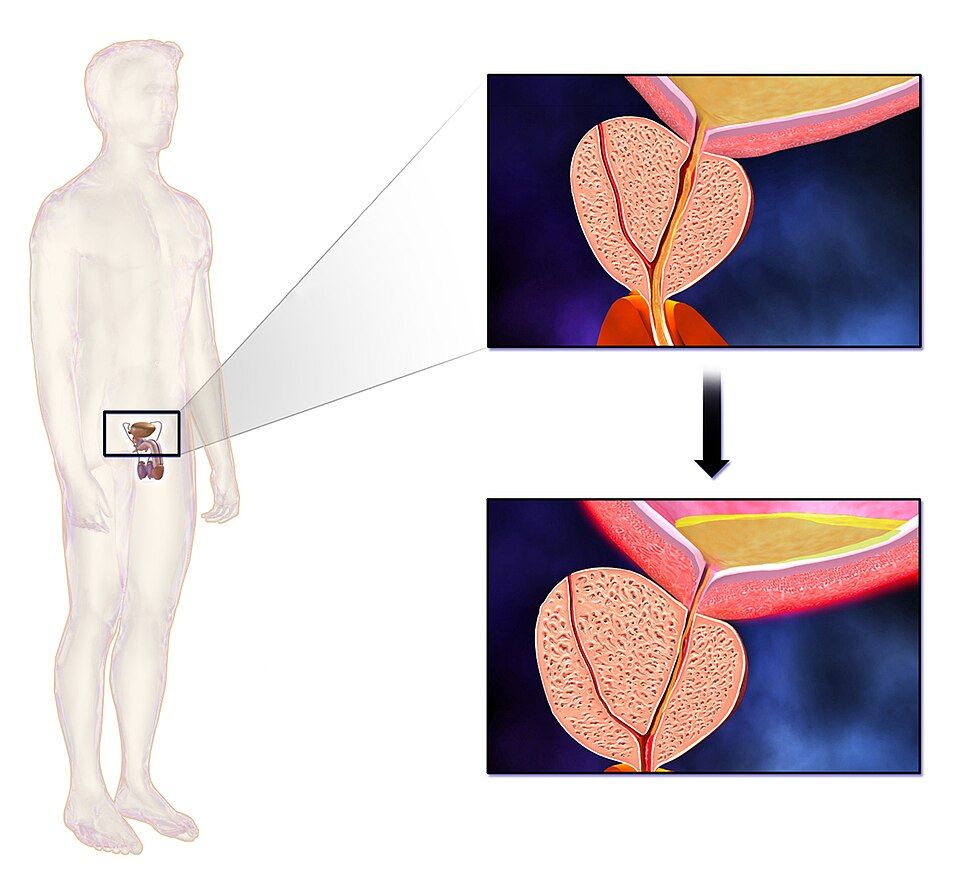
The male reproductive system is built around a simple plumbing problem: the same tube, the urethra, has to carry both urine out of the bladder and semen out during ejaculation. That urethra runs straight through the middle of the prostate, a walnut-sized gland that sits just below the bladder and adds fluid to semen. Blood flow matters too: an erection is really a vascular event, where arteries relax and let blood pool inside the penis. Keep those two ideas in mind, the tube running through the prostate and the blood-flow story, and most male reproductive disorders start to make sense.
Benign prostatic hyperplasia (BPH)
BPH is exactly what the name says: benign (not cancer), hyperplasia (too many cells). With age and lifelong androgen (mostly DHT, dihydrotestosterone) exposure, the prostate slowly enlarges. Because the urethra passes right through the gland, that growth squeezes the tube like a hand gently closing around a straw. Everything downstream flows from that pinch. The stream gets weak and hesitant because it is harder to push urine past the narrowing. The bladder muscle (detrusor) has to work harder, so it thickens and becomes irritable, which drives urinary frequency, urgency, and nocturia (waking at night to void). Over time the bladder cannot fully empty, leaving residual urine, and in the worst case flow stops completely, called acute urinary retention. These lower urinary tract symptoms are often lumped as LUTS.
Prostatitis
Prostatitis is inflammation of that same gland, and it feels very different from BPH. Acute bacterial prostatitis is usually caused by gut or urinary organisms (often E. coli) tracking into the prostate. Because it is an active infection, the man is genuinely sick: fever, chills, malaise, plus pelvic or perineal pain and painful, difficult urination (dysuria). On exam the prostate is boggy, swollen, and exquisitely tender. Chronic prostatitis is the slow-burn version, with lingering pelvic discomfort and recurrent urinary infections but few systemic signs.
| Feature | BPH | Acute prostatitis |
|---|---|---|
| Cause | Age-related growth (androgen-driven) | Bacterial infection |
| Fever / feeling sick | No | Yes, often high |
| Onset | Gradual, over years | Sudden, over hours to days |
| Prostate on exam | Enlarged, smooth, firm | Swollen, boggy, very tender |
| Main problem | Obstructed flow | Infection plus dysuria |
Erectile dysfunction (ED)
Erectile dysfunction is the consistent inability to get or keep an erection firm enough for intercourse. Since an erection depends on arteries dilating and filling the penis with blood, ED is most often a vascular problem. The same processes that clog coronary arteries, atherosclerosis, diabetes, hypertension, and smoking, also damage the small penile vessels and the endothelium that releases nitric oxide, the signal that tells smooth muscle to relax. This is why ED is often called an early warning sign: those small vessels narrow before the big ones cause a heart attack. Nerve damage (as in diabetes or after prostate surgery), low testosterone, certain medications, and psychological factors add to the picture.
Testicular torsion
Testicular torsion is a true surgical emergency. The testicle twists on its spermatic cord, kinking the blood supply exactly like stepping on a garden hose. Without arterial flow the testicle becomes ischemic and, if not fixed quickly, dies. It classically strikes adolescents with sudden, severe one-sided scrotal pain, often with nausea and vomiting. The affected testicle may sit high and horizontal, and the cremasteric reflex (stroking the inner thigh normally lifts the testicle) is typically absent. Time is the whole story: salvage rates are best within about 6 hours, so torsion goes straight to surgery, and often it is not imaged first if the diagnosis is clear.
Explore the interactive figure below to see how each of these disorders maps onto male reproductive anatomy.
- BPH is benign, age-related prostate growth that squeezes the urethra, causing weak stream, frequency, nocturia, and possible retention.
- Prostatitis is inflammation of the prostate; the acute bacterial form brings fever, pelvic pain, and a boggy, tender gland, unlike the painless enlargement of BPH.
- Erectile dysfunction is usually vascular and can be an early warning sign of underlying cardiovascular disease like atherosclerosis or diabetes.
- Testicular torsion is a surgical emergency: sudden severe scrotal pain with an absent cremasteric reflex, best salvaged within about 6 hours.
- Because the urethra runs through the prostate, most prostate disorders show up first as changes in the urinary stream.
Section 4Cancers of the male system

The male reproductive system runs on two organs that quietly do very different jobs. The testes sit outside the body in the scrotum, where the slightly cooler temperature lets them make sperm and pump out testosterone. The prostate is a walnut-sized gland wrapped around the urethra just below the bladder; its job is to add fluid to semen. Because the prostate hugs the urine tube, anything that makes it swell can squeeze that tube and change how a man pees. Keep that anatomy in mind, because it explains almost every symptom in this section.
Prostate cancer: common and often patient
Prostate cancer is the most common non-skin cancer in men, and the risk climbs steeply with age, especially after 65. It is also frequently slow-growing, so many men live for years with disease that never threatens them. The tumor usually starts in the peripheral (outer) zone of the gland, which is why it often causes no early symptoms at all, and why a clinician can sometimes feel a hard, irregular nodule on a digital rectal exam (DRE). Screening pairs the DRE with a blood test for PSA (prostate-specific antigen), a protein the prostate normally leaks in small amounts. When cancer cells disrupt the gland's architecture, more PSA escapes into the blood, so a rising level is a red flag, though not a diagnosis. Because prostate cancer likes to spread to bone, a later sign can be deep, unexplained back or hip pain.
Cancer versus BPH: same gland, different problem
Students mix these up constantly because both make the prostate bigger and both mess with urination. The key difference is where and what kind. Benign prostatic hyperplasia (BPH) is non-cancerous overgrowth of the inner (transitional) zone, the part closest to the urethra, so it squeezes the tube early and loudly. Cancer usually grows in the outer zone, so it stays quiet until it is more advanced.
| Feature | BPH (benign) | Prostate cancer |
|---|---|---|
| Nature | Overgrowth of normal cells | Malignant cells that can spread |
| Usual zone | Inner/transitional (near urethra) | Peripheral/outer |
| Early urinary symptoms | Common and prominent | Often none until advanced |
| DRE finding | Smooth, rubbery, symmetric | Hard, irregular nodule |
| PSA | Mildly elevated | Often higher or rising |
The urinary symptoms of BPH follow directly from the squeezed urethra: a weak stream, hesitancy (waiting for flow to start), dribbling, and nocturia (waking to pee) because the bladder never fully empties. These same symptoms can appear in advanced cancer, so symptoms alone cannot tell the two apart. That is exactly why the exam and PSA matter.
Testicular cancer: the young man's cancer
Testicular cancer flips the age script. It is the most common solid cancer in men roughly 15 to 35 years old, and the biggest risk factor is a history of an undescended testicle (cryptorchidism), even one surgically corrected in childhood. The classic presentation is a painless, firm lump or swelling in one testicle, often found by chance or during a shower. Because it is painless, men easily ignore it, which is dangerous because these tumors can grow quickly. Many arise from germ cells and can raise tumor markers such as AFP, beta-hCG, and LDH, which help with diagnosis and tracking. The reassuring news is that testicular cancer is highly treatable, with excellent cure rates even when it has spread, so early detection changes outcomes dramatically.
When to worry and refer
Refer promptly for evaluation when you see a hard or nodular prostate on exam, a rising or clearly elevated PSA, or new bone pain with a prostate history. For testes, any painless testicular mass, new firmness, or size change warrants urgent workup, usually a scrotal ultrasound, before assuming it is benign. Red flags such as blood in the urine or semen, unexplained weight loss, or difficulty urinating also deserve escalation. Your nursing role is not to diagnose but to notice, document, and route, and to normalize screening so men actually show up.
- Prostate cancer is common, often slow-growing, favors older men, grows in the gland's outer zone, and is screened with PSA plus digital rectal exam.
- BPH is benign inner-zone overgrowth that squeezes the urethra early (weak stream, hesitancy, nocturia); cancer stays outer and quiet until advanced, so exam and PSA distinguish them.
- PSA is a clue, not a diagnosis; infection, BPH, and recent exam can raise it, and a rising level earns a referral.
- Testicular cancer strikes young men (usually 15 to 35), typically a painless firm mass, and is highly curable, so monthly self-exam and early reporting matter.
- Refer promptly for a hard or nodular prostate, rising PSA, new bone pain, or any painless testicular mass.
Section 5Sexually transmitted infections
The reproductive tract has built-in defenses. The vagina keeps an acidic pH thanks to lactobacilli, cervical mucus forms a physical plug, and the mucosal lining of the urethra, cervix, and rectum is coated in immune cells ready to trap invaders. Sexually transmitted infections (STIs) happen when a pathogen — bacterial, viral, or protozoal — is passed through vaginal, anal, or oral contact and gets past those defenses to colonize mucosa or enter the bloodstream. The tricky part for nursing care is that many STIs are silent: the person feels fine while the organism quietly climbs the tract or spreads systemically.
Bacterial STIs: chlamydia and gonorrhea
Chlamydia trachomatis and Neisseria gonorrhoeae are the two most commonly reported bacterial STIs, and they often travel together. Both infect columnar epithelial cells of the cervix and urethra. When symptoms do appear, expect dysuria, mucopurulent discharge, or post-coital bleeding — but roughly 70% of women and many men have no symptoms at all, which is exactly why they spread. That silence is dangerous. If the organism ascends from the cervix into the uterus and fallopian tubes, it triggers pelvic inflammatory disease (PID). The resulting inflammation and scarring of the tubes can block egg transport, causing infertility and raising the risk of a life-threatening ectopic pregnancy. Gonorrhea can also disseminate into blood and joints (disseminated gonococcal infection). Both are treatable with antibiotics — chlamydia with doxycycline, gonorrhea with ceftriaxone — but treatment does not reverse scarring already done.
Syphilis and its stages
Syphilis, caused by the spirochete Treponema pallidum, is the great imitator because it moves through the body in distinct stages, each with different signs as the organism spreads from local tissue to the bloodstream and eventually the nervous system.
| Stage | What you see | Why |
|---|---|---|
| Primary | Single painless chancre at entry site; heals on its own | Local spirochete replication; often missed because it does not hurt |
| Secondary | Diffuse rash (often palms/soles), fever, lymphadenopathy | Organism has spread through blood, provoking a systemic immune response |
| Latent | No symptoms; positive serology | Bacteria persist quietly; still transmissible early on |
| Tertiary | Gummas, cardiovascular damage, neurosyphilis | Years of unchecked tissue and vessel destruction |
Penicillin remains the treatment. The painless, self-healing early lesions are exactly why untreated syphilis marches forward — the patient assumes it is gone.
Viral STIs
Viral STIs differ in a key way: antibiotics do nothing, and several are lifelong because the virus integrates into or hides within host cells.
- Human papillomavirus (HPV) — infects squamous epithelium. Low-risk strains cause genital warts; high-risk strains (16, 18) drive cervical, anal, and oropharyngeal cancers by disrupting tumor-suppressor genes. The HPV vaccine prevents the high-risk strains, and Pap/HPV screening catches precancerous change early.
- Herpes simplex virus (HSV) — causes painful vesicular genital lesions, then retreats into sacral nerve ganglia where it stays for life, reactivating under stress. Antivirals (acyclovir) suppress outbreaks but do not cure.
- HIV — attacks CD4 T-helper cells, progressively crippling immunity until opportunistic infections define AIDS. Modern antiretroviral therapy lets people live long lives and, when viral load is undetectable, prevents transmission.
- Hepatitis B — a bloodborne and sexually transmitted virus that inflames the liver and can cause chronic hepatitis, cirrhosis, and liver cancer. It is vaccine-preventable.
Why screening and prevention matter
Because so many STIs are asymptomatic, screening finds them before complications set in, and treating partners breaks the reinfection loop. Prevention layers together: barrier methods (condoms) reduce transmission, vaccines stop HPV and hepatitis B before exposure, and prompt treatment shortens the window of infectivity. For nurses, patient teaching is nonjudgmental, practical, and centered on the truth that catching an STI early is almost always easier than treating what it becomes.
- Chlamydia and gonorrhea are often silent but can ascend to cause PID, infertility, and ectopic pregnancy; treat the patient and partners.
- Syphilis progresses through primary (painless chancre), secondary (systemic rash), latent, and tertiary (neurosyphilis, cardiovascular) stages; penicillin treats it.
- Viral STIs (HPV, HSV, HIV, hepatitis B) resist antibiotics and are often lifelong; HPV and hepatitis B are vaccine-preventable.
- High-risk HPV drives cervical and other cancers; HIV destroys CD4 cells and immunity.
- Screening, partner treatment, barrier methods, and vaccination prevent the severe long-term complications of untreated STIs.
The chapter in one breath
- Endometriosis is ectopic endometrial tissue that still bleeds each cycle, causing chronic pelvic pain, dysmenorrhea, and infertility from adhesions and scarring.
- PID (usually ascending chlamydia or gonorrhea) inflames the upper tract, and its scarring is a leading cause of ectopic pregnancy and infertility.
- Cervical cancer is nearly always caused by persistent high-risk HPV (types 16 and 18), which is why HPV vaccination and Pap/HPV co-testing screening catch it early.
- Breast cancer risk climbs with lifetime estrogen exposure and BRCA1/BRCA2 mutations, and a new painless hard fixed lump or nipple change warrants mammography.
- BPH is a non-malignant enlargement of the prostate's inner zone that squeezes the urethra, causing hesitancy, weak stream, nocturia, and urinary retention.
- Prostate cancer usually arises in the peripheral zone (so it is felt on DRE and rarely obstructs early), often silent until an elevated PSA or bone metastases appear.
- Testicular cancer is the most common solid tumor in young men aged 15 to 35 and typically presents as a painless firm testicular lump.
- STIs often hide silently: chlamydia and gonorrhea may be asymptomatic yet cause PID and infertility, while untreated syphilis progresses through primary chancre, secondary rash, and tertiary organ damage.
ReferenceWords worth knowing
A quick reference for the terms in this chapter — skim it now, or circle back whenever a word trips you up.
- Amenorrhea
- The absence of menstrual periods, either because they never started by the expected age (primary) or because they stopped for three or more cycles in someone who previously menstruated (secondary), often signaling pregnancy, hormonal imbalance, or excessive stress or exercise.
- Benign prostatic hyperplasia (BPH)
- A non-cancerous enlargement of the prostate gland common in aging men that squeezes the urethra and causes urinary symptoms like weak stream, hesitancy, frequency, and nighttime urination (nocturia).
- BRCA
- The BRCA1 and BRCA2 tumor-suppressor genes whose inherited mutations sharply raise a person's lifetime risk of breast and ovarian (and some prostate) cancers because the damaged gene can no longer help repair DNA.
- Chlamydia
- A very common bacterial sexually transmitted infection caused by Chlamydia trachomatis that is often silent (asymptomatic) yet can quietly scar the reproductive tract and lead to pelvic inflammatory disease and infertility if untreated.
- Dysmenorrhea
- Painful menstrual cramping caused mainly by prostaglandin-driven uterine contractions, ranging from primary (no underlying disease) to secondary (from conditions like endometriosis or fibroids).
- Endometriosis
- A condition in which tissue resembling the uterine lining grows outside the uterus, bleeding cyclically with each period to cause chronic pelvic pain, painful intercourse, and often infertility.
- Fibroid
- A benign (non-cancerous) tumor of uterine smooth muscle, also called a leiomyoma, that can enlarge the uterus and cause heavy menstrual bleeding, pelvic pressure, and pain.
- Gonorrhea
- A bacterial sexually transmitted infection caused by Neisseria gonorrhoeae that inflames the urethra, cervix, or other mucous membranes and, if untreated, can spread to cause pelvic inflammatory disease or disseminated infection.
- HPV (human papillomavirus)
- A very common sexually transmitted virus whose high-risk strains can cause cervical, anal, and other cancers, while low-risk strains cause genital warts.
- HPV vaccine
- A vaccine (such as Gardasil) that protects against the high-risk human papillomavirus strains most likely to cause cervical and other cancers, given ideally before a person becomes sexually active.
- Pap smear
- A screening test that samples cells from the cervix to detect precancerous or cancerous changes early, often paired with HPV testing to catch problems before they become cancer.
- Pelvic inflammatory disease (PID)
- An infection of the upper female reproductive organs (uterus, fallopian tubes, ovaries), usually spreading upward from an untreated STI like chlamydia or gonorrhea, that can scar the tubes and cause infertility or ectopic pregnancy.
- Polycystic ovary syndrome (PCOS)
- A common hormonal disorder marked by irregular or absent ovulation, elevated androgens (causing acne and excess hair growth), and often insulin resistance, frequently seen with multiple small ovarian follicles on ultrasound.
- PSA (prostate-specific antigen)
- A protein made by the prostate and measured in the blood as a screening marker; higher levels can suggest prostate cancer but also rise with benign prostatic hyperplasia, infection, or inflammation.
- Syphilis
- A sexually transmitted infection caused by the bacterium Treponema pallidum that progresses in stages, starting with a painless sore (chancre) and, if untreated, eventually damaging the heart, brain, and other organs.
- Testicular torsion
- A surgical emergency in which the testicle twists on its spermatic cord, cutting off blood flow and causing sudden severe scrotal pain and swelling that must be relieved within hours to save the testicle.
DrillFlashcards for this unit
These are the Week 12 cards — the same deck as the hub, pulled in here so you can review the moment you finish reading. Tap the card to flip it; hit Shuffle to mix them up. To grade yourself and track mastery across the whole deck, open the full flashcards page.
Check yourself
Digestive: GI, Liver & Pancreas
The digestive tract is a single long tube with some powerful accessory organs, and trouble anywhere along it shows up fast. This chapter runs from the reflux and ulcers of the upper GI down through inflammatory bowel disease and obstruction, then into the liver, gallbladder, and pancreas — and the jaundice that signals something is backing up.
By the end of this chapter you can
- Explain GERD and peptic ulcer disease and their dangers.
- Contrast Crohn disease with ulcerative colitis.
- Recognize bowel obstruction and tell an upper from a lower GI bleed.
- Describe cirrhosis and its consequences (portal hypertension, varices, ascites, encephalopathy).
- Explain gallstones, pancreatitis, and the three types of jaundice.
Section 1Upper GI: GERD & peptic ulcers
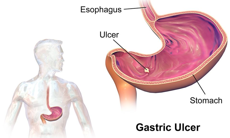
Your stomach makes acid on purpose. Special parietal cells in the stomach lining pump out hydrochloric acid (HCl) to start digesting food and to kill swallowed bacteria. That acid is powerful enough to burn skin, so the body has to protect itself in two ways. First, a muscular ring at the bottom of the esophagus called the lower esophageal sphincter (LES) stays tightly closed to keep acid down in the stomach where it belongs. Second, the stomach and duodenum are coated in a thick layer of mucus and bicarbonate that acts like a chemical raincoat, keeping acid from touching the delicate cells underneath. Trouble in the upper GI tract almost always comes down to one of these two defenses failing.
GERD: when the sphincter leaks
Gastroesophageal reflux disease (GERD) happens when the LES is weak or relaxes at the wrong time, letting stomach acid splash backward into the esophagus. The esophagus has no mucus raincoat, so the acid irritates its lining. That is why the classic symptom is heartburn (pyrosis) - a burning behind the breastbone, often worse after big meals or when lying down (gravity is no longer helping keep acid down). Patients may also describe regurgitation of sour fluid, a chronic cough, or a hoarse voice from acid reaching the throat.
Occasional reflux is normal; GERD is when it happens often enough to cause symptoms or damage. The real long-term worry is what repeated acid exposure does to the esophageal lining. Over years, the normal squamous cells can transform into intestinal-type columnar cells - a change called Barrett esophagus. This is the body adapting to survive the acid, but it is a form of metaplasia that raises the risk of esophageal adenocarcinoma. That is why chronic GERD is monitored, not just medicated.
Peptic ulcer disease: when the raincoat wears through
A peptic ulcer is an open sore that erodes through the protective mucosa into the wall of the stomach or duodenum. The two big culprits both attack the mucus defense. First is Helicobacter pylori, a spiral bacterium that burrows into the mucus layer and triggers inflammation that weakens it. Second is chronic use of NSAIDs (like ibuprofen and aspirin). NSAIDs block prostaglandins - the very chemicals that tell the stomach to make protective mucus and bicarbonate. Take away the prostaglandins and you take away the raincoat, so acid digests the tissue itself. Together, H. pylori and NSAIDs cause the large majority of peptic ulcers.
Gastric vs. duodenal: read the pain
Where the ulcer sits changes the timing of the pain, and this is a favorite exam point. The trick is to think about where the food goes.
| Feature | Gastric ulcer | Duodenal ulcer |
|---|---|---|
| Location | In the stomach lining | In the first part of the small intestine |
| Pain timing | Often worse with food (usually 30 min after eating) | Often relieved by food, then returns 2-3 hours later |
| Classic clue | May avoid eating, can lose weight | Night pain that eating settles; may gain weight |
| Typical cause lean | More often NSAID-related | More often H. pylori-related |
The logic: in a gastric ulcer, food entering the stomach means more acid production right on top of the sore, so eating hurts. In a duodenal ulcer, food buffers the acid and stimulates the stomach to close its outlet briefly, so eating soothes the pain until the meal moves through.
When an ulcer turns dangerous
Ulcers matter because they can go deeper. Two complications are emergencies. Bleeding occurs when the ulcer erodes into a blood vessel. Watch for hematemesis (vomiting blood, sometimes coffee-ground colored from digested blood) and melena (black, tarry, foul stool - blood that has traveled the whole gut). Slow chronic bleeding shows up as iron-deficiency anemia and fatigue. The second is perforation: the ulcer bores all the way through the wall, spilling acid and gut contents into the abdominal cavity. This causes sudden, severe pain and a rigid, board-like abdomen - a sign of peritonitis and a surgical emergency.
Take a look at the interactive figure below to see how a weak sphincter lets acid climb back into the esophagus.
- GERD is acid refluxing past a weak LES; chronic exposure can drive Barrett esophagus and raise esophageal cancer risk.
- Peptic ulcers form when the mucus defense fails - usually from H. pylori or NSAIDs (which block protective prostaglandins).
- Gastric ulcer pain is often worse with food; duodenal ulcer pain is often relieved by food and returns hours later.
- The dangerous complications are bleeding (hematemesis, melena, anemia) and perforation (sudden pain, rigid board-like abdomen, peritonitis).
Section 2Inflammatory bowel disease
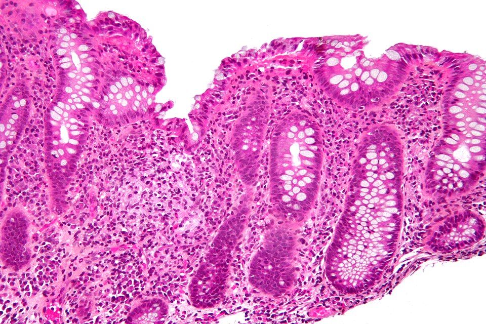
Your gut is a long tube that runs from your mouth to your anus, and its inner lining has a hard job: pull nutrients out of food while keeping trillions of bacteria on the inside of the tube and out of your bloodstream. That lining, the mucosa, sits on top of your immune system like a screen door, letting the good stuff through and holding the line against invaders. Normally your immune cells and your gut bacteria live in a quiet truce. In inflammatory bowel disease (IBD), that truce breaks down. The immune system starts attacking the bowel wall itself, causing chronic, immune-mediated inflammation that flares and settles, flares and settles, for the rest of a person's life.
Two diseases, one family
IBD is an umbrella term for two related conditions: Crohn disease and ulcerative colitis (UC). They share a lot. Both are chronic and relapsing, meaning they cycle between flares (active inflammation, misery) and remission (things calm down). Both involve an immune system that overreacts to the normal gut, driven by a mix of genetics and environmental triggers. And both can produce systemic misery beyond the belly, including fatigue, weight loss, and low-grade fever, because widespread inflammation is a whole-body event. What sets them apart is where they attack and how deep they go.
Crohn disease: patchy and deep
Crohn can strike anywhere from the mouth to the anus, though it loves the terminal ileum (the tail end of the small intestine) and the colon. Its signature is skip lesions: patches of angry, inflamed bowel sitting right next to perfectly healthy tissue, like a moth-eaten pattern. Crucially, the inflammation is transmural, meaning it burrows through all the layers of the bowel wall, not just the surface.
That depth explains Crohn's worst complications. When inflammation tunnels all the way through the wall, it can carve an abnormal connection between two structures, called a fistula (for example, bowel-to-skin or bowel-to-bladder). Chronic transmural damage also scars and narrows the tube, producing strictures that can block the bowel. Because the small intestine does most nutrient absorption, Crohn often causes malabsorption, weight loss, and vitamin deficiencies. The diarrhea is frequently non-bloody, and patients may have right-lower-quadrant pain that mimics appendicitis.
Ulcerative colitis: shallow but continuous
UC is more predictable in its footprint. It is limited to the colon only, it almost always starts in the rectum, and it spreads continuously upward with no skip areas, one unbroken zone of inflammation. It is also shallow: the damage stays in the mucosa (and submucosa), sparing the deeper muscle layers. Because it doesn't go transmural, UC rarely causes fistulas or strictures.
But that inflamed, ulcerated mucosal surface bleeds easily, so the hallmark symptom is bloody diarrhea mixed with mucus, often with crampy lower-abdominal pain and a constant urge to go (tenesmus). Losing blood and fluid stool many times a day can leave patients anemic and dehydrated. A feared emergency is toxic megacolon, where the colon dilates and can perforate. Long-standing UC also raises the risk of colorectal cancer, so patients need ongoing colonoscopy surveillance.
Side by side
| Feature | Crohn disease | Ulcerative colitis |
|---|---|---|
| Location | Mouth to anus (often terminal ileum) | Colon only, starts in rectum |
| Pattern | Skip lesions (patchy) | Continuous |
| Depth | Transmural (all layers) | Mucosa/submucosa only |
| Classic stool | Diarrhea, often non-bloody | Bloody diarrhea with mucus |
| Complications | Fistulas, strictures, malabsorption | Toxic megacolon, higher colon cancer risk |
IBD is not IBS
One last distinction that trips people up because the letters look alike. IBD is structural, real, visible inflammation and tissue damage you can see on scope and biopsy; IBS (irritable bowel syndrome) is functional, meaning the gut hurts and misbehaves but the tissue itself looks normal. Explore the interactive figure below to compare the Crohn and UC patterns side by side.
- IBD is chronic, immune-mediated inflammation of the bowel that relapses in flares and settles into remission; it splits into Crohn disease and ulcerative colitis.
- Crohn can hit anywhere mouth-to-anus with skip lesions and transmural inflammation, so it causes fistulas, strictures, and malabsorption.
- Ulcerative colitis stays in the colon only, spreads continuously from the rectum, and stays mucosal, so its hallmark is bloody diarrhea.
- Deep vs shallow explains the complications: Crohn tunnels through the wall (fistulas), UC ulcerates the surface (bleeding, toxic megacolon, cancer risk).
- Do not confuse IBD (structural inflammation, visible damage) with IBS (a functional disorder with normal-looking tissue).
Section 3Bowel obstruction & GI bleeding
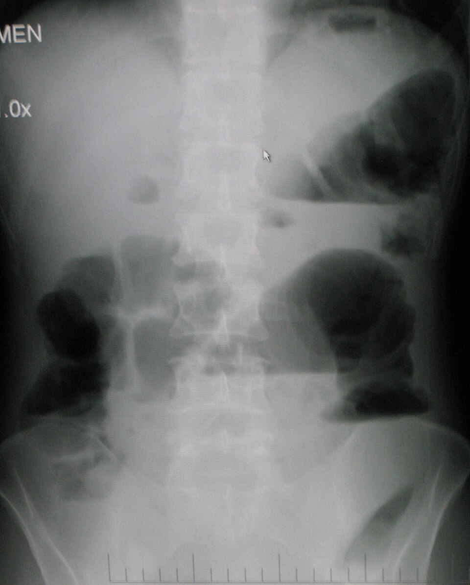
Picture your intestines as a long, muscular hose, roughly 20 feet of it, that never stops working. Normally the gut moves its contents forward with rhythmic squeezing waves called peristalsis, driven by smooth muscle and coordinated by the enteric nervous system (the gut's own built-in wiring). Food, fluid, gas, and eventually stool all travel one direction: down and out. When that forward flow stops, everything backs up behind the blockage, and the trouble begins.
Mechanical obstruction vs paralytic ileus
There are two very different reasons the highway can shut down. In a mechanical obstruction, something physically plugs the lumen. The most common culprits are adhesions (bands of internal scar tissue, usually from previous abdominal surgery), a hernia (a loop of bowel trapped where it pokes through the abdominal wall), and tumors (especially in the colon). The muscle still wants to push, so early on it squeezes harder against the wall it cannot pass.
In a paralytic ileus, nothing is physically blocking the tube, but the muscle simply stops moving. Peristalsis goes quiet. This happens after abdominal surgery (the gut is famously sluggish for a day or two), with peritonitis, with electrolyte problems (especially hypokalemia, since potassium is needed for smooth-muscle contraction), and with opioids, which slow the gut down. The distinction matters enormously: mechanical blockages often need a procedure to relieve them, while an ileus usually needs the underlying cause fixed and the bowel rested.
| Feature | Mechanical obstruction | Paralytic ileus |
|---|---|---|
| Problem | Physical blockage of the lumen | Gut muscle stops contracting |
| Common causes | Adhesions, hernia, tumor | Post-op state, hypokalemia, opioids, peritonitis |
| Bowel sounds (early) | High-pitched, hyperactive, tinkling | Diminished to absent from the start |
| Pain pattern | Crampy, wave-like (colicky) | Steady, diffuse discomfort |
| Usual fix | Often surgical or decompression | Treat the cause, rest the bowel |
The cardinal signs and why they happen
No matter the cause, obstruction produces a classic quartet. Abdominal distension develops because fluid and swallowed air pile up behind the block and the bowel balloons outward. Vomiting follows as the backed-up contents have nowhere to go but up; a low (distal) blockage often produces foul, feculent vomiting, while a high (proximal) small-bowel block causes earlier, more forceful emesis. Obstipation, the complete failure to pass any stool or gas, is a telling sign that nothing is getting through.
Bowel sounds tell a story over time. Early in a mechanical obstruction, listen for high-pitched, hyperactive, tinkling sounds as the muscle strains against the blockage. As hours pass and the muscle fatigues, or as an ileus sets in, those sounds fade to absent. The classic teaching: sounds go high-pitched then silent.
Why obstruction is an emergency
Trapped bowel does not just hurt, it dies. Fluid pours into the distended loop (third-spacing), dropping circulating volume and risking hypovolemic shock plus metabolic imbalances from all that lost fluid and electrolytes. Worse, if the swelling squeezes off the bowel's blood supply, the tissue becomes strangulated, turns ischemic, and can perforate, spilling gut contents into the abdomen and triggering life-threatening peritonitis and sepsis. Fever, rising heart rate, and sudden severe pain signal that a simple blockage has become a surgical emergency.
GI bleeding: upper vs lower
Bleeding anywhere in the digestive tract is graded by where it starts, because the appearance of the blood is a built-in clue. An upper GI bleed comes from above the ligament of Treitz, meaning the esophagus, stomach, or duodenum, and is often caused by peptic ulcers or esophageal varices. It shows up as hematemesis (vomited blood, sometimes coffee-ground in appearance once stomach acid has partly digested it) and melena (black, tarry, sticky stool). The stool is black because hemoglobin is chemically broken down during its long trip through the gut.
A lower GI bleed arises in the jejunum, ileum, or colon, commonly from diverticulosis, angiodysplasia, hemorrhoids, or colorectal cancer. Because the blood exits quickly without time to be digested, it appears as hematochezia: bright red or maroon blood mixed with or coating the stool.
Why a major bleed is an emergency
A significant GI bleed can quietly drain enough blood to cause hypovolemic shock before the source is even found. Watch for tachycardia, falling blood pressure, orthostatic dizziness, pallor, and a dropping hemoglobin and hematocrit. Early vital-sign changes often precede a visible drop in labs, so trending them and securing IV access is a nursing priority.
- Mechanical obstruction (adhesions, hernia, tumor) physically plugs the bowel, while paralytic ileus means the gut muscle simply stops moving.
- The cardinal signs are distension, vomiting, obstipation (no stool or gas), and bowel sounds that go high-pitched then absent.
- Upper GI bleed = hematemesis and melena (black, tarry, digested blood); lower GI bleed = hematochezia (bright red or maroon).
- Obstruction can progress to strangulation, perforation, and peritonitis; a major bleed can cause hypovolemic shock, so both are true emergencies.
- Monitor trending vital signs, look for tachycardia and hypotension, and watch for third-spacing and electrolyte loss.
Section 4The liver: hepatitis & cirrhosis
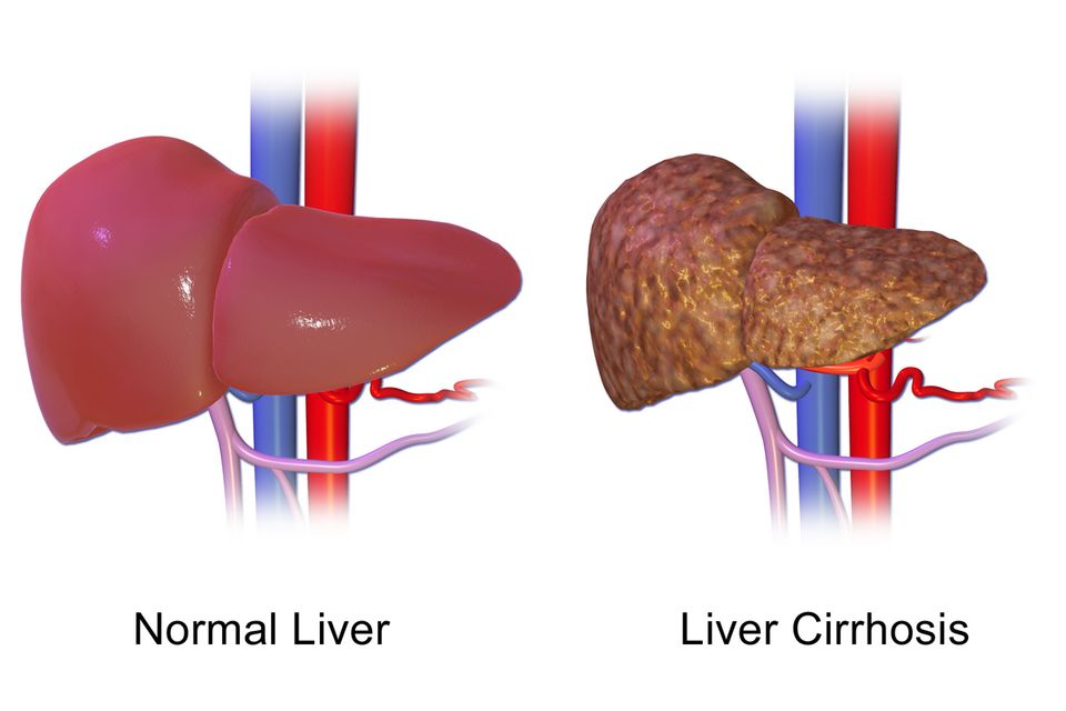
Think of your liver as the body's busiest chemistry lab. It sits under your right rib cage and quietly runs hundreds of jobs at once. Three of those jobs matter most for understanding what goes wrong. First, it is the body's detox center: it filters drugs, alcohol, and metabolic waste out of the blood, and it converts toxic ammonia (a byproduct of protein breakdown) into urea so the kidneys can excrete it. Second, it is a factory: it manufactures most of the body's clotting factors and albumin, the protein that holds fluid inside blood vessels. Third, it processes bilirubin, the yellow-orange pigment left over when old red blood cells are recycled, packaging it so it can leave the body in bile and stool. Hold onto those three jobs, because nearly every symptom of liver disease traces back to one of them failing.
Hepatitis: when the liver gets inflamed
Hepatitis simply means inflammation of the liver. The most common causes are viral, but alcohol and certain drugs (notably acetaminophen in overdose) can do the same damage. The three viral players behave differently, and that difference drives nursing care.
| Feature | Hepatitis A | Hepatitis B & C |
|---|---|---|
| How it spreads | Fecal-oral (contaminated food/water) | Blood & body fluids (needles, sex, birth) |
| Course | Acute, self-limiting; usually full recovery | Often becomes chronic; can smolder for years |
| Long-term risk | Rarely lasting damage | Cirrhosis & liver cancer |
| Prevention | Vaccine; hand hygiene | Hep B vaccine; no Hep C vaccine yet |
During acute hepatitis, patients often feel fatigued and nauseated, and many turn jaundiced (yellow skin and eyes) because the inflamed liver cannot keep up with bilirubin processing. Alcoholic and drug-induced hepatitis follow the same theme: the toxin injures liver cells (hepatocytes) faster than they can heal.
Cirrhosis: scarring that does not undo
When injury to the liver continues long enough, healing itself becomes the problem. Instead of regenerating normal tissue, the liver lays down fibrosis (scar tissue). Cirrhosis is the end-stage result: the once-soft organ becomes hard, nodular, and shrunken. Scar tissue does not do liver work, and it does not let blood flow through normally. Both of those facts explain the cascade of complications that follow.
Portal hypertension and its downstream chaos
Normally, blood from the intestines drains through the portal vein into the liver. Cirrhotic scar tissue squeezes those channels, so pressure backs up, a state called portal hypertension. High pressure in this system forces blood to find detours through smaller veins that were never built for the load. Where those veins swell, we get problems:
- Esophageal varices are ballooned, fragile veins at the base of the esophagus. They can rupture and cause sudden, massive, life-threatening bleeding.
- Ascites is fluid pooling in the abdomen. Two forces drive it: the high portal pressure pushes fluid out of vessels, and the failing liver makes too little albumin to hold fluid in. Low albumin means low oncotic pressure, so water leaks into the belly.
Why liver failure causes bleeding and jaundice
Remember the factory job. A failing liver stops making enough clotting factors, so patients bruise and bleed easily, and lab tests show a prolonged PT/INR. Pair that with fragile varices, and bleeding becomes a true emergency. Remember the bilirubin job, too: as the liver falters, bilirubin builds up in the blood and stains the skin and sclera yellow, the classic jaundice.
Hepatic encephalopathy: the toxin job fails
When the detox center goes down, ammonia from protein digestion is no longer converted to urea. It accumulates and crosses into the brain, causing hepatic encephalopathy: confusion, personality changes, and a flapping tremor of the hands called asterixis. Nurses often watch for early confusion as a warning sign, and the drug lactulose is given to trap ammonia in the gut and pull it out in stool.
- The liver detoxifies (converting ammonia to urea), builds clotting factors and albumin, and processes bilirubin — remember these jobs and the symptoms make sense.
- Hepatitis is liver inflammation; Hep A spreads fecal-oral and resolves, while Hep B and C spread through blood, often turn chronic, and lead to cirrhosis.
- Cirrhosis is irreversible scarring; the scar tissue blocks blood flow and causes portal hypertension, driving esophageal varices and ascites.
- Liver failure causes bleeding (too few clotting factors, high PT/INR) and jaundice (bilirubin buildup).
- Rising ammonia causes hepatic encephalopathy (confusion, asterixis), typically treated with lactulose.
Section 5Gallbladder & pancreas
Your gallbladder and pancreas are two quiet helpers that sit just under your liver and stomach. Normally, the gallbladder is a small green sac that stores bile (a soap-like fluid the liver makes to break up fat). When you eat a greasy meal, a hormone called cholecystokinin (CCK) tells the gallbladder to squeeze, sending bile down the common bile duct into the small intestine. The pancreas, meanwhile, pours powerful digestive enzymes down its own pancreatic duct, which usually joins the bile duct right before both empty into the duodenum. Those enzymes are supposed to stay switched off until they reach the gut. Trouble starts when a stone plugs the plumbing, or when those enzymes turn on too early.
Gallstones and cholecystitis
When bile has too much cholesterol or sits too long, it can harden into gallstones (cholelithiasis). Many stones are silent. But if a stone lodges in the gallbladder's neck or the cystic duct, the gallbladder keeps squeezing against a locked door, becomes trapped, inflamed, and infected. This is acute cholecystitis. Because fatty food is the trigger for gallbladder contraction, the classic story is right-upper-quadrant (RUQ) pain 30 minutes to a few hours after a fatty meal, often radiating to the right shoulder or scapula, plus nausea and vomiting. The pain can be severe and cramping (biliary colic).
A bedside clue is Murphy's sign. You place your hand under the right rib margin and ask the patient to take a deep breath. As the inflamed gallbladder descends onto your fingers, the patient catches their breath and stops from pain. A positive Murphy's sign points strongly to cholecystitis. Think of the classic risk profile as the 4 F's: female, forty, fertile, and overweight.
When a stone blocks the ducts
A stone that escapes the gallbladder can travel and wedge deeper. If it blocks the common bile duct (choledocholithiasis), bile backs up. Bilirubin spills into the blood, so the skin and eyes turn yellow (jaundice), the urine darkens, and stools turn clay-pale because bile pigment no longer reaches the gut. Trapped, stagnant bile invites infection of the whole biliary tree (cholangitis).
Here is the dangerous overlap: because the bile duct and pancreatic duct usually share a final channel, one stone can dam both. If it blocks where the pancreatic duct drains, enzymes cannot escape, pressure rises, and the pancreas is set up to attack itself.
Acute pancreatitis: the organ that digests itself
Acute pancreatitis happens when those digestive enzymes activate inside the pancreas instead of the intestine. Trypsin switches on prematurely and triggers the rest, so the gland begins to autodigest its own tissue (autodigestion). The result is intense inflammation, swelling, and sometimes bleeding.
The hallmark is severe, constant epigastric pain that bores straight through to the back. Patients often feel worse lying flat and better leaning forward. Nausea and vomiting are common, and the belly may be tender and distended. On labs, lipase rises sharply (usually more than three times the upper normal limit) and stays elevated longer than amylase, which makes lipase the more specific marker.
What causes it
By far the two leading causes are gallstones and alcohol (a helpful memory hook is "I GET SMASHED," where the first culprits are Idiopathic, Gallstones, and Ethanol). Gallstones block the shared duct; alcohol is directly toxic to pancreatic cells and thickens secretions.
| Feature | Cholecystitis | Acute pancreatitis |
|---|---|---|
| Core problem | Blocked, inflamed gallbladder | Pancreas digesting itself |
| Pain location | Right upper quadrant, to right shoulder | Epigastric, boring to the back |
| Trigger | Fatty meal | Alcohol or a gallstone |
| Key exam/lab | Positive Murphy's sign | High lipase |
- Gallstones can block bile flow; if the neck or cystic duct is trapped, the gallbladder inflames (cholecystitis) with RUQ pain after fatty meals and a positive Murphy's sign.
- A stone in the common bile duct causes jaundice, dark urine, and pale stools; because the ducts share a channel, one stone can block the pancreatic duct too.
- Acute pancreatitis is autodigestion: enzymes activate early and the pancreas attacks itself, causing severe epigastric pain that bores to the back.
- Lipase is the key lab, usually more than triple the upper limit, and is more specific than amylase.
- The top two causes of pancreatitis are gallstones and alcohol.
Section 6Jaundice & bilirubin
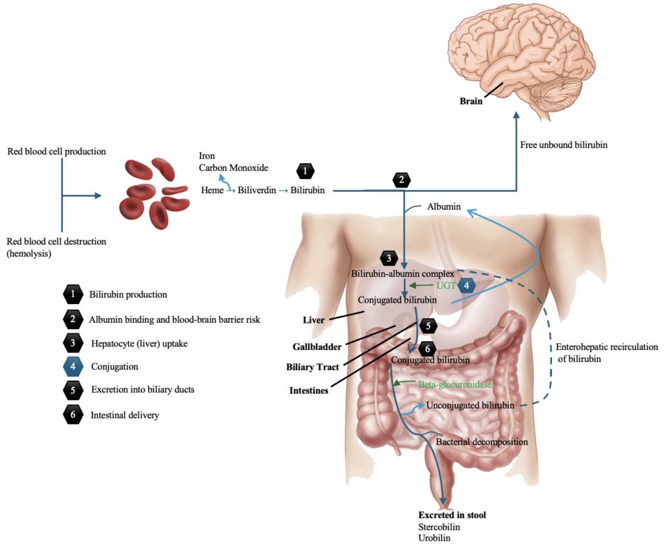
Every day, your body quietly retires about 1% of its red blood cells, mostly because they have worn out after roughly 120 days in circulation. When a red cell breaks down, the hemoglobin inside it is dismantled. The heme portion becomes a yellow-orange pigment called bilirubin. This first form is unconjugated (indirect) bilirubin, and it has a problem: it is fat-loving and not water-soluble, so it cannot simply be flushed out in urine. Instead it rides through the blood attached to albumin and is delivered to the liver.
How the liver handles bilirubin
The liver is the cleanup station. Liver cells (hepatocytes) take up unconjugated bilirubin and attach sugar molecules to it using an enzyme called UDP-glucuronyl transferase. This step is conjugation, and it converts the pigment into conjugated (direct) bilirubin, which is now water-soluble. The liver secretes conjugated bilirubin into bile, which drains through the bile ducts into the small intestine. There, gut bacteria change it into urobilinogen and then stercobilin, the pigment that gives stool its normal brown color. A small amount is reabsorbed and leaves in urine as urobilin, giving urine its yellow tint.
Jaundice (also called icterus) is the yellowing of skin, mucous membranes, and the sclera of the eyes. It appears when serum bilirubin rises above roughly 2 to 3 mg/dL. Because bilirubin loves fatty tissue and elastin-rich tissue, the sclera and skin show it first. Jaundice is never a disease by itself; it is a signal that one of the steps above has broken down. To find the break, nurses group jaundice by where in the pathway the problem sits.
The three types of jaundice
Prehepatic (hemolytic) jaundice happens before the liver, when red cells are destroyed faster than the liver can keep up. Causes include hemolytic anemias, sickle cell crisis, and transfusion reactions. The liver is healthy but overwhelmed, so unconjugated bilirubin piles up in the blood.
Hepatic (intrahepatic) jaundice comes from the liver itself being sick. In hepatitis, cirrhosis, or drug injury, damaged hepatocytes cannot conjugate and excrete bilirubin efficiently. Both unconjugated and conjugated forms often rise.
Posthepatic (obstructive) jaundice happens after the liver, when bile cannot drain. A gallstone in the common bile duct or a pancreatic head tumor blocks the flow. Conjugated bilirubin, already water-soluble, backs up and spills into the blood.
| Feature | Prehepatic (hemolytic) | Posthepatic (obstructive) |
|---|---|---|
| Problem | Too many red cells breaking down | Blocked bile drainage |
| Main bilirubin type | Unconjugated (indirect) | Conjugated (direct) |
| Urine color | Usually normal | Dark (tea-colored) |
| Stool color | Normal or dark | Pale/clay-colored |
| Itching (pruritus) | Uncommon | Common |
Why urine darkens and stool turns pale
This clue is one of the most useful at the bedside, and it makes sense once you follow the plumbing. In obstruction, conjugated bilirubin cannot reach the intestine, so no stercobilin is made and stool loses its brown pigment, turning pale or clay-colored. Meanwhile the water-soluble conjugated bilirubin backs up into the blood, and because the kidneys can filter this water-soluble form, it appears in urine, turning it dark, tea-colored. Bile salts also build up under the skin, often causing intense pruritus (itching).
Newborn jaundice in one line
Physiologic jaundice of the newborn is common and usually harmless: a baby's immature liver has not yet made enough UDP-glucuronyl transferase to conjugate bilirubin quickly, so unconjugated bilirubin rises for a few days and then falls on its own. It needs no treatment unless the level crosses the hour-specific threshold for the infant's age and risk factors, at which point phototherapy is started.
An interactive figure below lets you explore how rising bilirubin turns tissues yellow.
- Bilirubin is a pigment from broken-down red cells; the liver conjugates it (making it water-soluble) and excretes it in bile.
- Jaundice appears when serum bilirubin exceeds about 2 to 3 mg/dL and is classified as prehepatic (hemolysis, unconjugated), hepatic (liver disease, both), or posthepatic (obstruction, conjugated).
- Obstruction causes dark urine (water-soluble conjugated bilirubin spills into urine) and pale stool (no stercobilin reaches the gut), often with pruritus.
- Hemolytic jaundice usually leaves urine and stool color normal because the excess bilirubin is unconjugated and cannot enter urine.
- Newborn physiologic jaundice reflects an immature liver enzyme system and resolves on its own; phototherapy is added only when the bilirubin crosses the hour-specific threshold, and untreated extremes risk kernicterus.
The chapter in one breath
- GERD happens when a weak lower esophageal sphincter lets acidic stomach contents wash back up, burning the esophageal lining and causing heartburn, regurgitation, and (over time) Barrett esophagus from chronic acid injury.
- Peptic ulcers are breaks in the stomach or duodenal wall, usually from H. pylori infection or NSAIDs stripping the protective mucus barrier, so acid digests the tissue itself; watch for coffee-ground emesis or melena signaling a bleed.
- Crohn disease causes patchy, full-thickness (transmural) inflammation anywhere mouth-to-anus with skip lesions and fistulas, while ulcerative colitis is continuous, mucosa-only inflammation limited to the colon with bloody diarrhea.
- A bowel obstruction blocks intestinal flow, backing up gas and fluid to cause distension, vomiting, and often high-pitched then absent bowel sounds; strangulation cuts off blood supply and threatens ischemic necrosis.
- A GI bleed from an upper source shows as black tarry melena or hematemesis, while a lower source shows bright red hematochezia; either can drop hemoglobin and drive the patient toward hypovolemic shock.
- Hepatitis is liver inflammation (viral A-E, alcohol, or toxins) that impairs the liver's jobs, and repeated injury drives scarring into cirrhosis, where fibrosis raises portal hypertension and causes ascites, esophageal varices, and bleeding from lost clotting factors.
- Gallstones form when bile is oversaturated with cholesterol; a stone lodged in the cystic duct triggers cholecystitis with RUQ pain worsening after fatty meals, and a stone blocking the shared channel at the ampulla obstructs pancreatic drainage, so enzymes back up under pressure inside the gland and activate prematurely.
- Pancreatitis is auto-digestion of the pancreas by its own activated enzymes (usually from gallstones or alcohol), producing severe epigastric pain radiating to the back with sharply elevated lipase and amylase.
- Jaundice is yellow skin and sclera from bilirubin buildup, and its type points to the problem: unconjugated from excess hemolysis or an immature/failing liver, versus conjugated from blocked bile flow that also gives dark urine and clay-colored stools.
ReferenceWords worth knowing
A quick reference for the terms in this chapter — skim it now, or circle back whenever a word trips you up.
- Ascites
- The buildup of protein-rich fluid inside the peritoneal cavity (the belly), usually driven by portal hypertension and low blood albumin, which makes the abdomen swell and feel tight.
- Barrett esophagus
- A change in the lower esophageal lining from normal squamous cells to intestinal-type columnar cells (metaplasia) after years of acid reflux, which raises the risk of esophageal cancer.
- Bilirubin
- The yellow pigment made when the body breaks down old red blood cells; when it builds up in the blood it causes jaundice and turns the skin and whites of the eyes yellow.
- Cholecystitis
- Inflammation of the gallbladder, usually because a gallstone blocks the cystic duct, causing right-upper-quadrant pain, fever, and often a positive Murphy sign.
- Cirrhosis
- Permanent scarring (fibrosis) of the liver that replaces working tissue with hard nodules, so the liver slowly loses its ability to filter blood, make proteins, and process toxins.
- Crohn disease
- A chronic inflammatory bowel disease that can inflame any part of the GI tract from mouth to anus in patchy skip lesions, with inflammation going through the full thickness of the bowel wall (transmural).
- GERD
- Gastroesophageal reflux disease, the frequent backflow of stomach acid into the esophagus because the lower esophageal sphincter is weak or relaxes too easily, causing heartburn and regurgitation.
- Hematochezia
- The passage of bright red or maroon blood through the rectum, usually signaling bleeding low in the GI tract (colon, rectum, or anus) or a very rapid upper GI bleed.
- Hepatic encephalopathy
- A decline in brain function, ranging from confusion to coma, that happens when a failing liver can no longer clear ammonia and other toxins from the blood so they reach the brain.
- H. pylori
- Helicobacter pylori, a spiral-shaped bacterium that burrows into the stomach's protective mucus layer and is the leading cause of peptic ulcers and chronic gastritis.
- Ileus
- A temporary loss of the normal muscular wave motion (peristalsis) of the intestine, often after abdominal surgery, so contents stop moving even though nothing is physically blocking them.
- Melena
- Black, tarry, foul-smelling stool that forms when blood from an upper GI bleed is digested on its way through the gut, turning the hemoglobin dark.
- Pancreatitis
- Inflammation of the pancreas that occurs when its digestive enzymes activate too early and begin to digest the organ itself, usually triggered by gallstones or alcohol and causing severe epigastric pain that radiates to the back.
- Peptic ulcer
- An open sore in the lining of the stomach or duodenum where acid and pepsin have eroded through the protective mucosa, most often caused by H. pylori or NSAID use.
- Portal hypertension
- Abnormally high blood pressure in the portal venous system feeding the liver, usually from cirrhosis backing up blood flow, which drives ascites, esophageal varices, and an enlarged spleen.
- Ulcerative colitis
- A chronic inflammatory bowel disease limited to the colon and rectum that inflames only the innermost lining (mucosa) in one continuous stretch, causing bloody diarrhea and urgency.
DrillFlashcards for this unit
These are the Week 13 cards — the same deck as the hub, pulled in here so you can review the moment you finish reading. Tap the card to flip it; hit Shuffle to mix them up. To grade yourself and track mastery across the whole deck, open the full flashcards page.
Check yourself
Musculoskeletal & Integumentary
This final chapter covers the frame and the wrapping — the bones and joints that hold us up and the skin that keeps the world out. It runs from fractures and their dangerous complications through the common joint diseases, then across the skin’s own disorders, infections, and the whole-body emergency of a serious burn.
By the end of this chapter you can
- Describe fracture types and the stages of bone healing.
- Recognize compartment syndrome and fat embolism as emergencies.
- Tell osteoarthritis from rheumatoid arthritis, and recognize gout and osteoporosis.
- Stage pressure injuries and recognize melanoma (ABCDE).
- Assess burn depth and size and name the priorities.
Section 1Fractures & bone healing
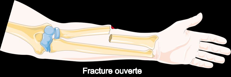
Bone looks solid and permanent, but it is actually living tissue that constantly rebuilds itself. A tough outer shell of compact (cortical) bone surrounds a spongy inner layer, and the whole thing is wrapped in a thin, blood-rich, nerve-rich membrane called the periosteum. Inside, bone cells work in a lifelong partnership: osteoblasts lay down new matrix, and osteoclasts break old matrix down. Because bone is loaded with blood vessels and nerves, a break bleeds and hurts a lot. A fracture is simply a break in the continuity of a bone, and it happens when the force applied is greater than the bone can absorb, whether from a fall, a crash, repetitive stress, or a bone already weakened by disease.
Types of fractures
Naming a fracture describes what actually happened to the bone, which guides how it is treated. The first question is always whether the skin is broken.
| Feature | Closed (simple) | Open (compound) |
|---|---|---|
| Skin | Intact over the break | Bone pierces skin or wound reaches the bone |
| Infection risk | Lower | High, an orthopedic emergency |
| Priority | Alignment and immobilization | Prevent infection (osteomyelitis), then repair |
Beyond that, we describe the break pattern. A complete fracture goes all the way through, splitting the bone into pieces. An incomplete fracture only cracks part way, and the classic example is a greenstick fracture, where a soft child's bone bends and splinters on one side like a young twig. Other common patterns include comminuted (the bone shatters into three or more fragments, often from high-energy trauma), transverse (a straight break across the shaft), oblique (an angled line), spiral (a twisting force, sometimes a red flag for abuse), compression (bone is crushed, typical in osteoporotic vertebrae), and pathologic (a break through bone already weakened by tumor or disease). A stress fracture is a tiny crack from repeated overuse.
How bone heals
Bone is one of the few tissues that can heal without a scar, rebuilding itself back to living bone. It moves through overlapping stages, usually taking weeks to months.
- Hematoma formation (first hours to days): torn vessels bleed, and a clot forms at the break. This fracture hematoma seals the ends and delivers inflammatory cells that clean up debris. This is why the area is swollen, warm, and painful early on.
- Soft callus (roughly days to 2-3 weeks): fibroblasts and chondroblasts build a soft bridge of fibrous tissue and cartilage between the fragments. It is fragile, which is exactly why the bone must stay still now.
- Hard callus (about 3 weeks to several months): osteoblasts replace the soft callus with woven bone, forming a bumpy but sturdy bony callus that unites the pieces.
- Remodeling (months to years): osteoclasts and osteoblasts reshape the callus along lines of mechanical stress, restoring the bone's original strength and contour.
Signs of a fracture, and why we align and immobilize
Suspect a fracture with pain that worsens with movement, swelling, bruising (ecchymosis), obvious deformity or shortening, loss of function, and sometimes crepitus, a grating sound or feel as bone ends rub. Alignment (reduction) puts the fragments back into their normal position so the callus bridges correctly and the limb heals straight. Immobilization with a cast, splint, or fixation holds that alignment so healing can proceed and, just as importantly, protects nearby nerves and vessels from being torn by sharp bone ends.
Why osteoporosis raises fracture risk
In a healthy adult, bone breakdown and rebuilding stay balanced. In osteoporosis, osteoclast activity outpaces osteoblast activity, so bone is resorbed faster than it is replaced. The result is low bone mass and deteriorated microarchitecture, leaving bone porous and brittle. It is often called a silent disease because there are no symptoms until a bone breaks. Falling estrogen after menopause is a major driver, since estrogen normally restrains osteoclasts. That is why postmenopausal women often suffer fragility fractures of the hip, wrist, and spine from forces that would never break healthy bone, sometimes just a minor bump or even a hard cough.
The interactive figure lets you compare fracture patterns and walk through the healing stages, so explore it below.
- A fracture is a break in bone continuity; describe it by skin status (open vs closed) and pattern (complete, greenstick, comminuted, and others) because those details guide treatment.
- Healing proceeds through hematoma, soft callus, hard callus, then remodeling, and the soft-callus stage is fragile, which is why immobilization matters.
- Classic signs include pain, swelling, deformity, loss of function, and crepitus; always check neurovascular status (the 5 P's) distal to the injury.
- Reduction restores alignment and immobilization holds it, allowing straight healing and protecting nearby nerves and vessels.
- Osteoporosis tips the balance toward resorption, creating porous, brittle bone and fragility fractures, especially in postmenopausal women.
Section 2Fracture complications
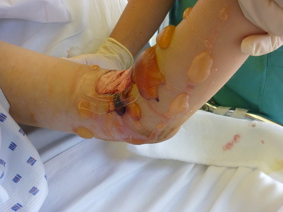
When a bone breaks, the body kicks off a beautifully organized repair project. Blood pools at the fracture site and clots into a hematoma, inflammatory cells clean up debris, a soft cartilage callus bridges the gap, and over weeks that callus is replaced by hard bone and finally remodeled back to shape. For this to work, the tissue around the break needs steady blood flow, oxygen, and room to swell. Most fracture complications happen when one of those requirements is violated: pressure builds where it cannot escape, clots or fat droplets travel where they should not, or the healing timeline stalls. Explore the interactive figure below as you read.
Compartment syndrome: pressure with nowhere to go
Muscles are packed into compartments wrapped by tough, inelastic fascia. After a fracture (classically the tibia or forearm), bleeding and swelling raise the pressure inside that closed box. Because the fascia will not stretch, rising pressure first collapses the thin-walled veins, so blood backs up and swelling worsens, then it squeezes capillaries until tissue no longer gets oxygen. This is acute compartment syndrome, and it is a surgical emergency: without prompt fasciotomy to open the compartment, muscle and nerve die within hours, sometimes costing the limb.
Bedside recognition rests on the 6 P's. The earliest and most reliable is pain out of proportion to the injury, especially pain on passive stretch of the fingers or toes. The others are paresthesia (pins-and-needles from nerve ischemia), pallor, poikilothermia (the limb drifts toward room temperature and feels cool), paralysis, and pulselessness. On palpation the compartment also feels tight and tense - a useful extra finding, but it is not one of the six P's. A crucial teaching point: pulselessness is a late sign. Waiting for the pulse to disappear means waiting until the muscle is already dying.
Fat embolism syndrome: droplets on the move
Long bones such as the femur and tibia, and the flat, irregular bones of the pelvis, all contain fatty yellow marrow. When they fracture, marrow fat can enter torn venous channels, travel to the lungs, and trigger an inflammatory cascade. Classic fat embolism syndrome (FES) shows up 12 to 72 hours after injury as a triad: respiratory distress (hypoxemia, tachypnea, dyspnea from fat lodging in pulmonary capillaries), neurologic changes (confusion, restlessness, decreased level of consciousness), and a petechial rash across the chest, axillae, and conjunctivae. Those petechiae are fairly specific, so watch for them. Care is largely supportive with oxygen; the best medicine is prevention through early fracture stabilization.
Clots from lying still: DVT and PE
Immobility after a fracture checks every box of Virchow's triad: venous stasis (no calf-muscle pumping), hypercoagulability (the injury itself is prothrombotic), and endothelial injury. The result is deep vein thrombosis (DVT), usually in the calf, with unilateral swelling, warmth, and tenderness. The feared sequel is a piece breaking off and lodging in the lung, a pulmonary embolism (PE), presenting as sudden dyspnea, pleuritic chest pain, tachycardia, and hypoxia. Prevention is nursing gold: early mobilization, sequential compression devices, and prophylactic anticoagulation.
| Feature | Fat embolism | Pulmonary embolism (from DVT) |
|---|---|---|
| Usual timing | 12 to 72 hours post-fracture | Days to weeks (immobility) |
| Embolic material | Marrow fat droplets | Blood clot |
| Telltale sign | Petechiae + confusion | Sudden pleuritic chest pain, leg DVT |
| Fracture link | Long-bone / pelvic | Any, from immobility |
When healing stalls: delayed union, nonunion, and infection
Healing needs blood supply, stability, and apposed bone ends. Delayed union is healing that is slower than expected but still progressing. Nonunion is a fracture that stops healing altogether, often from poor blood flow, too much motion, a wide gap, or infection, and typically requires surgery or bone grafting. Malunion heals in a crooked position. Osteomyelitis, a bone infection, is a special threat in open fractures where skin is broken and bacteria reach bone directly; it presents with localized pain, warmth, drainage, and fever, and is notoriously stubborn to clear.
- Compartment syndrome is a surgical emergency: pain out of proportion is the earliest sign and pulselessness is late, treat with fasciotomy, and never ice or elevate above the heart.
- The 6 P's: pain, paresthesia, pallor, poikilothermia (coolness), paralysis, pulselessness - with a tight, tense compartment as an extra palpation finding.
- Fat embolism follows long-bone or pelvic fractures at 12 to 72 hours with respiratory distress, neuro changes, and petechiae; prevent it with early stabilization.
- Immobility drives DVT and PE via Virchow's triad, prevent with early mobility, SCDs, and anticoagulation.
- Nonunion, malunion, and osteomyelitis stall recovery; frequent neurovascular checks are the nursing priority for catching complications early.
Section 3Musculoskeletal disorders
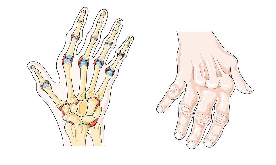
Bone can feel like the most lifeless part of us, but it is actually living, changing tissue. Throughout your whole life, two crews are at work: osteoblasts lay down new bone, and osteoclasts chew old bone away. This constant teardown-and-rebuild is called remodeling, and when the two crews stay balanced, bone stays strong. Joints, meanwhile, glide smoothly because the ends of bones are capped with slick articular cartilage and cushioned by synovial fluid. When that balance or that cushioning breaks down, you get the disorders below. Explore the interactive figure below to see each one.
Osteoporosis: quietly losing bone
In osteoporosis, the osteoclasts outpace the osteoblasts, so more bone is removed than replaced. The bone that remains is normal in chemistry but there is simply less of it - lower bone mineral density (BMD) and a thinner, more porous internal scaffold. The result is fragility: bones that snap under loads a healthy skeleton would shrug off. This is why a minor fall, or even a hard cough, can cause a fragility fracture, classically at the hip, wrist, and vertebrae. Vertebral compression fractures explain the height loss and stooped upper-back curve (kyphosis) often seen in older adults.
The scary part is that it is usually silent until something breaks - patients feel nothing while density slips away. Risk climbs with age, being female (estrogen normally restrains osteoclasts, so density often drops sharply after menopause), small body frame, smoking, low calcium and vitamin D, and long-term corticosteroid use. Screening is done with a DEXA scan, reported as a T-score; a value of -2.5 or lower usually defines osteoporosis.
Osteoarthritis vs rheumatoid arthritis
Both are called "arthritis," but they are completely different problems. Osteoarthritis (OA) is a mechanical, wear-and-tear process: over years, the articular cartilage cushioning a joint frays and thins until bone rubs on bone. It targets the weight-bearing joints - knees, hips, spine - plus the small joints at the fingertips. The pain is use-related: worse after activity and at the end of the day, better with rest. Because it is local mechanical damage, there are no systemic signs - no fever, no fatigue. Any morning stiffness is brief, usually under 30 minutes.
Rheumatoid arthritis (RA) is an autoimmune disease. The immune system attacks the synovium (the joint lining), inflaming it into a destructive tissue called pannus that erodes cartilage and bone from within. RA is symmetric (both hands, both wrists) and favors the small joints of the hands and feet. Its hallmark is morning stiffness lasting longer than an hour. Because it is a body-wide immune process, it brings systemic signs: fatigue, low-grade fever, and elevated inflammatory markers (ESR, CRP). Over time it can deform joints into patterns like ulnar deviation.
| Feature | Osteoarthritis | Rheumatoid arthritis |
|---|---|---|
| Cause | Wear-and-tear cartilage loss | Autoimmune synovial attack |
| Joints | Weight-bearing, often asymmetric | Small joints, symmetric |
| Pain pattern | Worse with use, better with rest | Stiff after rest, eases with movement |
| Morning stiffness | Usually < 30 min | Usually > 1 hour |
| Systemic signs | None | Fatigue, fever, high ESR/CRP |
Gout: crystals in the joint
Gout comes from too much uric acid, a waste product of purine breakdown. When blood uric acid stays high (hyperuricemia), needle-shaped monosodium urate crystals precipitate inside a joint. The immune system treats these crystals as invaders and mounts a fierce inflammatory response. The result is a sudden, severe attack - often overnight - with a joint that is red, hot, swollen, and so tender that even a bedsheet hurts. The classic first target is the big toe (the first metatarsophalangeal joint), a presentation called podagra. Attacks are often triggered by red meat, shellfish, alcohol, or dehydration.
Telling them apart at the bedside
A few quick questions usually sort these out. Ask about tempo: gout explodes over hours; OA and RA build over years. Ask about pattern: OA hits big weight-bearing joints and worsens with use; RA hits small joints symmetrically with long morning stiffness; gout usually strikes one joint, classically the toe. Ask about the whole body: fatigue and fever point toward RA (or an acute gout flare), while OA stays purely local. And remember osteoporosis lives in a different lane entirely - it is painless until a fragile bone fractures.
- Osteoporosis is silent low bone density (osteoclasts > osteoblasts) leading to fragility fractures of the hip, wrist, and spine; diagnosed by DEXA T-score.
- Osteoarthritis is mechanical cartilage wear in weight-bearing joints, worse with use, with brief stiffness and no systemic signs.
- Rheumatoid arthritis is autoimmune synovitis: symmetric small joints, morning stiffness > 1 hour, and systemic features (fatigue, fever, high ESR/CRP).
- Gout is urate crystal deposition causing a sudden, severe, single hot joint - classically the big toe (podagra).
- At the bedside, sort by tempo, pattern, and systemic signs: hours vs years, one joint vs symmetric, local vs whole-body.
Section 4Skin conditions
Your skin is the largest organ you have, and its main job is to be a barrier - a smart, self-repairing wall between your insides and the outside world. It keeps water in, keeps microbes out, senses touch and temperature, and helps regulate body heat. When you understand that barrier job, most skin diseases start to make sense: they are what happens when the wall gets inflamed, overbuilt, broken down, or invaded.
Skin has three layers. The epidermis is the thin, tough outer layer that makes the waterproof keratin shell and constantly sheds and regrows. Beneath it, the dermis holds collagen, blood vessels, nerves, hair follicles, and sweat glands. Under that sits the subcutaneous (hypodermis) layer of fat and connective tissue for insulation and cushioning. Damage that stays in the epidermis usually heals cleanly; damage that reaches the dermis or deeper is where scarring and serious injury begin.
Dermatitis (eczema) and psoriasis
Dermatitis, often called eczema in its atopic form, is inflammation of the epidermis. The skin barrier is leaky, so it loses water and lets irritants and allergens in. The immune system overreacts, and the result is red, itchy, sometimes weepy or crusted skin. Because it is inflammatory and often allergic, it tends to flare with triggers - soaps, stress, dry air - and improves with moisturizers and topical steroids that calm the inflammation.
Psoriasis looks different because the mechanism is different. Here the problem is rapid epidermal turnover: skin cells that normally take about a month to rise and shed do so in just days, driven by an overactive immune (T-cell) response. The cells pile up faster than they can mature, forming thick plaques topped with silvery scales, classically on elbows, knees, and scalp. It is chronic and autoimmune in nature, so it is managed, not cured.
| Feature | Eczema/Dermatitis | Psoriasis |
|---|---|---|
| Core mechanism | Barrier defect + allergic inflammation | Rapid cell turnover (autoimmune) |
| Look | Red, weepy, crusted, very itchy | Thick plaques with silvery scale |
| Typical sites | Flexural folds, hands, face | Elbows, knees, scalp (extensor) |
Pressure injuries
A pressure injury (formerly pressure ulcer or bedsore) happens when sustained pressure - usually over a bony spot like the sacrum, heels, or hips - squeezes blood vessels shut. Without blood flow, tissue is starved of oxygen (ischemia) and begins to die. Shear and moisture make it worse. This is why immobile patients are so vulnerable, and why prevention is a nursing priority. Standard staging:
- Stage 1: intact skin with non-blanchable redness.
- Stage 2: partial-thickness loss into the dermis; a shallow open sore or blister.
- Stage 3: full-thickness loss with visible fat.
- Stage 4: full-thickness loss exposing muscle, tendon, or bone.
- Unstageable (base obscured by slough/eschar) and deep tissue injury (persistent maroon/purple area) are separate categories.
Prevention centers on relieving pressure: reposition usually every 2 hours, offload heels, use support surfaces, keep skin clean and dry, and maintain nutrition and hydration so tissue can repair.
Skin cancers and the ABCDE rule
Most skin cancers trace back to UV-damaged DNA in epidermal cells that then grow uncontrolled. The three big ones differ sharply in danger.
- Basal cell carcinoma: the most common and least aggressive. Arises from basal keratinocytes; often a pearly, pink, or waxy bump that may ulcerate. It rarely spreads (metastasizes) but destroys local tissue.
- Squamous cell carcinoma: from upper keratinocytes; a scaly, crusted, or firm red nodule. More likely than basal cell to metastasize if ignored.
- Melanoma: the deadliest, arising from pigment-making melanocytes. It grows and metastasizes quickly, so early recognition saves lives.
Screen suspicious moles with ABCDE: Asymmetry, Border irregularity, Color variation, Diameter larger than about 6 mm (pencil eraser), and Evolving - any change in size, shape, color, or new bleeding/itching.
Take a look at the interactive figure below to compare how these common skin conditions actually appear.
- The skin's three layers (epidermis, dermis, subcutaneous) form a barrier, and most skin disease is that barrier inflamed, overbuilt, broken down, or invaded.
- Eczema is a leaky-barrier allergic inflammation (red, weepy, itchy); psoriasis is rapid autoimmune turnover producing thick, silvery plaques.
- Pressure injuries come from ischemia under sustained pressure; stage 1 (non-blanchable redness) through stage 4 (muscle/bone exposed), prevented by repositioning, offloading, skin care, and nutrition.
- Basal cell is common and local, squamous cell can metastasize, and melanoma is the deadliest because it spreads early.
- Use ABCDE - Asymmetry, Border, Color, Diameter >6 mm, Evolving - to flag suspicious lesions for evaluation.
Section 5Skin infections
Your skin is your body's largest organ and its first wall of defense. Two layers do the heavy lifting: the tough outer epidermis, waterproofed by a protein called keratin, and the deeper dermis, which holds blood vessels, nerves, and hair follicles. On top of all this lives a healthy crowd of harmless bacteria and fungi called the normal skin flora, plus an oily, slightly acidic film (the acid mantle) that keeps troublemakers in check. Infection happens when that barrier is breached, a cut, a scratch, cracked skin, a bug bite, or a weakened immune system, and organisms slip past the wall and multiply faster than your defenses can clear them.
Bacterial infections: cellulitis, impetigo, and MRSA
Cellulitis is a deep infection of the dermis and the fat below it, usually caused by Streptococcus or Staphylococcus aureus. Bacteria enter through a break in the skin and your immune system floods the area, which is exactly why you see the classic signs: redness, warmth, swelling, and pain. Those aren't the bacteria, they're inflammation, dilated vessels bringing white blood cells and fluid. The border is often poorly defined and spreading; nurses frequently outline the edge with a pen and note the time so the team can see if it advances.
Impetigo is a superficial infection of the epidermis, most common in children. It produces oozing sores that dry into a signature honey-colored crust. It is highly contagious by direct contact and easily spreads with scratching. MRSA (methicillin-resistant Staphylococcus aureus) is not a different disease but a resistant strain, it shrugs off common penicillin-family antibiotics because of an altered penicillin-binding protein. MRSA often looks like an angry, painful abscess or boil that patients swear is a "spider bite." It demands specific antibiotics and, for abscesses, drainage.
Fungal infections: tinea and candida
Fungi love warm, dark, moist places, which tells you exactly where to look. Tinea (dermatophyte infection) feeds on keratin in the dead outer skin. As the fungus grows outward and the center clears, it forms the ring-with-raised-scaly-edge that gives ringworm its name (there is no worm involved). Tinea gets body-part names: tinea pedis (athlete's foot), tinea cruris (jock itch), tinea capitis (scalp).
Candida is a yeast that normally lives quietly on us but overgrows in skin folds, under breasts, in the groin, in diaper areas, producing a beefy-red rash with satellite lesions (small separate spots at the edges). It thrives after antibiotics wipe out competing bacteria and in patients with diabetes or immune suppression, a reason high blood sugar and recurrent yeast infections often travel together.
Viral infections: herpes simplex and herpes zoster
Herpes viruses share a signature trick: after the first infection they hide in nerve cells for life and reactivate under stress, illness, sunlight, or immune suppression. Herpes simplex virus (HSV) causes painful, grouped, fluid-filled vesicles, cold sores around the mouth (HSV-1) or genital lesions (HSV-2).
Herpes zoster (shingles) is the reactivation of the chickenpox virus, varicella-zoster, which had been lying dormant in a sensory (dorsal root) ganglion since childhood. When it wakes, it travels down that single nerve and erupts as a painful blistering rash in a dermatome, the strip of skin one nerve supplies. That's why shingles classically appears as a stripe on one side of the body that stops abruptly at the midline. Burning pain often precedes the rash, and pain can linger for months as postherpetic neuralgia.
| Feature | Herpes simplex | Herpes zoster (shingles) |
|---|---|---|
| Virus | HSV-1 / HSV-2 | Varicella-zoster (reactivated) |
| Pattern | Grouped vesicles, mouth or genitals | Single dermatome, one side, stops at midline |
| Recurs? | Yes, same site | Usually once; pain can persist |
When skin infection turns dangerous: necrotizing fasciitis
Necrotizing fasciitis, sometimes called "flesh-eating" infection, is a surgical emergency. Aggressive bacteria race along the fascia (the sheet beneath the skin and fat), releasing toxins that kill tissue and clot off its blood supply. The tell-tale clue is pain out of proportion to what you see, the surface may look only mildly red while the patient is in agony. Later signs include rapid spread, dusky or purple skin, blisters, crepitus (crackling from gas in tissue), and a patient who looks systemically sick. Treatment is emergent surgical removal of dead tissue plus IV antibiotics; delay costs limbs and lives. Explore the infection types in the interactive figure below.
- Skin infection starts when the epidermal barrier is breached; redness, warmth, and swelling are inflammation, not the microbe itself.
- Cellulitis is deep and spreading (mark the border); impetigo shows honey-colored crusts; MRSA is antibiotic-resistant staph, often a painful abscess.
- Fungi (tinea, candida) favor warm, moist areas; candida overgrows after antibiotics and in diabetes.
- HSV and zoster hide in nerves and reactivate; shingles follows a single dermatome and stops at the midline.
- Necrotizing fasciitis is a surgical emergency, pain out of proportion to the exam is the warning sign.
Section 6Burns
Your skin is the largest organ you have, and it does a lot of quiet work you never think about. It holds fluid in, keeps germs out, senses touch and temperature, and helps regulate your body heat. A burn injures this barrier, and once enough of it is gone, the problems ripple far beyond the skin itself. That is the single most important idea in burn care: a large burn is a whole-body emergency, not just a wound.
Burn depth: how deep did the injury go?
Skin has layers. The thin outer epidermis sits on top of the thicker dermis, which holds blood vessels, nerve endings, hair follicles, and sweat glands. Below that is subcutaneous fat, then muscle and bone. How deep a burn reaches determines how it looks, how much it hurts, and whether it can heal on its own.
| Feature | Superficial | Partial-thickness | Full-thickness |
|---|---|---|---|
| Layers involved | Epidermis only | Epidermis + part of dermis | All of epidermis and dermis (may reach fat, muscle, bone) |
| Old name | First-degree | Second-degree | Third-degree |
| Appearance | Red, dry, no blisters | Red, moist, blisters, weeping | White, leathery, waxy, or charred; dry |
| Pain | Painful | Very painful | Often painless in the deepest areas |
| Healing | Days, no scar | Weeks, may scar | Cannot self-heal; needs grafting |
The pain pattern surprises students. A superficial burn (think mild sunburn) hurts because the nerve endings are irritated but intact. A full-thickness burn can be eerily painless in its center because the sensory nerves themselves were destroyed. So a quiet, leathery, white patch is often worse than a screaming red blistered one. Deeper full-thickness burns can also form a tight, inelastic band of dead tissue called an eschar, which may need to be surgically released (an escharotomy) if it constricts a limb or the chest.
Estimating burn size: the rule of nines
Depth tells you how bad; size tells you how big the whole-body threat is. Clinicians estimate the total body surface area (TBSA) burned using the rule of nines, which divides the adult body into sections worth about 9% each (or multiples of 9): head and neck 9%, each arm 9%, the front of the torso 18%, the back 18%, each leg 18%, and the genital area 1%. For scattered small burns, the patient's own palm is roughly 1% TBSA. Children have proportionally larger heads, so pediatric charts differ. TBSA matters because it drives fluid resuscitation math and decisions about transfer to a burn center.
Why a big burn threatens the whole body
Once TBSA climbs past roughly 20-25%, the injury stops being local. Damaged tissue releases inflammatory mediators that make capillaries everywhere leaky. Plasma, protein, and fluid pour out of the bloodstream into the tissues, causing massive third-spacing and edema. The result is burn shock, a form of hypovolemic shock: blood volume drops even though total body water has not, so blood pressure falls and organs are underperfused. This is why we prioritize the ABCs.
- Airway first. If someone was burned in an enclosed space, watch for inhalation injury. Clues include facial burns, singed nose hairs, soot in the mouth, hoarseness, and carbonaceous sputum. Hot gases and swelling can close the airway fast, so early intubation is often chosen before swelling makes it impossible.
- Breathing and circulation. Suspect carbon monoxide poisoning in smoke exposure; give high-flow oxygen. A pulse oximeter can read falsely normal because it cannot tell oxygen from carbon monoxide.
- Fluid resuscitation. Large burns need aggressive IV fluids, usually lactated Ringer's, guided by a formula such as the Parkland formula and titrated to urine output (often about 0.5 mL/kg/hr in adults). Too little fluid worsens shock; too much causes dangerous edema.
The delayed threat: infection
The burned skin barrier is gone, and the burn wound is a warm, protein-rich surface that bacteria love. After the first day or two, infection and sepsis become the leading cause of death in major burns. This is why burn wounds get meticulous cleaning, topical antimicrobials, and strict aseptic technique. The immune disruption plus the huge open surface make these patients extraordinarily vulnerable.
- Depth ranges from superficial (red, dry, painful) to partial-thickness (blistered, weeping, very painful) to full-thickness (white/leathery, often painless because nerves are destroyed).
- The rule of nines estimates TBSA (head 9%, each arm 9%, front torso 18%, back 18%, each leg 18%, genitals 1%; palm about 1%) and drives fluid and transfer decisions.
- Large burns cause leaky capillaries and third-spacing, producing hypovolemic burn shock despite intact total body water.
- Priorities follow time: airway first (inhalation injury), then fluid resuscitation (lactated Ringer's, titrated to urine output), then infection/sepsis prevention.
- A large burn is a whole-body emergency, not just a skin wound.
The chapter in one breath
- Fracture healing moves through hematoma, soft then hard callus, and remodeling, so pain, swelling, and deformity are the body walling off and rebuilding bone over weeks to months.
- Compartment syndrome is the can't-miss fracture emergency: rising pressure in a closed muscle compartment is diagnosed EARLY by pain out of proportion to injury and pain with passive stretch, with paresthesia as an early sensory sign; pulselessness, pallor, paralysis, and poikilothermia are LATE, ominous signs that tissue is already dying, so although the classic list is the 6 P's (pain, paresthesia, pallor, pulselessness, paralysis, poikilothermia), do not wait for the late P's — a palpable pulse does not rule it out — and it demands a fasciotomy before tissue dies.
- Fat embolism usually follows long-bone or pelvic fractures within 24 to 72 hours, classically showing dyspnea, confusion, and a petechial rash as marrow fat lodges in the lungs and brain.
- Osteoporosis is silent low bone density from bone resorption outpacing formation, so the first sign is often a fragility fracture of the hip, wrist, or a vertebra rather than pain.
- Tell the arthritides apart: osteoarthritis is asymmetric wear-and-tear joint pain worse with use, while rheumatoid arthritis is symmetric autoimmune inflammation with prolonged morning stiffness, and gout is monosodium urate crystals causing a hot, red, exquisitely painful great-toe (podagra) attack.
- Learn skin cancer by ABCDE (Asymmetry, Border, Color, Diameter >6 mm, Evolving), because melanoma is the deadliest due to early metastasis, unlike the more common but rarely fatal basal and squamous cell carcinomas.
- Cellulitis is a deeper dermal and subcutaneous bacterial infection (often Staph or Strep) that is warm, red, swollen, and tender with poorly defined borders, and it can turn systemic with fever.
- Estimate burn extent with the rule of nines and depth by tissue: superficial reddens and blanches, partial-thickness blisters and hurts intensely, and full-thickness is leathery, painless, and needs grafting.
- For major burns the killer is hypovolemic (burn) shock from massive capillary fluid leak, so fluid resuscitation (often Parkland formula) and airway watch for inhalation injury come first.
ReferenceWords worth knowing
A quick reference for the terms in this chapter — skim it now, or circle back whenever a word trips you up.
- ABCDE rule
- A quick screening checklist for suspicious skin lesions, prompting you to watch for Asymmetry, irregular Borders, uneven Color, Diameter greater than about 6 mm, and any Evolving change in size, shape, or color over time.
- Callus
- The soft, new woven bone and cartilage that forms as a bridge around a healing fracture, which the body later remodels into stronger, mature lamellar bone.
- Cellulitis
- A deeper bacterial skin infection (usually Staphylococcus or Streptococcus) that spreads through the dermis and subcutaneous tissue, producing warmth, redness, swelling, and tenderness with poorly defined edges.
- Compartment syndrome
- A limb-threatening emergency in which swelling or bleeding inside a closed muscle compartment raises pressure high enough to cut off circulation, classically signaled by pain out of proportion and worsening with passive stretch.
- Fat embolism
- A complication, most often after long-bone or pelvic fractures, in which fat globules from the marrow enter the bloodstream and lodge in the lungs or brain, typically causing shortness of breath, confusion, and a petechial rash.
- Fracture
- A break or disruption in the continuity of a bone, ranging from a tiny hairline crack to a complete separation, caused when force exceeds what the bone can absorb.
- Gout
- A form of inflammatory arthritis caused by uric acid crystals depositing in a joint (classically the big toe), triggering sudden severe pain, redness, warmth, and swelling.
- Herpes zoster
- Also called shingles, a painful blistering rash that erupts along a single dermatome when the dormant varicella-zoster (chickenpox) virus reactivates in a nerve root.
- Melanoma
- The most dangerous skin cancer, arising from pigment-producing melanocytes, which can spread (metastasize) early and is often first spotted by applying the ABCDE rule to a changing mole.
- Osteoarthritis
- A degenerative "wear-and-tear" joint disease in which cartilage gradually breaks down, causing pain and stiffness that worsens with use and typically eases with rest.
- Osteoporosis
- A condition of low bone mass and weakened bone microstructure that leaves bones porous and fragile, greatly raising the risk of fractures, especially in the hip, spine, and wrist.
- Partial-thickness burn
- A burn that damages the epidermis and part of the dermis, usually appearing red, moist, and blistered, and remaining painful because sensory nerve endings are still intact.
- Pressure injury
- Localized damage to skin and underlying tissue over a bony prominence caused by sustained pressure that cuts off blood flow (ischemia), often developing in immobile patients over the sacrum, heels, or hips.
- Psoriasis
- A chronic autoimmune skin disorder in which skin cells turn over far too quickly, building up into well-defined red plaques covered by silvery scales, commonly on the elbows, knees, and scalp.
- Rheumatoid arthritis
- A chronic autoimmune disease in which the body attacks the joint lining (synovium), producing symmetric pain, swelling, and morning stiffness that typically improves with movement.
- Rule of nines
- A rapid method for estimating the percentage of total body surface area burned by dividing the adult body into regions worth about 9% each (or multiples of nine), used to guide fluid resuscitation.
DrillFlashcards for this unit
These are the Week 14 cards — the same deck as the hub, pulled in here so you can review the moment you finish reading. Tap the card to flip it; hit Shuffle to mix them up. To grade yourself and track mastery across the whole deck, open the full flashcards page.