Hematology: The Blood
Blood is a liquid organ — it carries oxygen, stops leaks, and defends the body — so when it goes wrong, the whole body feels it. This chapter covers how blood cells are made, the anemias that starve tissues of oxygen, the clotting and bleeding disorders, the rules of blood typing and transfusion, and the cancers of the blood itself.
By the end of this chapter you can
- Describe hematopoiesis and the role of erythropoietin.
- Classify the common anemias and match each to its cause.
- Explain hemostasis and the major bleeding and clotting disorders.
- Apply ABO/Rh compatibility and respond to a transfusion reaction.
- Distinguish leukemia, lymphoma, and multiple myeloma.
Section 1Blood, marrow & how cells are made
Think of blood as a river that touches every cell you own. It looks like a simple red liquid, but it is actually a connective tissue - living cells suspended in fluid. Blood delivers oxygen and nutrients, hauls away carbon dioxide and waste, carries hormones and immune defenders, helps clot a wound, and even balances your temperature and pH. When something goes wrong with blood, the effects show up everywhere at once, because everything depends on it.
What blood is made of
If you spin a tube of blood in a centrifuge, it separates into layers. The top ~55% is plasma - a straw-colored fluid that is mostly water plus proteins (albumin, which holds fluid inside vessels; clotting factors; and antibodies), electrolytes, glucose, and wastes. The bottom ~45% is packed red cells; that percentage is the hematocrit. A thin middle layer, the buffy coat, holds white cells and platelets.
- Red blood cells (erythrocytes) - carry oxygen. By far the most numerous.
- White blood cells (leukocytes) - the immune army (neutrophils, lymphocytes, monocytes, eosinophils, basophils).
- Platelets (thrombocytes) - cell fragments that plug leaks and start clotting.
| Feature | Red cells | Platelets |
|---|---|---|
| Main job | Carry oxygen | Stop bleeding (hemostasis) |
| Has a nucleus? | No (mature RBCs) | No - they are cell fragments |
| Typical lifespan | About 120 days | About 8 to 10 days |
| Made from | Bone marrow stem cells | Megakaryocytes in marrow |
Hematopoiesis - where blood cells are born
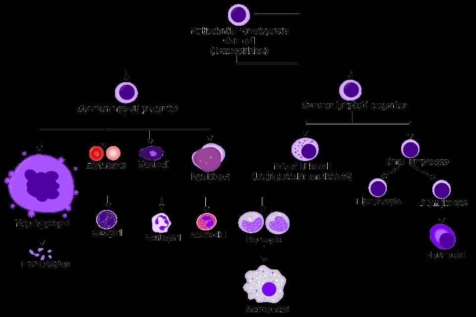
Every blood cell traces back to one ancestor: the hematopoietic stem cell (HSC) living in the red bone marrow (in adults, mainly the pelvis, sternum, vertebrae, and ends of long bones). This process is called hematopoiesis. A stem cell is pluripotent - it can become any blood cell - and it makes copies of itself so the factory never runs out. As daughter cells mature, they commit down one of two big lines: the myeloid line (red cells, platelets, and most white cells) or the lymphoid line (B and T lymphocytes). Because the marrow constantly replaces cells that wear out, anything that damages the marrow - chemotherapy, radiation, leukemia crowding it out, or aplastic anemia - can drop all the cell lines at once, a state called pancytopenia.
Erythropoietin - the kidney's oxygen alarm
How does the body know to make more red cells? The kidneys act as oxygen sensors. When they detect low oxygen reaching their own tissue (hypoxia) - from blood loss, high altitude, or lung disease - they release a hormone called erythropoietin (EPO). EPO travels to the bone marrow and tells it to ramp up red cell production. This is a beautiful negative-feedback loop: more red cells means more oxygen delivery, which quiets the alarm and lowers EPO again.
This explains a classic clinical picture. In chronic kidney disease, the damaged kidneys stop making enough EPO, so the marrow never gets the signal - the patient develops anemia of chronic kidney disease even though the marrow itself is healthy. That is why these patients are often treated with synthetic EPO. It also explains why people living at high altitude naturally run higher hemoglobin and hematocrit: chronic low oxygen keeps EPO switched on. Endurance athletes are the instructive exception - training does raise total red cell mass, but it expands plasma volume even more, so the measured hematocrit often reads normal or slightly low, a dilutional picture called athlete's pseudoanemia.
Hemoglobin - how red cells carry oxygen
A red cell is essentially a bag stuffed with hemoglobin (Hgb), the iron-containing protein that actually binds oxygen. Each hemoglobin molecule has four heme groups, and each heme holds one iron atom that grabs one oxygen molecule - so one hemoglobin carries up to four oxygen molecules. The red cell's flexible biconcave disc shape gives it lots of surface area and lets it squeeze through tiny capillaries.
This is why the pieces have to line up. You need enough iron (to build heme), enough vitamin B12 and folate (to build the cell), and a healthy marrow responding to EPO. Take away any one and oxygen delivery falls. Low hemoglobin is anemia, and its symptoms flow directly from the mechanism: fatigue and pallor because tissues are starved of oxygen, and tachycardia and shortness of breath because the heart speeds up to move the remaining oxygen faster. Nurses usually watch the hemoglobin and hematocrit together as the fastest read on a patient's oxygen-carrying capacity.
- Blood is a connective tissue: ~55% plasma (water plus albumin, clotting factors, antibodies) and ~45% cells - red cells, white cells, and platelets.
- Hematopoiesis makes all blood cells from pluripotent stem cells in the red bone marrow, splitting into myeloid and lymphoid lines; marrow damage can cause pancytopenia.
- The kidneys sense low oxygen and release erythropoietin to drive red cell production - so chronic kidney disease causes anemia from lost EPO.
- Hemoglobin is the iron-based protein that binds oxygen; each molecule carries up to four O2, and building it needs iron, B12, and folate.
- Low hemoglobin (anemia) starves tissues of oxygen, producing fatigue, pallor, and compensatory tachycardia - watch Hgb and hematocrit together.
Section 2Anemias
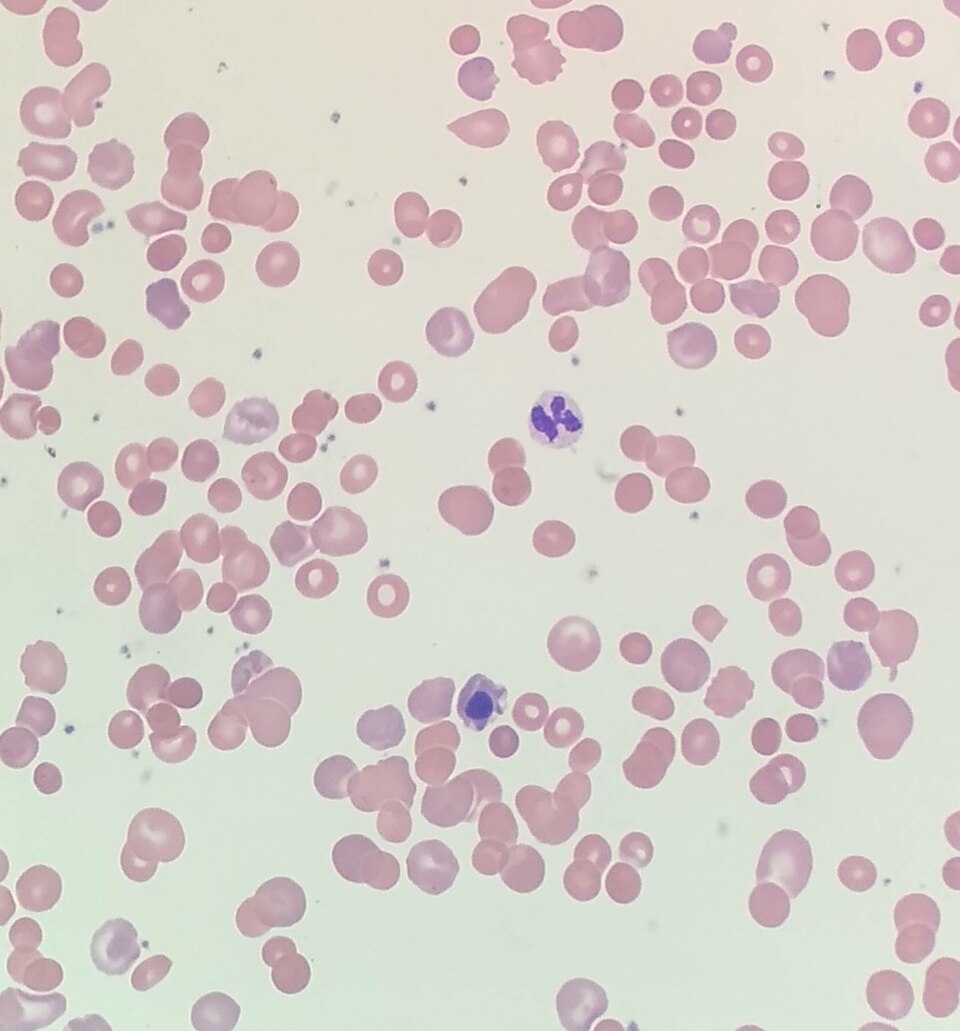
Every one of your cells needs a steady supply of oxygen to make energy, and oxygen does not travel through blood on its own. It rides on hemoglobin, the iron-rich protein packed inside your red blood cells (RBCs). Healthy bone marrow constantly builds new RBCs, stuffs them with hemoglobin, and releases them to circulate for about 120 days. When there is not enough functional hemoglobin to carry the oxygen your tissues need, we call that anemia. Notice the wording: anemia is not simply "low blood." It is low oxygen-carrying capacity, whether from too few RBCs, too little hemoglobin per cell, or hemoglobin that does not work.
Because the problem is tissue oxygen delivery, the classic symptoms make sense once you trace the mechanism. Tissues starved of oxygen produce less energy, so patients feel fatigue and weakness. With fewer red cells reflecting light through the skin and mucous membranes, you see pallor (pale skin, conjunctiva, nail beds). The heart tries to compensate by pumping faster to move what little oxygen there is, causing tachycardia. And because the blood is oxygen-poor, patients breathe faster and feel short of breath, especially with exertion, which is dyspnea. These are the shared symptoms of almost every anemia; the differences lie in why the hemoglobin is missing.
Iron-deficiency anemia (microcytic)
Iron is the core of the heme molecule that binds oxygen. Without enough iron, the marrow makes red cells that are small (microcytic, low MCV) and pale (hypochromic) because they simply cannot fill with hemoglobin. This is the most common anemia worldwide. Causes usually fall into three buckets: blood loss (heavy menstrual periods, or slow GI bleeding from ulcers or colon cancer, which quietly drains iron), increased demand (pregnancy, infancy, rapid growth), and poor intake or absorption (diets low in iron, or gut conditions). Lab clues include low ferritin (the body's iron storage form) and low serum iron.
B12 and folate deficiency (macrocytic)
Vitamin B12 and folate are required to build DNA. When they run short, developing red cells cannot divide properly, so they grow large and fragile instead, producing macrocytic (high MCV) megaloblastic anemia. Folate deficiency comes from poor diet (few leafy greens), alcohol use, or pregnancy demand. B12 deficiency has a special cause worth memorizing: pernicious anemia. Absorbing B12 from food requires intrinsic factor, a protein made by the stomach's parietal cells. In pernicious anemia, an autoimmune attack destroys those cells, intrinsic factor disappears, and ordinary dietary B12 cannot be absorbed no matter how much is eaten. Replacement is therefore given by injection, or as high-dose oral B12 (1,000 to 2,000 mcg daily), which works because about 1 percent of a swallowed dose crosses the gut by passive diffusion without any intrinsic factor at all. One key distinction: because B12 also maintains the nervous system, its deficiency adds neurologic signs (numbness, tingling, balance problems) that folate deficiency does not.
| Feature | Iron deficiency | B12 / folate deficiency |
|---|---|---|
| Cell size (MCV) | Small (microcytic) | Large (macrocytic) |
| Core problem | Too little hemoglobin | Faulty DNA / cell division |
| Typical cause | Blood loss, low intake | Poor diet, pernicious anemia |
| Neuro symptoms | No | Yes (B12 only) |
Hemolytic anemias and sickle cell disease
Hemolytic anemias occur when red cells are destroyed faster than the marrow can replace them (hemolysis means RBC breakdown). Because dying cells spill their contents, you often see jaundice from the released bilirubin. The classic inherited example is sickle cell disease. A single genetic change produces abnormal hemoglobin called HbS. When oxygen levels drop, or with dehydration, infection, or cold, HbS molecules stick together and warp the soft round RBC into a rigid, crescent (sickle) shape. These stiff cells jam small vessels, blocking blood flow in what is called a vaso-occlusive crisis - episodes of severe, sudden pain as tissues downstream are starved of oxygen. The sickled cells are also fragile and are destroyed early, driving chronic anemia.
Finally, aplastic anemia is a marrow-failure state where the bone marrow stops producing blood cells altogether, so RBCs, white cells, and platelets all fall together (pancytopenia). Explore the different anemia types in the figure below.
- Anemia is low functional hemoglobin, reducing oxygen delivery; shared signs (fatigue, pallor, tachycardia, dyspnea) all stem from tissue hypoxia and cardiac compensation.
- Iron-deficiency anemia is microcytic and most common; a new case in an older adult means rule out GI bleeding.
- B12 and folate deficiencies are macrocytic; only B12 deficiency causes neurologic signs, and pernicious anemia is loss of intrinsic factor blocking B12 absorption.
- Sickle cell disease (HbS) sickles under low oxygen, causing painful vaso-occlusive crises; manage with hydration and pain control, adding oxygen when the patient is hypoxic.
- Aplastic anemia is bone marrow failure producing pancytopenia.
Section 3Bleeding & clotting disorders
Every time you get a paper cut, a beautifully choreographed emergency response kicks in to stop the bleeding within minutes. This process is called hemostasis (literally "blood standing still"), and it happens in overlapping steps. Understanding the normal sequence is the key to understanding what goes wrong in bleeding and clotting disorders.
Normal hemostasis: how the body plugs a leak
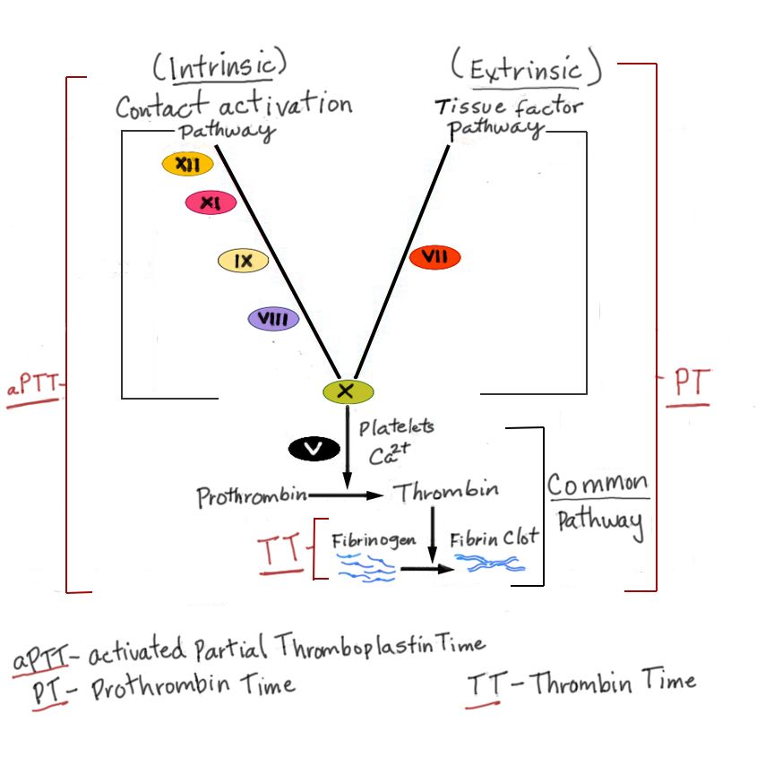
The moment a blood vessel is injured, three things happen in quick succession. First, vasoconstriction: the vessel clamps down to slow blood flow to the area. Second, the platelet plug forms. Platelets are tiny cell fragments that normally glide past smooth vessel walls, but when they touch exposed collagen at an injury, they stick, activate, change shape, and clump together, helped by a bridging protein called von Willebrand factor (vWF). This soft plug is a quick fix. Third comes the coagulation cascade, a chain reaction of clotting proteins (factors) that activate one another in sequence. The cascade has an intrinsic and extrinsic pathway that converge, ultimately turning prothrombin into thrombin. Thrombin then converts fibrinogen into fibrin, tough protein threads that weave through the platelet plug like rebar in concrete, creating a stable clot. Later, the clot is dissolved by fibrinolysis once healing is underway.
Thrombocytopenia and ITP: too few platelets
Thrombocytopenia means a low platelet count (usually below 150,000/mm3, with serious bleeding risk often under 20,000). With too few platelets, the first plug is weak. Patients show petechiae (pinpoint red spots), purpura, easy bruising, nosebleeds, and gum bleeding. In immune thrombocytopenic purpura (ITP), the immune system makes antibodies that tag the patient's own platelets, and the spleen destroys them. It often follows a viral illness in children and is more chronic in adults. Treatment targets the immune attack, using corticosteroids, IVIG, and sometimes splenectomy.
Hemophilia and von Willebrand disease: the factor problems
Hemophilia is an inherited deficiency of a clotting factor, so the platelet plug forms fine but the fibrin mesh never sets properly. Both common types are X-linked recessive, which is why they classically affect males, whose mothers are carriers. Hemophilia A is a deficiency of factor VIII; hemophilia B (Christmas disease) is a deficiency of factor IX. Clinically they look identical: hemarthrosis (deep bleeding into joints), muscle bleeds, and prolonged bleeding after surgery or trauma. Lab work shows a prolonged aPTT with a normal platelet count. Treatment is replacing the missing factor.
Von Willebrand disease is the most common inherited bleeding disorder and is usually autosomal (affecting both sexes equally). Missing or defective vWF means platelets cannot stick well, and because vWF also carries factor VIII, that factor drops too. So it blends both worlds: mucosal, platelet-type bleeding (heavy menstrual periods, nosebleeds) plus a mild factor picture.
| Feature | ITP | Hemophilia A/B | von Willebrand |
|---|---|---|---|
| Core defect | Platelets destroyed | Factor VIII or IX missing | vWF missing/defective |
| Inheritance | Acquired (autoimmune) | X-linked recessive | Usually autosomal |
| Bleeding type | Petechiae, mucosal | Deep joints, muscles | Mucosal, menstrual |
| Key lab | Low platelet count | Prolonged aPTT | Low vWF, sometimes low VIII |
DIC and DVT: when clotting itself becomes the disease
Disseminated intravascular coagulation (DIC) is a dangerous paradox. Triggered by sepsis, trauma, obstetric complications, or cancer, the cascade fires everywhere at once, throwing tiny clots throughout the microcirculation. This consumes platelets and clotting factors faster than the body can replace them. The result is a patient who is both clotting and bleeding: microclots damage organs while the depleted supply leaves them oozing from IV sites, gums, and incisions. Labs show low platelets, prolonged PT/aPTT, low fibrinogen, and elevated D-dimer. DIC is always secondary, so treatment means fixing the underlying cause.
Clotting can also go wrong in a single vessel. A deep vein thrombosis (DVT) is a clot in a deep vein, usually of the leg. Virchow's triad names the three ingredients that promote it: venous stasis (immobility), endothelial injury (vessel-wall damage), and hypercoagulability (blood that clots too readily). The feared complication is a piece breaking off and traveling to the lungs as a pulmonary embolism.
You can explore hemostasis, bleeding, and clotting in the interactive figure below.
- Hemostasis runs in order: vasoconstriction, platelet plug (needs vWF), then the coagulation cascade making thrombin and fibrin.
- Platelet/vWF problems cause immediate superficial bleeding (petechiae, mucosal); factor problems cause delayed deep bleeding (joints, muscles).
- ITP is autoimmune platelet destruction; hemophilia A (factor VIII) and B (factor IX) are X-linked; von Willebrand disease is the most common inherited bleeding disorder.
- DIC consumes platelets and factors, so the patient clots AND bleeds at once, with a high D-dimer; it is always secondary to another problem.
- DVT forms via Virchow's triad (stasis, endothelial injury, hypercoagulability) and can embolize to the lungs.
Section 4Blood types & transfusion
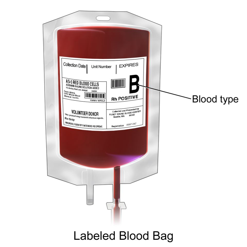
Every drop of blood carries a kind of ID badge. On the surface of your red blood cells sit tiny protein and sugar markers called antigens, and floating in your plasma are antibodies, immune proteins that attack any antigen your body does not recognize as its own. Your immune system learns early in life to leave your own red cells alone while staying armed against foreign ones. This self-versus-nonself recognition is the whole reason transfusions must be matched. Give someone blood carrying an antigen their plasma has antibodies against, and those antibodies clamp onto the donor cells and destroy them, sometimes with life-threatening speed.
The ABO system
The ABO system sorts blood into four types based on which antigens sit on the red cells. Type A cells carry the A antigen, type B carry B, type AB carry both, and type O carry neither. Here is the elegant, slightly counterintuitive part: your plasma automatically makes antibodies against whatever antigen you lack. So type A blood has anti-B antibodies, type B has anti-A, type O has both anti-A and anti-B, and type AB has neither.
Because type O cells carry no A or B antigen, there is nothing on them for a recipient's antibodies to attack, which is why O-negative is called the universal donor, the go-to in a trauma emergency before typing is done. Because type AB plasma carries no anti-A or anti-B antibodies, an AB person can receive any ABO type, making AB-positive the universal recipient.
| Blood type | Antigen on red cell | Antibody in plasma |
|---|---|---|
| A | A | anti-B |
| B | B | anti-A |
| AB | A and B | none |
| O | none | anti-A and anti-B |
The Rh factor and hemolytic disease of the newborn
On top of ABO sits a second marker, the Rh factor (also called the D antigen). If your red cells carry it, you are Rh-positive; if not, Rh-negative. Unlike ABO, Rh-negative people do not come with ready-made anti-Rh antibodies. They only build them after being exposed to Rh-positive blood, a process called sensitization.
This matters most in pregnancy. If an Rh-negative mother carries an Rh-positive baby, a small leak of fetal blood, often during delivery, can sensitize her. The first baby is usually fine, but in a later Rh-positive pregnancy her anti-Rh antibodies can cross the placenta and destroy the fetal red cells. This is hemolytic disease of the newborn (HDN), also called erythroblastosis fetalis, causing fetal anemia, jaundice, and heart failure.
The fix is RhoGAM (Rh immune globulin), given around 28 weeks and again within 72 hours of delivery. RhoGAM works by mopping up any stray fetal Rh-positive cells before the mother's immune system can notice them and start making its own permanent antibodies, so sensitization never happens.
Type, screen, and crossmatch
Before any transfusion, the lab types the blood (identifies ABO and Rh), screens the plasma for unexpected antibodies, and crossmatches by mixing donor cells with recipient plasma to confirm they do not clump. Clumping, called agglutination, is the visible warning that antibodies are attacking, and it means that unit is unsafe. This careful matching is why transfusion is normally so safe.
When a transfusion goes wrong
Reactions still happen, and telling them apart guides your response.
- Acute hemolytic reaction is the feared one, usually from an ABO mismatch (often a clerical or ID error). Recipient antibodies destroy donor cells, releasing hemoglobin that can trigger fever, chills, flank or back pain, dark urine, hypotension, and kidney failure.
- Febrile non-hemolytic reaction is the most common: fever and chills from the recipient reacting to donor white blood cells, uncomfortable but usually not dangerous.
- Allergic reaction is a response to donor plasma proteins, ranging from hives and itching up to anaphylaxis.
For any suspected reaction, the priority is the same: stop the transfusion immediately. Keep the IV line open with normal saline (using fresh tubing so no more blood enters), then check vital signs, notify the provider and blood bank, and send the blood bag and tubing back for analysis.
Explore the ABO and Rh types in the figure below.
- ABO type is set by antigens on red cells; plasma carries antibodies against the antigens you lack, so O-negative is the universal donor and AB-positive the universal recipient.
- Rh-negative people make anti-Rh antibodies only after exposure; RhoGAM prevents maternal sensitization and hemolytic disease of the newborn.
- Blood is typed, screened, and crossmatched to catch agglutination before it reaches the patient.
- Acute hemolytic reactions (ABO mismatch) are the most dangerous; febrile and allergic reactions are more common but usually milder.
- For any suspected transfusion reaction, stop the transfusion first, then keep the line open with saline and notify the provider and blood bank.
Section 5Blood cancers
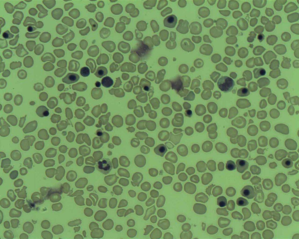
Your bone marrow is a busy blood factory. Deep inside your big bones, hematopoietic stem cells divide and mature into three great families of blood cells: red cells that carry oxygen, platelets that plug leaks, and white cells that fight infection. Normally this factory is tightly controlled. Every cell knows when to grow, when to stop, and when to die. Blood cancers happen when one line of blood cells loses that control and multiplies without a brake. The trouble is not just the abnormal cells themselves, but the fact that they take up space, resources, and function that healthy cells need. Understanding which cell went rogue and where it piles up explains almost every symptom your patient will have.
Leukemia: the marrow gets crowded out
Leukemia is cancer of the white-cell-forming lines in the bone marrow. Immature, useless white cells (often called blasts) reproduce so fast that they physically crowd out normal production. Picture a factory floor jammed with defective units so nothing else can be built. That single mechanism explains the classic triad: too few red cells means anemia (fatigue, pallor, shortness of breath), too few platelets means thrombocytopenia (easy bruising, petechiae, bleeding gums), and too few functional white cells means dangerous infection risk, even though the total white count on the lab report may look high.
Leukemias are sorted two ways. Acute vs chronic describes tempo: acute leukemias explode with immature blasts and make patients sick in days to weeks, while chronic leukemias smolder for months to years with more mature-looking cells. Myeloid vs lymphocytic describes the parent line: myeloid cells give rise to red cells, platelets, and granulocytes, while lymphocytic cells give rise to lymphocytes. Combine them and you get the four big names.
| Type | Who it usually affects | Key feature |
|---|---|---|
| ALL (acute lymphocytic) | Children most often | Most common childhood cancer; responds well to treatment |
| AML (acute myeloid) | Older adults | Blasts may show Auer rods |
| CLL (chronic lymphocytic) | Older adults | Often found by accident on routine bloodwork |
| CML (chronic myeloid) | Middle-aged adults | Linked to the Philadelphia chromosome |
Lymphoma: cancer that builds tumors in lymph tissue
Where leukemia floods the marrow and blood, lymphoma is a solid cancer of lymphocytes that grows into masses inside lymph nodes and lymphoid organs like the spleen. The hallmark sign is painless lymphadenopathy: a firm, swollen, non-tender node in the neck, armpit, or groin. Patients often also have B symptoms, which is the textbook name for the trio of drenching night sweats, unexplained fever, and weight loss.
Lymphomas split into two branches. Hodgkin lymphoma is defined by a specific giant abnormal cell, the Reed-Sternberg cell, on biopsy. It usually starts in one node region and spreads in an orderly, predictable pattern, which helps make it one of the more curable cancers. Non-Hodgkin lymphoma is a large, diverse group without Reed-Sternberg cells; it tends to be more scattered and unpredictable at diagnosis and is far more common overall.
Multiple myeloma: rogue plasma cells attack bone and kidney
Plasma cells are the mature antibody-factory version of B lymphocytes. In multiple myeloma, one plasma cell line turns malignant and churns out a single useless antibody, called a monoclonal (M) protein, while crowding the marrow. Its damage spells out neatly with the mnemonic CRAB: Calcium elevated, Renal (kidney) injury, Anemia, and Bone lesions.
- Bone lesions: myeloma cells activate osteoclasts that eat away bone, creating punched-out lytic lesions, deep bone pain, and pathologic fractures.
- Hypercalcemia: the dissolving bone dumps calcium into the blood, causing confusion, constipation, and thirst.
- Kidney injury: the flood of M protein and calcium clogs and damages the renal tubules.
- Anemia and infection: marrow crowding plus antibodies that do not actually work.
Explore the interactive figure below to see how leukemia fills the marrow, how lymphoma builds masses in nodes, and how myeloma eats into bone.
- Leukemia crowds the bone marrow with abnormal white cells, causing anemia, bleeding (thrombocytopenia), and infection; classified acute vs chronic and myeloid vs lymphocytic (ALL, AML, CLL, CML).
- Lymphoma forms solid masses in lymph nodes: Hodgkin shows Reed-Sternberg cells and spreads predictably; non-Hodgkin is more common and more scattered. Watch for painless lymphadenopathy plus B symptoms.
- Multiple myeloma is malignant plasma cells; remember CRAB: hyperCalcemia, Renal injury, Anemia, Bone lytic lesions, driven by a monoclonal M protein.
- The shared red flags across blood cancers are fatigue, frequent infections, and easy bruising; a high WBC in leukemia still means the patient is immunocompromised.
The chapter in one breath
- Hematopoiesis from a single bone-marrow stem cell makes all blood cells; erythropoietin from the kidney drives red-cell production, so kidney failure often causes anemia.
- Blood splits into plasma (fluid, proteins, clotting factors) and formed elements: RBCs carry oxygen via hemoglobin, WBCs fight infection, and platelets plug leaks.
- Iron-deficiency anemia makes RBCs small and pale (microcytic, hypochromic) with low ferritin, causing fatigue, pallor, and pica.
- Vitamin B12/folate deficiency causes large immature cells (macrocytic, megaloblastic anemia); only B12 loss adds neuro signs, and B12 needs stomach intrinsic factor to absorb.
- Sickle cell anemia is inherited hemoglobin S that warps RBCs when deoxygenated, triggering painful vaso-occlusive crises, while hemolytic anemias destroy cells early and raise bilirubin.
- Clotting fails from too few platelets (thrombocytopenia), missing factors (hemophilia), or widespread clotting-then-bleeding (DIC); watch for petechiae, bruising, and prolonged bleeding.
- Transfusion safety hinges on ABO and Rh typing: type O is the universal RBC donor, AB the universal recipient, and a mismatch triggers a life-threatening hemolytic reaction.
- Leukemias flood the marrow with abnormal white cells (crowding out normal lines), lymphomas grow in lymph nodes (Hodgkin vs non-Hodgkin), and multiple myeloma is malignant plasma cells eating bone.
ReferenceWords worth knowing
A quick reference for the terms in this chapter — skim it now, or circle back whenever a word trips you up.
- ABO system
- The main red-cell blood-group classification (A, B, AB, O) based on which sugar antigens sit on the red-cell surface, so a patient's plasma carries antibodies against the antigens they lack and a mismatched transfusion triggers a dangerous hemolytic reaction.
- Anemia
- A reduced number of red blood cells or amount of hemoglobin, which lowers the blood's oxygen-carrying capacity and usually causes fatigue, pallor, and shortness of breath.
- DIC (disseminated intravascular coagulation)
- A life-threatening emergency in which widespread clotting is switched on throughout the small vessels, using up clotting factors and platelets so fast that the patient paradoxically both clots and bleeds at the same time.
- Erythropoietin
- A hormone released mainly by the kidneys in response to low oxygen that signals the bone marrow to make more red blood cells.
- Hematopoiesis
- The ongoing process, occurring mostly in red bone marrow, by which stem cells mature into all the formed elements of blood: red cells, white cells, and platelets.
- Hemoglobin
- The iron-containing protein inside red blood cells that binds oxygen in the lungs and releases it to the tissues, and also gives blood its red color.
- Hemophilia
- An inherited (usually X-linked) bleeding disorder caused by a missing or deficient clotting factor (factor VIII in type A, factor IX in type B), leading to prolonged bleeding, often into joints and muscles.
- Hemostasis
- The body's coordinated process of stopping bleeding, moving from blood-vessel spasm to a platelet plug and finally a stable fibrin clot.
- Intrinsic factor
- A protein made by the stomach's parietal cells that binds vitamin B12 so it can be absorbed in the small intestine, without which B12 deficiency develops.
- Leukemia
- A cancer of the blood-forming tissue in which the marrow overproduces abnormal, immature white blood cells that crowd out healthy cells, leaving the patient prone to infection, bleeding, and anemia.
- Macrocytic
- Describing red blood cells that are abnormally large, a pattern often seen in vitamin B12 or folate deficiency.
- Microcytic
- Describing red blood cells that are abnormally small, a pattern classically seen in iron-deficiency anemia.
- Pernicious anemia
- A type of macrocytic anemia caused by loss of intrinsic factor (often from autoimmune damage to the stomach lining), which prevents vitamin B12 absorption and impairs red-cell maturation.
- Sickle cell disease
- An inherited disorder in which abnormal hemoglobin S makes red cells stiffen into a crescent shape under low-oxygen stress, so the rigid cells block small vessels and cause painful crises, hemolysis, and organ damage.
- Thrombocytopenia
- An abnormally low platelet count, which raises the risk of easy bruising and prolonged or spontaneous bleeding.
- Virchow's triad
- The three conditions that together promote abnormal clot formation: sluggish blood flow (stasis), vessel-wall injury, and increased blood clottability (hypercoagulability).
DrillFlashcards for this unit
These are the Week 8 cards — the same deck as the hub, pulled in here so you can review the moment you finish reading. Tap the card to flip it; hit Shuffle to mix them up. To grade yourself and track mastery across the whole deck, open the full flashcards page.