The Cardiovascular System: Blood
Blood is the only tissue in the body you can pour into a test tube, and it is easy to underestimate — it looks like a plain red liquid. But that liquid is a connective tissue, a living fleet of cells drifting in a protein-rich sea, and it touches every other cell you own. In this chapter we take a tube of blood apart layer by layer: the straw-colored plasma on top, the packed red cells below, and the thin white line between them. You will learn what each part carries, where blood cells are born, how a red cell ferries oxygen, how a cut seals itself in seconds, and why matching a patient's blood type before a transfusion is a matter of life and death.
By the end of this chapter you can
- List the major functions of blood — transport, regulation, and protection — and explain why blood counts as a connective tissue.
- Describe what a spun tube of blood separates into, and define plasma, buffy coat, and hematocrit.
- Name the three formed elements and the five kinds of white blood cell, and give the main job of each.
- Trace hematopoiesis from a single stem cell down the myeloid and lymphoid lines, and name the hormone that drives red-cell production.
- Explain how hemoglobin loads oxygen in the lungs and unloads it in the tissues, and how blood carries carbon dioxide.
- Walk through the three steps of hemostasis and describe how the clotting cascade builds a fibrin mesh.
- Predict ABO and Rh compatibility and identify the universal donor and universal recipient.
Section 1What blood is made of
A tissue you can pour
It surprises most students to hear that blood is a connective tissue. Connective tissues are defined by scattered cells sitting in an extracellular matrix, and blood fits the definition perfectly — its "cells" are the red cells, white cells, and platelets, and its matrix is the liquid plasma they float in. What makes blood unusual is that the matrix is a fluid rather than something solid like bone or rubbery like cartilage. An average adult carries about 5 liters of it, a little less in women and a little more in men, and it makes up roughly 8 percent of body weight. Blood is also heavier and stickier than water — about five times as viscous — it runs warm at about 38 °C, and it is slightly alkaline, held in the narrow range of pH 7.35 to 7.45.
Three jobs: transport, regulation, protection
Everything blood does falls into three categories. First is transport: blood delivers O₂ and nutrients to cells, hauls CO₂ and metabolic wastes away, and carries hormones from glands to their distant targets. Second is regulation: by shifting where it flows and how much heat it carries to the skin, blood helps stabilize body temperature; its dissolved buffers steady the body's pH; and its proteins help control fluid balance between blood and tissues. Third is protection: the white cells and antibodies defend against invaders, and the clotting system seals leaks so you do not bleed out from a small wound.
Spin it down and it splits in three
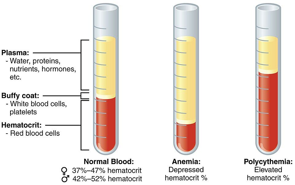
Put a tube of blood in a centrifuge and the heavier parts sink. You end up with three visible layers, and their proportions tell a clinical story:
| Layer | What it contains | Fraction of blood |
|---|---|---|
| Plasma (top) | Water, plasma proteins, nutrients, gases, hormones, wastes, ions | About 55% |
| Buffy coat (middle) | Leukocytes (white cells) and platelets | Less than 1% |
| Erythrocytes (bottom) | Red blood cells — this fraction is the hematocrit | About 45% |
That bottom fraction has its own name: the hematocrit is the percentage of whole-blood volume made up of red cells. Normal values run roughly 37–47 percent in women and 42–52 percent in men. The plasma itself is about 92 percent water; most of the rest is plasma proteins — albumin (the most abundant, which holds water in the vessels and ferries fatty molecules), globulins (which include antibodies and transport proteins), and fibrinogen (the raw material for clots). One term worth pinning down now: if you let blood clot and remove the clot, the pale fluid left behind is serum — plasma minus its clotting proteins.
Spin your own tube and pull apart the layers in the interactive below.
- Blood is a fluid connective tissue — cells (formed elements) suspended in a liquid matrix (plasma) — about 5 L and 8% of body weight.
- Its jobs sort into transport, regulation, and protection.
- Spun blood gives plasma (~55%), a thin buffy coat of white cells and platelets, and red cells (hematocrit, ~45%).
- Serum is plasma with the clotting proteins removed.
Section 2The formed elements
Three kinds of passenger
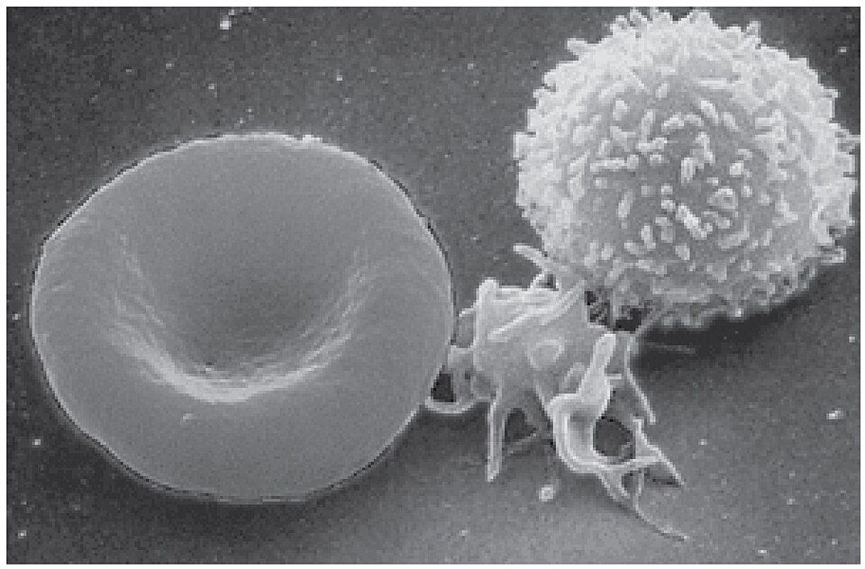
The living cargo of blood — the formed elements — comes in three types: erythrocytes (red blood cells), leukocytes (white blood cells), and platelets (also called thrombocytes). They are wildly unequal in number. Red cells dominate, at roughly 5 million per microliter; platelets number a few hundred thousand; and white cells are the rarest, at only about 5,000 to 10,000 per microliter. That lopsidedness is exactly why the spun tube from Section 1 is almost all red on the bottom with only a whisper of buffy coat.
Red cells: oxygen with everything stripped away
An erythrocyte is a design study in doing one job well. It is a biconcave disc — a flattened circle pinched in on both faces — a shape that gives it lots of surface area for gas exchange and lets it fold to squeeze through capillaries narrower than itself. To make room for as much hemoglobin as possible, the mature red cell throws almost everything else overboard: it has no nucleus and no organelles. That sacrifice has a cost — with no nucleus it cannot repair itself, so it wears out in about 120 days and is recycled by the spleen and liver. We will follow its oxygen work in Section 4.
White cells: the mobile defense force
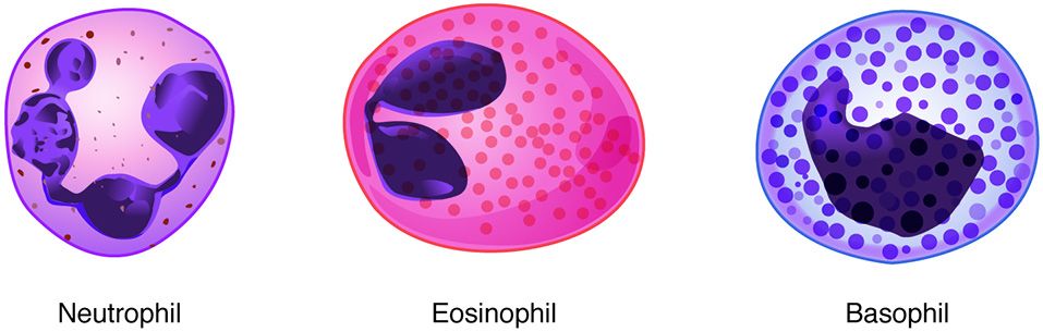
Unlike red cells, every leukocyte keeps its nucleus, and all of them are soldiers of the immune system. They split into two families by whether they carry visible granules. The granulocytes — neutrophils, eosinophils, and basophils — have grainy cytoplasm; the agranulocytes — lymphocytes and monocytes — do not. Learning them in order of abundance is easiest with a mnemonic: Never Let Monkeys Eat Bananas (Neutrophils, Lymphocytes, Monocytes, Eosinophils, Basophils).
| White cell | Group | Rough share | Main job |
|---|---|---|---|
| Neutrophil | Granulocyte | 50–70% | First responder; phagocytoses bacteria |
| Lymphocyte | Agranulocyte | 20–30% | Adaptive immunity — T, B, and NK cells |
| Monocyte | Agranulocyte | 2–8% | Becomes a macrophage; big-appetite phagocyte |
| Eosinophil | Granulocyte | 1–4% | Attacks parasites; roles in allergy |
| Basophil | Granulocyte | <1% | Releases histamine and heparin; inflammation |
Platelets: fragments that plug leaks
Platelets are not whole cells at all — they are cell fragments pinched off from giant marrow cells called megakaryocytes. They have no nucleus, live only about 10 days, and exist for one purpose: to rush to a wound, pile up, and start a clot. We will watch them do it in Section 5.
Meet all three formed elements — and the five white cells — in the interactive.
- The three formed elements are erythrocytes (oxygen), leukocytes (defense), and platelets (clotting); red cells vastly outnumber the rest.
- The red cell is a biconcave disc with no nucleus, packed with hemoglobin, lasting ~120 days.
- Five white cells, most to least: neutrophils, lymphocytes, monocytes, eosinophils, basophils (Never Let Monkeys Eat Bananas).
- Platelets are megakaryocyte fragments that trigger clotting.
Section 3Where blood cells are born
One stem cell, every blood cell
Red cells wear out in four months and neutrophils in a matter of days, so the body must replace blood cells constantly — on the order of hundreds of billions each day. That factory is the red bone marrow, and the whole process of making blood is called hematopoiesis. Astonishingly, every one of those cells traces back to a single kind of ancestor: the hematopoietic stem cell (also called a hemocytoblast). It is multipotent, meaning it can become any blood cell — but only blood cells, not liver or nerve or muscle — and it renews itself so the supply never runs dry. (Reserve the word pluripotent for embryonic and induced pluripotent stem cells, which can give rise to tissues of all three germ layers.)
Two family lines: myeloid and lymphoid
Early on, the stem cell's descendants commit to one of two branches. The myeloid line is the larger one — it produces red cells, platelets (by way of megakaryocytes), and most white cells: neutrophils, eosinophils, basophils, and monocytes. The lymphoid line is dedicated to the lymphocytes (the T, B, and NK cells of adaptive immunity). Knowing which line a cell comes from is more than trivia — leukemias are classified as myeloid or lymphoid precisely by which branch of this tree went wrong.
| Line | Cells it produces | Notice |
|---|---|---|
| Myeloid | Erythrocytes, platelets, neutrophils, eosinophils, basophils, monocytes | The broad branch — nearly everything |
| Lymphoid | Lymphocytes (T cells, B cells, NK cells) | The specialist branch — adaptive immunity |
Hormones set the pace
Production is not left to chance; hormones and growth factors tune it to demand. The star is erythropoietin (EPO), released by the kidneys when they sense low oxygen — EPO tells the marrow to make more red cells. Thrombopoietin (from the liver) drives platelet production, and a family of colony-stimulating factors and interleukins ramps up white-cell output during infection. Building a red cell also needs raw materials: iron for hemoglobin, plus vitamin B₁₂ and folate for DNA synthesis. Run short on any of these and you become anemic, which is why the freshly released, not-quite-finished red cells — the reticulocytes — are counted as a gauge of how hard the marrow is working.
Climb the family tree node by node in the interactive.
- Hematopoiesis happens in red bone marrow; every blood cell descends from a multipotent hematopoietic stem cell.
- The stem cell branches into the myeloid line (red cells, platelets, most white cells) and the lymphoid line (lymphocytes).
- Erythropoietin from the kidney drives red-cell production; iron, B₁₂, and folate are the raw materials.
Section 4How blood carries oxygen
The molecule that does the carrying
Red cells matter because of one molecule: hemoglobin. Each hemoglobin is built from four protein chains (two alpha and two beta globins), and every chain cradles a heme group with an atom of iron at its center. Oxygen binds to that iron. Since one hemoglobin has four heme groups, it can carry four O₂ molecules at once — and because a single red cell is stuffed with roughly 270 million hemoglobins, one cell can shuttle over a billion oxygen molecules. Iron is the actual seat oxygen sits on, which is why iron deficiency so directly starves the tissues of oxygen.
Load in the lungs, unload in the tissues
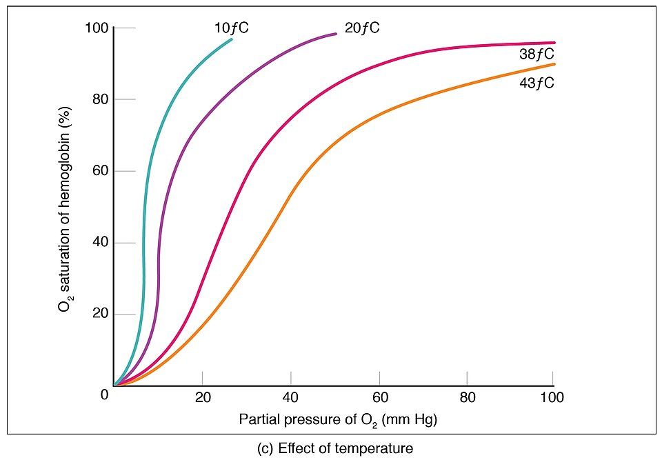
Hemoglobin's genius is that it grabs oxygen where oxygen is plentiful and lets go where it is scarce. In the lungs, where the partial pressure of oxygen is high, hemoglobin binds O₂ to become oxyhemoglobin — and this bright, oxygen-rich blood is a vivid scarlet. Out in the tissues, where cells have burned through their oxygen, the partial pressure is low and hemoglobin releases its cargo, becoming deoxyhemoglobin, a darker brick red. That color swing is why arterial blood looks so much brighter than venous blood. The binding is also cooperative: once the first O₂ is on, the next bind more easily, so loading and unloading both happen fast. Warmth, extra CO₂, and a more acidic environment — exactly the conditions in a hardworking muscle — nudge hemoglobin to release more oxygen right where it is needed.
And the return trip: carbon dioxide
Blood does not just deliver oxygen; it also clears the CO₂ that cells produce. That waste rides home three ways, and the proportions are worth knowing:
| Gas | How it travels | Share |
|---|---|---|
| O₂ | Bound to hemoglobin as oxyhemoglobin | ~98.5% |
| O₂ | Dissolved in plasma | ~1.5% |
| CO₂ | As bicarbonate (HCO₃⁻) in plasma | ~70% |
| CO₂ | Bound to hemoglobin (carbaminohemoglobin) | ~23% |
| CO₂ | Dissolved in plasma | ~7% |
Watch a red cell load up and drop off, on a loop, in the interactive.
- Hemoglobin = four globin chains, each with an iron-containing heme; it carries up to four O₂ per molecule.
- It forms oxyhemoglobin (bright red) in the lungs and releases O₂ as deoxyhemoglobin (darker) in the tissues.
- CO₂ travels home mostly as bicarbonate (~70%), some on hemoglobin, a little dissolved.
- Carbon monoxide outcompetes oxygen for hemoglobin, blocking delivery.
Section 5How bleeding stops
Three fast steps
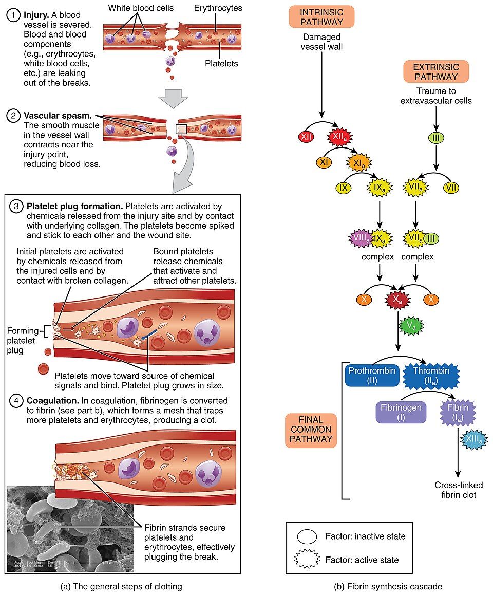
Cut a vessel and the body has to seal the leak before too much blood escapes — a process called hemostasis (literally "blood standstill"). It unfolds in three overlapping steps, each buying time for the next. The whole thing can begin within seconds.
| Step | What happens | Result |
|---|---|---|
| 1. Vascular spasm | Smooth muscle in the vessel wall contracts near the injury | The vessel narrows, slowing blood loss |
| 2. Platelet plug | Platelets stick to exposed collagen, activate, and pile up | A soft plug fills the hole |
| 3. Coagulation | A cascade of clotting factors builds a fibrin mesh | A firm clot locks the plug in place |
The platelet plug snowballs
When the smooth inner lining of a vessel is torn, the tough collagen underneath is exposed. Platelets grab onto it (with the help of a bridging protein, von Willebrand factor), and contact flips them from smooth discs into spiky, sticky activated platelets. Activated platelets then release chemicals — ADP, thromboxane A₂, and serotonin — that summon and activate still more platelets. This is a textbook positive feedback loop: each recruited platelet recruits others, so the plug grows quickly until the breach is covered. This is also exactly the step that aspirin blunts — it blocks thromboxane, which is why low-dose aspirin makes platelets less sticky.
Coagulation: turning liquid into gel
A platelet plug alone is fragile; coagulation reinforces it. A relay of plasma proteins called clotting factors triggers one another in a cascade, along an intrinsic and an extrinsic pathway that merge into a common pathway. The finale has two moves: the enzyme thrombin is switched on, and thrombin converts the soluble plasma protein fibrinogen into threads of insoluble fibrin. Those threads weave a mesh that traps red cells and platelets — the actual clot. Two behind-the-scenes players make it possible: calcium (clotting factor IV) is required at several steps, and vitamin K is needed by the liver to build several of the factors in the first place. Once the wound heals, the enzyme plasmin dissolves the clot (fibrinolysis).
Step through the seal-and-reinforce sequence in the interactive.
- Hemostasis has three steps: vascular spasm → platelet plug → coagulation.
- The platelet plug grows by positive feedback as activated platelets recruit more; aspirin blunts this.
- Coagulation ends with thrombin converting fibrinogen to a fibrin mesh; it needs calcium and vitamin K.
Section 6Blood types and transfusion
Antigens on the cell, antibodies in the plasma
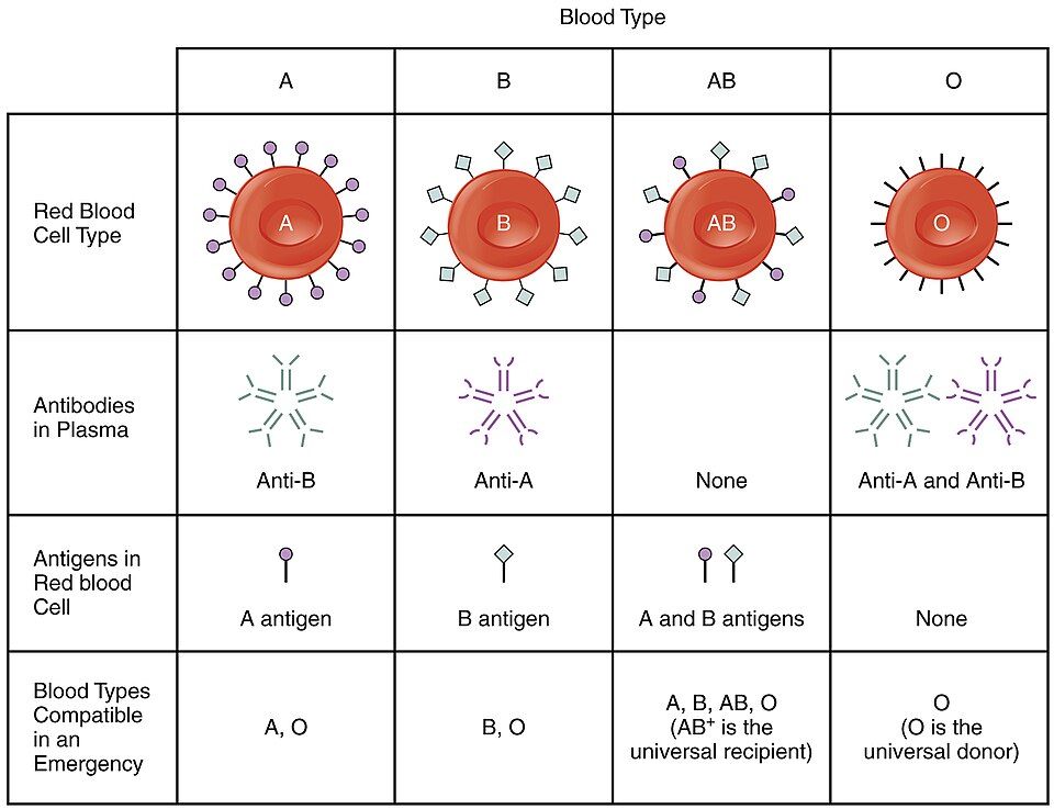
Your blood type is decided by molecular flags called antigens (agglutinogens) that stud the surface of your red cells. In the ABO system there are two possible flags, A and B, and your genes decide which you display. Here is the crucial rule: your plasma carries antibodies (agglutinins) against whichever antigen you lack. So a type A person has A antigens and anti-B antibodies; a type B person has B antigens and anti-A; a type AB person has both antigens and no ABO antibodies; and a type O person has neither antigen but both anti-A and anti-B.
Why a mismatch is dangerous
If a patient receives red cells carrying an antigen their plasma has antibodies against, those antibodies clamp onto the donor cells and clump them together — agglutination — and then rupture them (hemolysis). The debris can clog small vessels and overwhelm the kidneys; a serious transfusion reaction can be fatal. This is why blood is always typed and cross-matched before it is given.
| Type | Antigen on red cells | Antibody in plasma | Can receive from |
|---|---|---|---|
| A | A | Anti-B | A, O |
| B | B | Anti-A | B, O |
| AB | A and B | None | A, B, AB, O — universal recipient |
| O | None | Anti-A and Anti-B | O only — universal donor of red cells |
The Rh factor
Layered on top of ABO is the Rh system, named for the rhesus monkey used in the experiments that uncovered it. It carries several antigens — C, c, D, E, and e — but one of them, D, is by far the most immunogenic and the only one routinely typed. If you carry D you are Rh-positive; if not, Rh-negative. Unlike the ABO antibodies, which are present from early life, an Rh-negative person only makes anti-D antibodies after being exposed to Rh-positive blood. That detail drives one of the most important scenarios in obstetrics: hemolytic disease of the newborn. If an Rh-negative mother carries an Rh-positive baby, she can become sensitized to the D antigen, and in a later Rh-positive pregnancy her antibodies may cross the placenta and attack the fetus's red cells. The fix is elegant — a shot of Rh immune globulin (RhoGAM) clears the fetal cells before the mother's immune system can react.
Pick a blood type and see exactly who it can give to and take from in the interactive.
- Blood type = antigens on your red cells; your plasma holds antibodies against the antigens you lack.
- Mismatched transfusion causes agglutination and hemolysis, so blood is typed and cross-matched.
- O-negative is the universal red-cell donor; AB-positive is the universal recipient.
- Rh-negative mothers with Rh-positive babies risk hemolytic disease of the newborn, prevented by RhoGAM.
The chapter in one breath
- Blood is a fluid connective tissue — formed elements suspended in plasma — that handles transport, regulation, and protection.
- Spun blood separates into plasma (~55%), a thin buffy coat, and red cells (hematocrit, ~45%); plasma is mostly water plus albumin, globulins, and fibrinogen.
- The formed elements are erythrocytes (oxygen), five kinds of leukocyte (defense), and platelets (clotting).
- Hematopoiesis in red marrow starts from one stem cell that branches into the myeloid and lymphoid lines; the kidney's erythropoietin drives red-cell output.
- Hemoglobin carries four O₂ on iron-heme seats, loading in the lungs and unloading in the tissues; CO₂ rides home mostly as bicarbonate.
- Hemostasis seals a wound in three steps — vascular spasm, platelet plug, and coagulation — ending in a fibrin mesh.
- Blood type is set by red-cell antigens (A, B, Rh); plasma holds antibodies against the ones you lack, making O-negative the universal donor and AB-positive the universal recipient.
ReferenceWords worth knowing
A quick reference for the terms in this chapter — skim it now, or circle back whenever a word trips you up.
- Agglutination
- The clumping of red blood cells when antibodies bind matching antigens, as in a mismatched transfusion.
- Albumin
- The most abundant plasma protein; it holds water in the vessels (osmotic pressure) and transports fatty molecules and drugs.
- Antibody (agglutinin)
- A plasma protein that recognizes and binds a specific antigen; ABO antibodies target the A or B antigen a person lacks.
- Antigen (agglutinogen)
- A surface molecule on red cells (A, B, or Rh D) that defines blood type and can trigger an immune response.
- Buffy coat
- The thin layer of white blood cells and platelets sitting between plasma and red cells in a spun sample.
- Coagulation
- The clotting cascade in which factors activate one another to convert fibrinogen into a fibrin mesh.
- Erythrocyte
- A red blood cell — a biconcave, anucleate cell packed with hemoglobin to carry oxygen.
- Erythropoietin (EPO)
- A hormone from the kidney that stimulates red bone marrow to produce more red blood cells when oxygen is low.
- Fibrin
- The insoluble protein thread, formed from fibrinogen by thrombin, that meshes to trap cells and form a clot.
- Formed elements
- The cellular components of blood: erythrocytes, leukocytes, and platelets.
- Hematocrit
- The percentage of whole-blood volume made up of red blood cells (about 37–52%, varying by sex).
- Hematopoiesis
- The production of blood cells in red bone marrow, all descending from a multipotent hematopoietic stem cell.
- Hemoglobin
- The iron-containing protein in red cells (four globin chains, four heme groups) that binds and transports oxygen.
- Hemostasis
- The stopping of bleeding through vascular spasm, platelet plug formation, and coagulation.
- Leukocyte
- A white blood cell; the nucleated defenders of the body, divided into granulocytes and agranulocytes.
- Plasma
- The liquid matrix of blood — about 92% water plus proteins, nutrients, gases, hormones, and wastes.
- Platelet (thrombocyte)
- A cell fragment from a megakaryocyte that adheres to wounds and initiates clotting.
- Rh factor
- The D antigen of the Rh blood-group system; present in Rh-positive blood, absent in Rh-negative.
- Serum
- Plasma with its clotting proteins removed; the fluid left after blood clots.
- Thrombin
- The enzyme that converts fibrinogen into fibrin, the final step in building a clot.