The Cellular Level of Organization
The last chapter looked at the whole body. This one zooms all the way in, to the smallest thing that is still alive. The cell is where the body's work really happens. It is where oxygen is used to make energy. It is where genes are read to make proteins. It is where the choice to grow or divide is made. In this chapter you will meet the membrane, the thin wrap around every cell that picks what gets in and out. You will tour the organelles, the tiny parts that share the work inside. You will follow a gene, a set of instructions, as it becomes a working protein. Then you will watch one cell become two. Once you know the cell, the rest of the body makes more sense. Every tissue, organ, and system is just cells working together.
By the end of this chapter you can
- Describe the fluid mosaic model. Explain the jobs of the phospholipid bilayer, membrane proteins, cholesterol, and the sugar coat.
- Tell passive transport from active transport. Put diffusion, facilitated diffusion, osmosis, the Na⁺/K⁺ pump, and bulk transport in the right group.
- Name the main organelles and give the job of each one.
- Explain how the nucleus stores DNA. Explain how that DNA is packed into chromatin and chromosomes.
- Follow protein synthesis from gene to protein, through transcription and translation.
- Put the phases of the cell cycle in order. Describe what happens in each stage of mitosis.
- Predict what happens to a cell in isotonic, hypotonic, and hypertonic fluids. Link this to how an IV fluid is chosen.
Section 1The cell membrane
A smart wall
Every cell in your body is wrapped in a plasma membrane. This is the thin outer skin of the cell. It is so thin that you cannot see it with a normal light microscope. Yet it is smart enough to decide what comes in and what goes out. Without it, a cell would just fall apart into the fluid around it.
The membrane does three big jobs at once. First, it separates the inside of the cell from the outside. Second, it controls traffic across that border. It is selectively permeable, which means it lets some things through and blocks others. Third, it communicates. It carries receptors and ID tags, so the cell can sense signals and be known by other cells. Everything else in this chapter depends on this one wall doing its job.
The fluid mosaic model
Scientists picture the membrane with the fluid mosaic model. Both words matter. Mosaic means it is a patchwork of many pieces. Fats, proteins, and sugars sit side by side like tiles. Fluid means the pieces are not glued down. They drift and wiggle sideways in the sheet. So the membrane acts more like a thin film of oil than a hard wall.
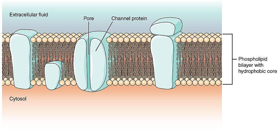
The base of the membrane is the phospholipid bilayer. It is two layers of fat molecules called phospholipids, lined up tail to tail. Each phospholipid is amphipathic, which means it has two very different ends. It has a hydrophilic ("water-loving") head and two hydrophobic ("water-fearing") fatty tails. Put these molecules in water and they line up on their own. The heads face the watery fluid on both sides. The oily tails hide in the middle, away from water.
That oily middle is the secret to how picky the membrane is. Small molecules that dissolve in fat slip right through it. Ions and other water-loving molecules get turned away. They need a protein doorway to get across.
Set into this fat sheet are the membrane proteins, and they do most of the work. Integral (transmembrane) proteins pass all the way through the membrane. They form the channels, carriers, and pumps that move things across. Peripheral proteins stick to just one surface. They often act as enzymes, which speed up reactions, or as anchors. Some proteins are receptors that catch hormones and other signals. Others are identity markers that label the cell as "self," part of your own body.
Tucked between the fats are molecules of cholesterol. Cholesterol keeps the membrane just right. It stops the membrane from getting too stiff when cold or too runny when warm. Last, short sugar chains stick to the outside of some proteins and fats. Together they form the glycocalyx, a fuzzy sugar coat on the cell's outer face. It helps cells know each other, stick together, and tell "self" from "not self."
| Part | What it is | Main job |
|---|---|---|
| Phospholipid bilayer | Two layers of amphipathic phospholipids, tails inward | The basic wall. Blocks water-loving molecules and lets small fat-soluble ones pass. |
| Integral proteins | Proteins that span the whole membrane | Channels, carriers, and pumps that move things; receptors |
| Peripheral proteins | Proteins stuck to one surface | Enzymes, anchors, and support for cell shape |
| Cholesterol | Fat molecules tucked between phospholipids | Keeps the membrane from getting too stiff or too runny as temperature changes |
| Glycocalyx | Sugar chains on glycoproteins and glycolipids | Helps cells know each other and stick together; "self" ID tags |
Tap each part of the membrane below to see it up close.
- The plasma membrane separates, controls traffic (it is selectively permeable), and communicates.
- The fluid mosaic model: a fluid phospholipid bilayer with proteins, cholesterol, and sugars set into it. The water-loving (hydrophilic) heads face out. The water-fearing (hydrophobic) tails face in.
- Small fat-soluble molecules cross the oily middle easily. Ions and other water-loving molecules need protein channels or carriers.
- Water seeps through the membrane slowly on its own. It moves much faster through aquaporin channels.
- Cholesterol keeps the membrane from getting too stiff or too runny. The glycocalyx helps cells know each other and mark "self."
Section 2Moving things across
The one question to ask: does it cost energy?
A cell must always bring in food, push out waste, and keep the right mix of salts and water. It has many ways to move things across its membrane. Every one of them falls into one of two groups. The dividing line is energy.
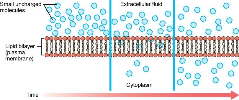
In passive transport, things move down their concentration gradient. That means they go from where there is a lot of them to where there is less. This costs the cell no ATP, because it just goes with the natural flow. In active transport, the cell moves things up a gradient. They go against the flow, from low amounts to high amounts. That always takes energy, most often ATP. So before you learn the names, ask one thing. Is it rolling downhill for free (passive)? Or is it being pushed uphill at a cost (active)?
Passive transport: rolling downhill
The simplest kind is simple diffusion. Molecules are always moving around at random. Over time, they spread out from crowded places to less crowded places, until they are evenly mixed. Small molecules with no charge, like oxygen and carbon dioxide, slip right through the fat layer. That is how O₂ gets into every cell and CO₂ gets out.
Some molecules are too big or too charged to cross the fat layer alone. They use facilitated diffusion. They still move downhill for free. But they go through a protein channel or carrier that gives them a way in. Glucose, a sugar, getting into a cell is the classic example.
One special and vital kind of passive transport is osmosis. Osmosis is the diffusion of water across a selectively permeable membrane. It is so important that it gets its own section at the end of this chapter.
Active transport: pushing uphill
Sometimes a cell needs more of a substance on one side than the other. That means moving it against its gradient, which takes energy. In primary active transport, a pump uses ATP directly. The best known is the sodium–potassium pump (Na⁺/K⁺ pump). For every ATP it uses, it pushes 3 Na⁺ out of the cell and brings 2 K⁺ in. This pump runs in every cell you have. It sets up the electrical charge that lets nerves send signals and muscles squeeze.
In secondary active transport, the cell uses the gradient that a primary pump already built. One ion rushes downhill, and it drags another substance uphill along with it. It is a bit like a water wheel that uses falling water to lift something else.
Some cargo is far too big for any channel, like whole droplets or bits of matter. For these the cell uses bulk transport. It wraps the cargo in a piece of membrane. Endocytosis brings cargo in. This includes phagocytosis, "cell eating," and pinocytosis, "cell drinking." Exocytosis sends cargo out.
| Method | Energy? | Direction and example |
|---|---|---|
| Simple diffusion | No (passive) | Downhill, right through the fat layer: O₂, CO₂ |
| Facilitated diffusion | No (passive) | Downhill, through a protein: glucose, ions |
| Osmosis | No (passive) | Water moves toward the saltier side |
| Primary active transport | Yes, uses ATP directly | Uphill, by a pump: Na⁺/K⁺ pump (3 out, 2 in) |
| Secondary active transport | Yes, uses an ion gradient | Rides another ion's gradient: glucose and Na⁺ moving in together in the gut |
| Bulk transport | Yes (active) | Cargo wrapped in membrane: endocytosis (in), exocytosis (out) |
Pick a transport method below and watch the molecules cross.
- Passive transport moves things down their gradient for free. Active transport moves them up a gradient, and it costs energy.
- Passive types: simple diffusion, facilitated diffusion (through a protein), and osmosis (water).
- The Na⁺/K⁺ pump moves 3 Na⁺ out and 2 K⁺ in for each ATP. It is primary active transport. It powers the electrical signals of nerves and muscles.
- Bulk transport moves the biggest cargo: endocytosis in, exocytosis out.
Section 3Inside the cell: the organelles
A factory with departments
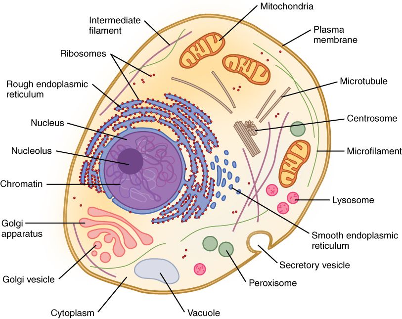
Look inside the membrane and you will not find an empty bag. You will find a busy, well-run workshop. The cell is filled with a jelly-like fluid called the cytosol. Floating in it are the organelles, which means "little organs." Each one is a department with its own job.
Splitting up the work lets the cell run many chemical jobs at once without them getting in each other's way. It is like the walls that separate the rooms of a factory. Many organelles have their own membrane that seals off what happens inside. A few, like ribosomes, do not. To learn the cell, you learn who does what.
The control room and the assembly line
The largest organelle is usually the nucleus, the cell's control center. It is wrapped in a double membrane called the nuclear envelope. The envelope has small holes, or pores, that let messages in and out. The nucleus holds the cell's DNA. DNA is the master set of instructions for making every protein.
Most of the time, DNA is spread out as a loose tangle called chromatin. When the cell gets ready to divide, the DNA packs tightly into chromosomes that you can see. Inside the nucleus is a dark spot called the nucleolus. This is where ribosomes are built.
Ribosomes are the machines that build proteins. Some float free in the cytosol. Others dot the rough endoplasmic reticulum (rough ER). The rough ER is a folded network of membrane. It makes and packs proteins that will be sent out of the cell or placed in the membrane. Next to it is the smooth ER, which has no ribosomes. The smooth ER makes fats, breaks down harmful substances, and stores calcium.
Proteins and fats then move to the Golgi apparatus. The Golgi is the cell's shipping center. It changes, sorts, and packs them into small bubbles called vesicles. Each vesicle is sent to where it is needed.
Power, cleanup, and shape
All this work needs energy. That energy comes from the mitochondria, the "powerhouses" of the cell. Here food and oxygen are turned into ATP, the energy the cell can spend. Cells that work hard, like muscle and liver cells, are packed with mitochondria.
Two organelles handle cleanup. Lysosomes are sacs full of digestive enzymes. They break down worn-out parts, germs, and junk. Peroxisomes make harmful by-products safe.
The cytoskeleton holds the whole cell together. It is a frame of protein threads. It gives the cell its shape, holds organelles in place, and helps things move. The centrosome, with its pair of centrioles, organizes this frame. It also builds the fibers that pull chromosomes apart when the cell divides.
| Organelle | Job | Memory hook |
|---|---|---|
| Nucleus | Stores DNA; runs the cell | The control room or library |
| Nucleolus | Builds ribosomes | Ribosome factory inside the nucleus |
| Ribosome | Builds proteins by reading mRNA | The 3-D printer |
| Rough ER | Makes and packs proteins (has ribosomes) | Protein assembly line |
| Smooth ER | Makes fats, breaks down harmful substances, stores Ca²⁺ | Fat and cleanup lab |
| Golgi apparatus | Changes, sorts, and ships proteins in vesicles | The post office |
| Mitochondrion | Makes ATP from food + O₂ | The powerhouse |
| Lysosome | Breaks down junk and worn-out parts | The recycling and wrecking crew |
| Cytoskeleton | Shape, support, moving things, movement | The inside frame |
Tap an organelle to light it up, or press Tour to walk the whole factory floor.
- Organelles are departments that float in the cytosol. Their membranes keep their jobs apart.
- The nucleus stores DNA as chromatin, which packs into chromosomes before division. The nucleolus builds ribosomes.
- The protein path is ribosomes → rough ER → Golgi → vesicles. The smooth ER makes fats and stores calcium.
- Mitochondria make ATP. Lysosomes break down junk. The cytoskeleton gives shape and moves cargo.
Section 4From gene to protein
The recipe and the problem
Your DNA is like a recipe book. Each gene is one recipe for one protein. But there is a problem. The DNA is locked inside the nucleus. Proteins are built by ribosomes out in the cytoplasm, the space outside the nucleus. The master book can never leave the library.
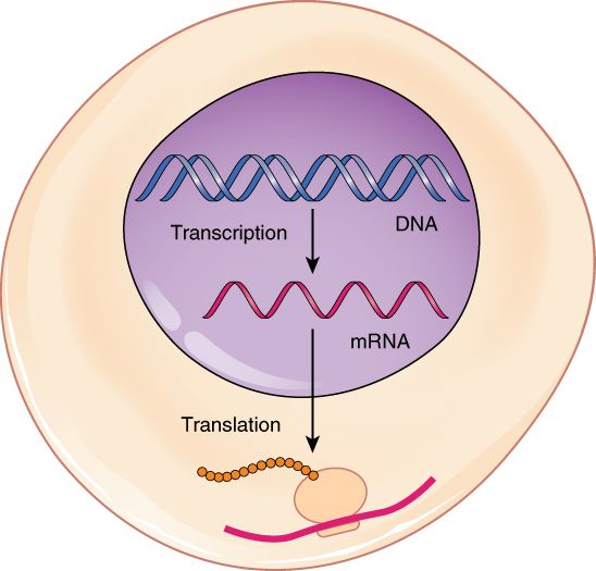
The fix is to make a throwaway copy of just the one recipe you need. That copy is carried out to the workshop. This is the heart of protein synthesis, and it takes two steps. Transcription copies the gene. Translation reads the copy and builds the protein. This flow of information, from DNA to RNA to protein, is so basic that it is called the central dogma of biology.
Transcription: copying the gene
Transcription happens inside the nucleus. An enzyme called RNA polymerase unzips the DNA at the gene it needs. It reads one strand and builds a matching strand of messenger RNA (mRNA). RNA is a lot like DNA. But it has only one strand, and some of its parts are a little different. For example, it uses the base uracil (U) in every place where DNA would use thymine.
The matching rules are strict. DNA's C pairs with G. DNA's A calls for a U in the RNA. The finished mRNA is a copy of the gene you can carry around. It is the working copy. It slips out through a pore in the nucleus and goes off to find a ribosome.
Translation: reading the copy
Translation happens at a ribosome. Here the mRNA is read three bases at a time. Each group of three is called a codon. Each codon stands for one amino acid, a building block of proteins.
The amino acids are brought in by transfer RNA (tRNA). Each tRNA carries one kind of amino acid. It also has a three-base anticodon that matches one codon. The ribosome moves along the mRNA one codon at a time. The tRNAs line up in order, and the ribosome links their amino acids into a growing chain.
When the ribosome reaches a "stop" codon, it lets the finished chain go. The chain folds into its working shape and becomes a protein. The whole path is DNA → mRNA → protein. It turns a stored instruction into a real molecule that does a job.
| Step | Where | What happens |
|---|---|---|
| Transcription | Nucleus | RNA polymerase copies a gene from DNA into mRNA |
| mRNA leaves | Nuclear pore | The working copy moves out to the cytoplasm |
| Translation | Ribosome | Codons are read three bases at a time. tRNA anticodons bring the matching amino acids. |
| Product | Cytoplasm or ER | Amino acids link into a chain that folds into a working protein |
Step through the whole trip from gene to protein below.
- A gene is the recipe for one protein. The central dogma is DNA → RNA → protein.
- Transcription happens in the nucleus. It copies a gene into mRNA. RNA uses uracil in place of thymine.
- Translation happens at a ribosome. It reads mRNA in three-base codons. tRNA anticodons bring the matching amino acids.
- A change in one base can change one amino acid and break the protein, as in sickle cell disease.
Section 5The cell cycle & mitosis
Living, then dividing
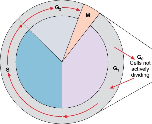
Your body makes billions of new cells every day. It needs them to grow, to heal a cut, and to replace skin and blood cells that wear out. Every new cell comes from an old cell that grew and split in two. The steps a cell follows to do this are called the cell cycle.
The cell cycle has two very unequal parts. The long part is interphase. This is the cell's normal working life. It grows, does its job, and quietly gets ready to divide. The short, dramatic ending is mitosis, when the nucleus actually splits. A cell spends most of its life in interphase. Mitosis is over quickly.
Interphase: grow, copy, get ready
Interphase has three stages. In G₁ (first gap), the cell grows, makes proteins, and does its normal work. In S phase (synthesis), the cell copies all of its DNA. That way, each new cell will get a full set. This is the most important step in getting ready to divide. In G₂ (second gap), the cell finishes growing. It builds the organelles it will need and checks the copied DNA for mistakes.
Only after all this does the cell commit to dividing. Checkpoints guard each step. They are quality-control stops, like a safety check before a plane takes off. At each one, the cell makes sure everything is in order before it moves on.
Mitosis: splitting the nucleus, then the cell
Mitosis splits the copied DNA evenly into two nuclei. It has four stages. You can remember them as PMAT.
In prophase, the copied chromatin packs into chromosomes you can see, and spindle fibers start to form. In metaphase, the chromosomes line up single file across the middle of the cell. In anaphase, the spindle fibers pull the two copies of each chromosome apart, toward opposite ends. In telophase, a new nuclear envelope forms around each of the two sets.
Mitosis splits the nucleus. A separate step, cytokinesis, pinches the rest of the cell in two. The result is two genetically identical daughter cells. Each has the same DNA, and each is ready to start its own interphase.
| Phase | Part of cycle | What happens |
|---|---|---|
| G₁ | Interphase | Cell grows and does its normal work |
| S | Interphase | All the DNA is copied |
| G₂ | Interphase | Last growth; DNA is checked for mistakes |
| Prophase | Mitosis | Chromatin packs into chromosomes; spindle forms |
| Metaphase | Mitosis | Chromosomes line up across the middle |
| Anaphase | Mitosis | Chromosome copies are pulled to opposite ends |
| Telophase | Mitosis | Two new nuclei form; cytokinesis splits the cell |
Step through interphase and the four phases of mitosis below.
- The cell cycle is a long interphase (G₁ → S → G₂) plus a short mitosis. DNA is copied in S phase.
- The order of mitosis is PMAT: prophase, metaphase, anaphase, telophase. Then cytokinesis splits the rest of the cell.
- The result is two genetically identical daughter cells.
- Checkpoints, guarded by genes like p53, stop damaged cells from dividing. When they fail, cancer can follow.
Section 6Osmosis & tonicity
Water chases salt
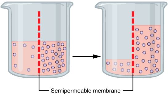
We saved the kind of transport that matters most in hospitals for last. Osmosis is the movement of water across a selectively permeable membrane. It follows one simple rule. Water moves toward the side with more dissolved particles. We call that side the one with more solute, or the "saltier" side.
The membrane keeps the solute from moving across to even things out. So water moves instead. It flows toward the saltier side until the two sides are balanced. A cell's membrane lets water pass freely but holds many solutes in place. So the amount of solute outside the cell decides which way water flows. That decides whether the cell gains or loses water.
Tonicity: three fluids, three results
Tonicity tells you how a fluid will change a cell's water balance. It compares the solute outside the cell to the solute inside. There are three cases.
An isotonic fluid has the same amount of solute as the inside of the cell. Water moves in and out equally, so the cell stays the same. This is its normal, healthy state.
A hypotonic fluid is more watery than the cell, with less solute outside. So water rushes in. The cell swells. If enough water gets in, the cell can burst. This is called lysis. In red blood cells it is called hemolysis.
A hypertonic fluid is saltier than the cell, with more solute outside. So water is pulled out. The cell shrivels. This shrinking is called crenation. Here is the trick to keep them straight. Water always follows the solute. Find the saltier side, and water goes there.
| Fluid | Outside vs. cell | Water moves | What happens to the cell |
|---|---|---|---|
| Isotonic | Same amount of solute | In = out (no overall change) | Stays normal |
| Hypotonic | Less solute outside (watery) | Into the cell | Swells and may burst (lysis) |
| Hypertonic | More solute outside (salty) | Out of the cell | Shrivels (crenation) |
Drop a cell into each fluid below and see what happens to it.
- Osmosis is water moving across a membrane toward the side with more solute.
- Isotonic → no overall change. Hypotonic → water goes in, and the cell swells or bursts (lysis). Hypertonic → water goes out, and the cell shrivels (crenation).
- Water always follows the solute. Find the saltier side.
- IV fluids use this rule. 0.9% normal saline is isotonic. Never give pure water into a vein.
The chapter in one breath
- Every cell is wrapped in a selectively permeable plasma membrane. The fluid mosaic model describes it as a fluid phospholipid bilayer with proteins, cholesterol, and sugars set into it.
- Things cross by passive transport or active transport. Passive means downhill and free: diffusion, facilitated diffusion, and osmosis. Active means uphill and costs energy: the Na⁺/K⁺ pump and bulk transport.
- Inside, organelles share the work. The nucleus stores DNA. Ribosomes → ER → Golgi build and ship proteins. Mitochondria make ATP. Lysosomes clean up.
- Protein synthesis flows from DNA → RNA → protein. Transcription copies a gene into mRNA in the nucleus. Translation reads its codons at a ribosome to build the protein.
- The cell cycle is a long interphase (G₁/S/G₂, with DNA copied in S). Then comes a short mitosis (PMAT) and cytokinesis. The result is two identical daughter cells. Broken checkpoints lead to cancer.
- Osmosis moves water toward the saltier side. Tonicity decides what happens to a cell. Isotonic keeps it normal. Hypotonic makes it swell or burst. Hypertonic makes it shrivel. This is exactly why IV fluids are chosen with care.
ReferenceWords worth knowing
Here are the key words from this chapter. Skim them now, or come back any time a word trips you up.
- Active transport
- Moving a substance up its concentration gradient, from low to high. It needs energy, such as ATP.
- Codon
- A group of three mRNA bases. It stands for one amino acid during translation.
- Crenation
- The shriveling of a cell that loses water in a hypertonic fluid.
- Cytokinesis
- The splitting of the cytoplasm after mitosis. It turns one cell into two.
- Cytoskeleton
- The inside frame of protein threads. It gives a cell shape and support and moves things around.
- Diffusion
- The free spreading of molecules from where there are many to where there are few, until they are evenly mixed.
- Fluid mosaic model
- The picture of the cell membrane as a fluid phospholipid bilayer. Proteins, cholesterol, and sugars sit in it like tiles.
- Golgi apparatus
- The organelle that changes, sorts, and packs proteins and fats into vesicles for delivery.
- Hypertonic
- A fluid with more solute than the cell. Water leaves the cell, and the cell shrivels.
- Hypotonic
- A fluid with less solute than the cell. Water enters the cell, and the cell swells or bursts.
- Interphase
- The long part of the cell cycle (G₁, S, G₂). The cell grows, copies its DNA, and gets ready to divide.
- Isotonic
- A fluid with the same amount of solute as the cell. Water does not move in or out overall, so the cell keeps its normal shape.
- Lysosome
- A sac of digestive enzymes wrapped in membrane. It breaks down junk, worn-out organelles, and things the cell takes in.
- Mitochondrion
- The organelle that makes ATP from food and oxygen. It is the cell's powerhouse.
- Mitosis
- The splitting of the nucleus into two nuclei with identical DNA. It goes through prophase, metaphase, anaphase, and telophase.
- Nucleus
- The control center of the cell, wrapped in membrane. It stores the cell's DNA and directs what the cell does.
- Osmosis
- The diffusion of water across a selectively permeable membrane. Water moves toward the side with more solute.
- Passive transport
- Moving a substance down its concentration gradient. It needs no energy from the cell.
- Phospholipid bilayer
- The double layer of amphipathic phospholipids that forms the core of the membrane. The water-loving heads face out, and the water-fearing tails face in.
- Sodium–potassium pump
- A primary active transport protein. For each ATP, it pushes 3 Na⁺ out and brings 2 K⁺ in. This keeps the cell's ion gradients in place.
- Transcription
- Copying a gene from DNA into messenger RNA. It happens inside the nucleus.
- Translation
- Reading mRNA codons at a ribosome to join amino acids into a protein.