Muscle Tissue
Muscle is the tissue that turns a choice into a movement. Every heartbeat, every blink, and every step is muscle at work. So is every push of food through your gut. Each one is a muscle cell doing the one thing muscle does best: shortening on command.
In this chapter we take one skeletal muscle apart, layer by layer. We keep going until we reach the tiny engine that actually pulls: the sarcomere. Then we watch that engine run. You will see how a nerve signal crosses into the muscle and how calcium flips the switch. You will see how threads of protein slide past each other. And you will see how the body turns the force up from a gentle flicker to an all-out heave. By the end, muscle action will not seem like magic. It will be a neat chain of events you can follow from thought to motion.
By the end of this chapter you can
- Compare the three types of muscle tissue: skeletal, cardiac, and smooth. Compare them by build, by control, and by where they are found.
- Trace the connective-tissue wrappings of a skeletal muscle, from the epimysium down to a single fiber. Name the parts inside that fiber.
- Draw a sarcomere and label its bands, zones, and lines. Explain why skeletal muscle looks striped.
- Explain the sliding filament theory and the steps of the cross-bridge cycle. Include the roles of ATP and calcium.
- Describe the neuromuscular junction. Follow a signal from a motor neuron to a muscle action potential.
- Define a motor unit. Explain how recruitment and summation raise muscle tension from a twitch to tetanus.
- Tell apart the three skeletal muscle fiber types. Match each one to the kind of work it does best.
Section 1How a muscle is built
Three kinds of muscle tissue
Before we open up one muscle, it helps to know that the body makes three kinds of muscle. Each is tuned to a different job.
Skeletal muscle is attached to bones and moves the body. It is under voluntary control, which means you choose when it works. Under the microscope its cells are long and striped (striated), and each has many nuclei.
Cardiac muscle is found only in the wall of the heart. It is striated too, but it is involuntary. That means it works without you thinking about it. Its cells are branched and most often have one nucleus. They are joined end to end by intercalated discs. These links let the whole heart squeeze as one.
Smooth muscle lines the walls of hollow organs, such as blood vessels, the gut, the bladder, and the airways. There it squeezes slowly and never seems to tire. It is involuntary and has no stripes. Its cells taper at both ends and have one nucleus in the middle.
This chapter focuses on skeletal muscle, because it shows every idea most clearly. But keep the other two in mind. Most of the machinery for squeezing is shared.
| Feature | Skeletal | Cardiac | Smooth |
|---|---|---|---|
| Location | Attached to bones | Heart wall only | Walls of hollow organs & vessels |
| Striations | Striated | Striated | Not striated |
| Control | Voluntary | Involuntary | Involuntary |
| Cell shape | Long tube shape, many nuclei | Branched, most often 1 nucleus | Spindle shape with tapered ends, 1 nucleus |
| Special feature | Controlled by motor neurons | Intercalated discs; sets its own rhythm | Slow and long-lasting; can trigger itself |
Bundles inside bundles
A whole skeletal muscle, such as your biceps, is not one solid block. It is a set of bundles inside bundles. Each bundle is wrapped in its own sheet of connective tissue. Let's go from the outside in.
The whole muscle is wrapped in the epimysium. Inside that, fibers are grouped into bundles called fascicles. Each fascicle is wrapped in perimysium. Inside each fascicle, every single muscle cell (a muscle fiber) is wrapped in a thin endomysium.
These three sheets are all joined to each other. At the ends of the muscle, they merge and reach past it. They form a tendon (a cord) or an aponeurosis (a flat sheet) that ties the muscle to bone. This setup is more than neat packing. The connective tissue carries blood vessels and nerves to each fiber. It also passes the pull of every cell to the tendon, so the whole muscle moves as one.
Inside a single fiber
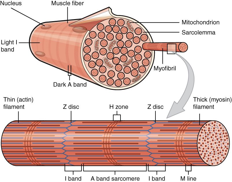
Zoom into one muscle fiber and you find a cell built for pulling. Its cell membrane is called the sarcolemma. Its cytoplasm, the jelly inside, is the sarcoplasm. The word part sarco- means “flesh.” The sarcolemma dives deep into the cell as tunnels called transverse (T) tubules. They carry the electrical signal into the core of the fiber.
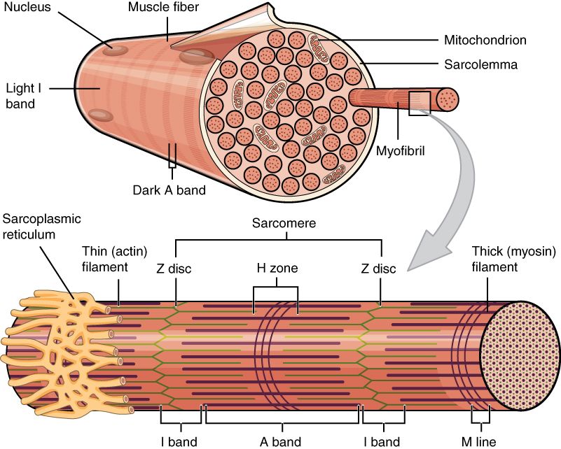
Wrapped around the working parts is the sarcoplasmic reticulum (SR). It is a special kind of smooth endoplasmic reticulum. It stores and releases the calcium that sets off a contraction. Most of the cell is filled with hundreds of long threads called myofibrils. Each myofibril is a chain of the units that shorten, the sarcomeres. These are made of myofilaments of two proteins, actin and myosin.
A muscle fiber forms when many cells fuse together as the body develops. So it holds many nuclei, pushed to the edge just under the sarcolemma. Nearby sit spare satellite cells that help repair damage.
Follow all of this in the interactive. Start at the whole muscle and zoom in, one wrapping at a time, down to the sarcomere.
- Three muscle types: skeletal (voluntary, striated, on bones), cardiac (involuntary, striated, in the heart), smooth (involuntary, not striated, in hollow organs).
- A skeletal muscle has bundles inside bundles: epimysium → fascicle (perimysium) → fiber (endomysium). All of these join the tendon.
- Inside a fiber: the sarcolemma, T-tubules, sarcoplasmic reticulum, and myofibrils of actin and myosin. There are also many nuclei at the edge.
Section 2The sarcomere
Stripes with a purpose
Skeletal and cardiac muscle have stripes. These stripes are not just for show. They are the outward sign of very orderly machinery inside. In every myofibril, the proteins that pull are lined up in the same repeating segments. Each segment is a sarcomere. It is the functional unit of contraction, the smallest part that can do the job. One sarcomere runs from one Z-disc to the next Z-disc.
When millions of these little units all shorten at once, lined up, the whole muscle shortens. Each unit has dark and light parts. Stacked side by side across the fiber, they look like stripes to our eyes. These are the striations.
The bands, zones, and lines
A sarcomere is built from two kinds of filament, or thread. The thick filaments are made of myosin. Each has a hinged head that sticks out. The thin filaments are made of actin. Where the two overlap, the sarcomere looks dark. Where only one kind is found, it looks lighter. It is worth learning this map of bands. In the next section you will watch these parts change size as the muscle contracts.
| Region | What it contains | Appearance |
|---|---|---|
| A band | The full length of the thick filaments (with overlap at its edges) | Dark; does not change length during contraction |
| I band | Thin filaments only, reaching into two sarcomeres side by side | Light; gets narrower during contraction |
| H zone | Thick filaments only, in the center of the A band (no overlap) | Lighter center; gets narrower during contraction |
| Z-disc (Z-line) | Holds the thin filaments in place; the border of the sarcomere | Dark line; Z-discs are pulled together |
| M-line | Holds the thick filaments at the center of the sarcomere | Thin line in the center |
Thick, thin, and the switch proteins
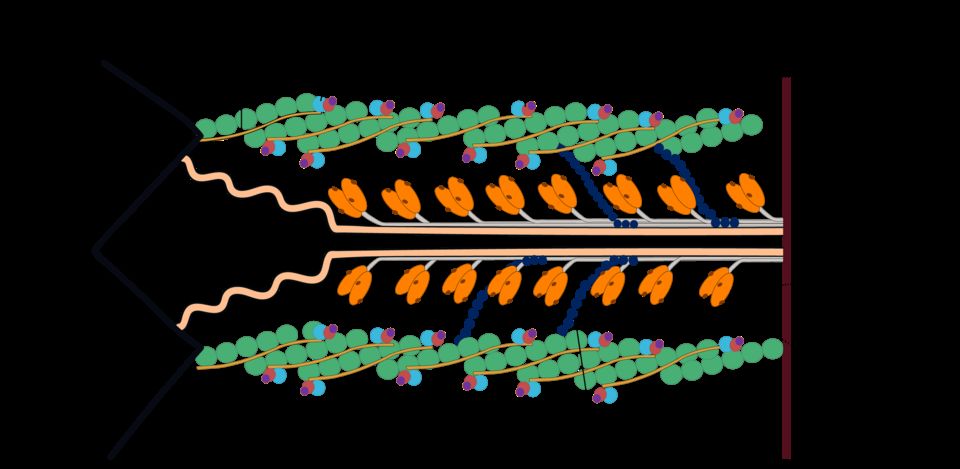
The thin filament is more than plain actin. A rope-like protein called tropomyosin lies along it. In a resting muscle, it covers the spots where myosin would grab on. These spots are the myosin-binding sites. A three-part protein group called troponin holds tropomyosin in place. One part of it, troponin C, senses calcium.
This is the switch. When calcium binds to troponin, the whole group shifts. Tropomyosin slides aside, and the binding sites open up.
Two more proteins keep the sarcomere in order. Titin is a huge, springy protein. It runs from the Z-disc to the M-line. It keeps the thick filament centered and lets muscle spring back when stretched. Nebulin runs along the thin filament and sets its length. Once again, the build is made exactly for the job.
Tap each part in the interactive to see how the filaments make the stripes.
- The sarcomere (Z-disc to Z-disc) is the functional unit of contraction. Sarcomeres lined up in order make the stripes you can see, the striations.
- Thick filaments are myosin, with heads. Thin filaments are actin. They are guarded by tropomyosin and by troponin, which senses calcium.
- The A band stays the same, while the I band and H zone narrow during contraction. This is the telltale sign of sliding filaments.
Section 3The sliding filament theory
Sliding, not shrinking
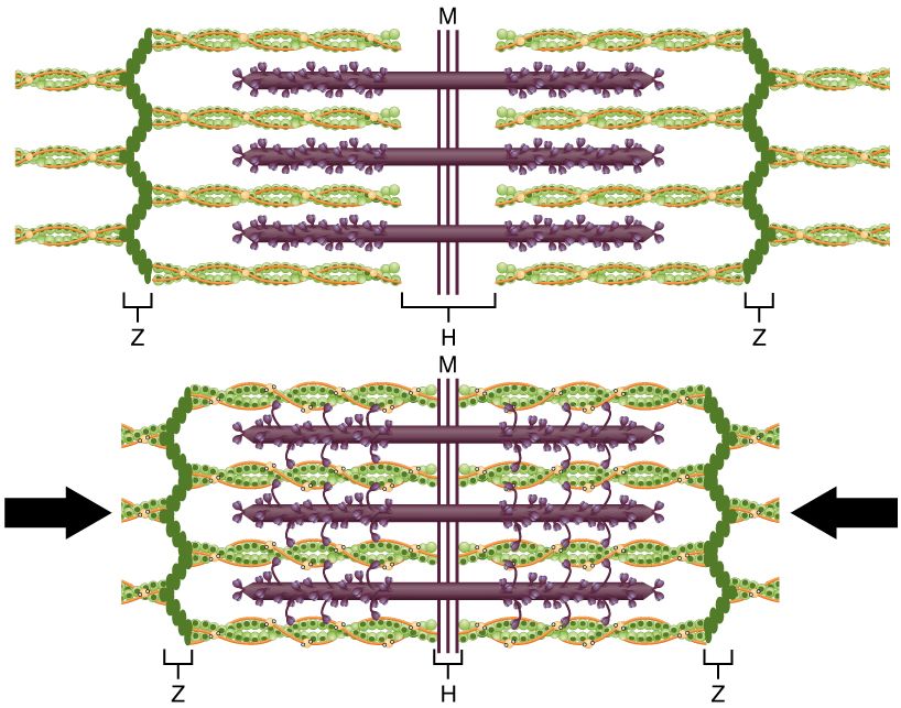
Here is the most important idea in this chapter. It surprises almost everyone the first time: the filaments do not get shorter. When a muscle contracts, the thick and thin filaments slide past each other. They overlap more and drag the Z-discs closer together. Each filament keeps its exact length. It is the sarcomere that shortens.
That is why the A band never changes in the reference figure. The A band is the length of the thick filament. Meanwhile the I band and H zone shrink, and the Z-discs move inward. Now add up that tiny slide across millions of sarcomeres lined up end to end. The whole muscle clearly shortens and pulls on its tendon.
The cross-bridge cycle
The myosin heads power the sliding. They act like tiny oars. Each head reaches out and grabs actin, forming a cross-bridge. Then it swivels. This is the power stroke. It pulls the thin filament a few nanometers toward the center. Then the head lets go, gets cocked again, and grabs again farther along. The cycle repeats many times each second. It runs on ATP, the cell's energy molecule:
| Step | What happens | Role of ATP / Ca²⁺ |
|---|---|---|
| 1. Cross-bridge forms | The charged-up myosin head binds to an open site on actin | Needs Ca²⁺ to have opened the site |
| 2. Power stroke | The head swivels and slides the thin filament inward; ADP & Pi (inorganic phosphate) are released | Uses energy stored from an earlier ATP |
| 3. Detachment | A new ATP binds the myosin head, and the head lets go of actin | ATP binding breaks the cross-bridge |
| 4. Re-cocking | ATP is split into ADP + Pi, which charges up and resets the head | ATP hydrolysis (splitting ATP) cocks the head again |
Notice that ATP has two jobs. It is needed to release the head (step 3). It is also needed to re-cock it (step 4). This leads to something you may have heard of. After death, the body stops making ATP. The myosin heads bind actin and can no longer let go. So the body stiffens. This is called rigor mortis.
Calcium throws the switch: excitation–contraction coupling
Cross-bridges can form only if the binding sites are uncovered. That is calcium's job. The link between an electrical signal and a pull is called excitation–contraction coupling. Here is how it works.
A muscle action potential, an electrical signal, races along the sarcolemma. It dives down the T-tubules into the fiber. That change in voltage tells the sarcoplasmic reticulum to release its stored Ca²⁺ into the sarcoplasm. Calcium binds to troponin. Troponin pulls tropomyosin off the actin. The binding sites open, and cross-bridges begin to form. The filaments start to slide.
Relaxing is just the reverse. When the signal stops, a pump called SERCA moves Ca²⁺ back into the SR. Tropomyosin covers the sites again. Cross-bridges can no longer form, and the muscle goes slack. No calcium, no contraction.
In the interactive, watch the Z-lines pull together while the filaments keep their length.
- In the sliding filament theory, the filaments keep their length. The sarcomere shortens as the thick and thin filaments overlap more.
- The cross-bridge cycle has four steps: bind, power stroke, detach, re-cock. It runs on ATP, which is needed to release and to re-charge the myosin head.
- Excitation–contraction coupling: an action potential → T-tubules → SR releases Ca²⁺ → troponin/tropomyosin uncover the binding sites. To relax, SERCA pumps the calcium back.
Section 4The neuromuscular junction
From nerve to muscle
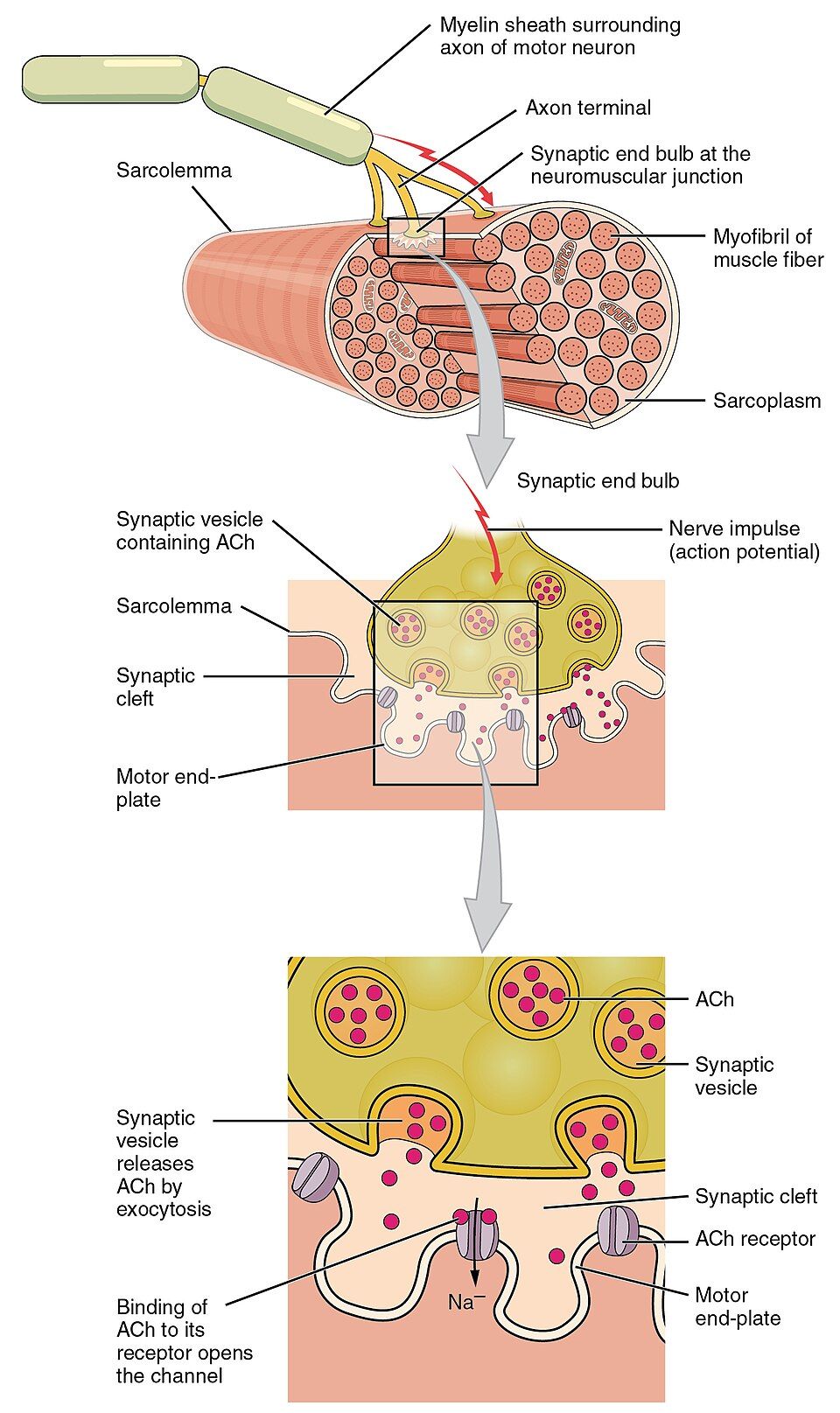
A skeletal muscle fiber will not contract on its own. It waits for a command from a nerve. That command is delivered at a special synapse, or meeting point, called the neuromuscular junction (NMJ). A motor neuron reaches out from the spinal cord. The end of its axon rests just above a folded patch of sarcolemma. This patch is the motor end plate.
The two never touch. A tiny gap, the synaptic cleft, separates them. To cross that gap, the neuron uses a chemical messenger. This is the neurotransmitter acetylcholine (ACh).
The handoff, step by step
Passing the signal from nerve to muscle is one of the most reliable events in the body. A single nerve impulse almost always sets off a muscle impulse. Follow the chain:
| Step | Event |
|---|---|
| 1 | A nerve action potential reaches the end of the axon (the axon terminal). |
| 2 | Voltage-gated Ca²⁺ channels open. Calcium rushes into the axon terminal. |
| 3 | Calcium makes small sacs (vesicles) release acetylcholine into the synaptic cleft. |
| 4 | ACh crosses the cleft and binds to nicotinic receptors on the motor end plate. |
| 5 | The receptor channels open. Na⁺ floods in and makes an end-plate potential. |
| 6 | This change in charge sets off a muscle action potential. It spreads along the sarcolemma and down the T-tubules. Then contraction follows. |
See how this fits with the last section. Step 6 is the very same muscle action potential that starts excitation–contraction coupling. The NMJ is the doorway. The sliding filaments are the room it opens into.
Switching the signal off, and where it breaks
A signal that never ended would leave the muscle stuck in contraction. So the cleft holds an enzyme called acetylcholinesterase (AChE). It chops ACh apart within milliseconds. This neat on/off system is also where many drugs and diseases act. That makes the NMJ one of the most important spots in medicine.
In myasthenia gravis, the immune system destroys ACh receptors. So muscles tire quickly and grow weak. Curare, an arrow poison, blocks the receptors and causes paralysis. Organophosphate bug sprays and nerve agents block AChE. Then ACh piles up and muscles seize. Botulinum toxin (Botox) blocks the release of ACh, so muscle relaxes. This is useful for treating spasms and wrinkles.
Step through the chemical handoff yourself in the interactive.
- The neuromuscular junction is the synapse where a motor neuron signals a muscle fiber. It uses acetylcholine to cross the synaptic cleft.
- ACh binds nicotinic receptors on the motor end plate. This lets Na⁺ in, which triggers a muscle action potential. Then acetylcholinesterase ends the signal.
- NMJ problems follow the messenger. Blocking ACh → weakness (myasthenia gravis, curare, botulinum toxin). Letting ACh last too long → spasm (AChE blockers, nerve agents).
Section 5Motor units & muscle tension
The motor unit
One motor neuron rarely controls just one muscle fiber. It branches and connects to a group of fibers. The neuron plus all the fibers it controls is a motor unit. When that neuron fires, every fiber in its unit contracts at once. They act as an all-or-nothing team.
The size of the unit sets how finely a muscle can be controlled. Muscles that need a gentle touch, like those that move the eye or the fingers, have small motor units. Each neuron links to only a few fibers. Muscles built for power, like the quadriceps or the calf, have large motor units. Each neuron links to hundreds of fibers. Small units mean fine control. Large units mean strong, rough force.
Twitch, summation, tetanus
One short stimulus gives a single quick contraction and relaxation. This is a twitch. It has three phases. First is a latent period, a hidden delay while calcium is released. Next is a contraction phase, then a relaxation phase. A twitch is a flick, not a useful pull.
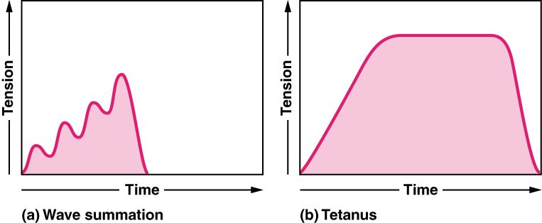
To get smooth, strong movement, the nervous system fires the motor neuron again before the fiber fully relaxes. So the second contraction stacks on top of the first. This stacking is wave (temporal) summation. Fire even faster, and the twitches blur into a partly smooth pull. This is unfused (incomplete) tetanus. Faster still, and you get a steady, perfectly smooth, all-out pull. This is fused (complete) tetanus. Almost all normal movements use tetanus, not single twitches.
| Response | Stimulus pattern | Result |
|---|---|---|
| Twitch | One stimulus | One short contraction, then full relaxation |
| Wave summation | A second stimulus before relaxation ends | The second contraction adds to the first, giving more tension |
| Unfused tetanus | Fast, repeated stimuli | Strong, lasting pull with slight wobbles |
| Fused tetanus | Stimuli at a very high rate | All-out, perfectly smooth, lasting contraction |
Two dials for force: recruitment and rate
The body controls how hard a muscle pulls with two dials at once. The first dial is recruitment, which means turning on more motor units. It follows the size principle. Small, weak units that resist tiring are turned on first. Larger, stronger ones join only when more force is needed. So movements start smooth and easy to control.
The second dial is rate coding. This means firing each active unit faster, to climb from summation up to tetanus. With both dials, the same muscle can lift a feather or a heavy weight. Even at rest, a few units are always active, taking turns. They keep muscles firm and ready. This steady, low tension is muscle tone.
Pick a stimulus pattern in the interactive. Watch tension climb from twitch to tetanus.
- A motor unit is one motor neuron plus all the fibers it controls. Small units give fine control. Large units give power.
- Tension builds from a twitch through wave summation to tetanus as the stimuli come faster. Real movement uses tetanus.
- Force is set by recruitment (adding units, smallest first; this is the size principle) and rate coding (firing faster). The steady activity at rest is muscle tone.
Section 6The three fiber types
Speed versus stamina
Not every skeletal muscle fiber is the same. There are three types. They trade speed against endurance, or staying power. No one design can be both as fast as possible and as tireless as possible.
Slow oxidative (Type I) fibers contract slowly and make only modest force. But they can keep going for hours. They are the endurance experts. Fast glycolytic (Type IIx) fibers contract quickly and with great power. But they tire within minutes. They are the experts at sprinting and lifting. In between are fast oxidative (Type IIa) fibers. They are fast and fairly slow to tire, a useful middle ground.
Aerobic versus anaerobic engines
The difference comes down to how each fiber makes its ATP. Oxidative fibers use aerobic respiration, which needs oxygen. They are packed with mitochondria, the cell's power plants. They have lots of capillaries, the tiniest blood vessels. They are also loaded with myoglobin, a pigment that holds oxygen and makes them look red. This red, oxygen-fed system is slow but can keep going.
Glycolytic fibers rely on anaerobic glycolysis, which does not need oxygen. They have few mitochondria and little myoglobin, so they look pale. They store lots of glycogen, a sugar fuel, for quick bursts. They give explosive power. But they fill up with lactate and tire fast.
| Property | Slow oxidative (I) | Fast oxidative (IIa) | Fast glycolytic (IIx) |
|---|---|---|---|
| Contraction speed | Slow | Fast | Fast |
| Fatigue resistance | High | Moderate | Low |
| Main ATP source | Aerobic (oxidative) | Both | Anaerobic (glycolytic) |
| Mitochondria / capillaries | Many | Many | Few |
| Myoglobin / color | High / red | High / red-pink | Low / white |
| Best for | Posture, endurance | Walking, steady effort | Sprinting, lifting |
Everybody is a mixture, plus a word on the other two muscles
Almost every real muscle has all three fiber types. The mix is set partly by your genes and partly by how you train. Endurance training moves fibers toward the oxidative end, where they resist tiring. Strength and sprint training push toward bigger, more powerful fibers. That is why a marathon runner's calves and a sprinter's thighs are built for different feats.
Now recall the two muscle types we set aside at the start. Cardiac muscle acts like tissue that is always tuned for endurance. It sets its own rhythm, and intercalated discs join its cells, so the heart never truly rests. Smooth muscle has no sarcomeres at all. It can hold tension for a long time using very little energy. This “latch” state is perfect for keeping a blood vessel or a sphincter squeezed all day.
Tap each fiber type in the interactive to compare speed, color, and stamina.
- Three fiber types trade speed for stamina. Slow oxidative (I) = endurance. Fast glycolytic (IIx) = power. Fast oxidative (IIa) = in between.
- Oxidative fibers are red (many mitochondria, lots of myoglobin and capillaries) and use oxygen. Glycolytic fibers are pale and do not use oxygen. They are powerful but tire quickly.
- Most muscles are a mix shaped by genes and training. Cardiac and smooth muscle are separate designs, built for work that never stops.
The chapter in one breath
- Muscle comes in three types. They are skeletal (voluntary, striated), cardiac (involuntary, striated, sets its own rhythm), and smooth (involuntary, not striated). All share the same basic machinery for contraction.
- A skeletal muscle is bundles inside bundles. They are wrapped in epimysium, perimysium, and endomysium, which all join the tendon. Inside each fiber are the sarcolemma, T-tubules, sarcoplasmic reticulum, and myofibrils.
- The sarcomere (Z-disc to Z-disc) is the functional unit. Overlapping thick (myosin) and thin (actin) filaments make the stripes. Tropomyosin and troponin guard the binding sites.
- In the sliding filament theory, the filaments keep their length while the sarcomere shortens. The cross-bridge cycle runs on ATP. Ca²⁺ released by the SR uncovers the binding sites.
- The neuromuscular junction passes the signal from nerve to muscle using acetylcholine. Then acetylcholinesterase clears it away. Toxins here cause weakness or spasm.
- A motor unit is one neuron and its fibers. Tension rises from a twitch to tetanus. It is controlled by recruitment and rate coding.
- Three fiber types trade speed for endurance. Most muscles blend all three, in a mix shaped by genes and training.
ReferenceWords worth knowing
Here is a quick list of the terms in this chapter. Skim it now, or come back when a word trips you up.
- A band
- The dark part of a sarcomere that spans the full length of the thick filaments. It does not change length during contraction.
- Acetylcholine (ACh)
- The neurotransmitter a motor neuron releases at the neuromuscular junction. It sets off a muscle action potential.
- Actin
- The protein of the thin filament. It has the myosin-binding sites where cross-bridges attach.
- Cross-bridge
- The link made when a myosin head binds an open site on actin. Its cycle of binding, power stroke, and release makes the filaments slide.
- Endomysium
- The thin sheet of connective tissue that wraps each single muscle fiber.
- Epimysium
- The sheet of connective tissue that wraps a whole skeletal muscle.
- Excitation–contraction coupling
- The chain of events that links a muscle action potential to contraction. The signal travels down the T-tubules and makes the sarcoplasmic reticulum release Ca²⁺.
- Fascicle
- A bundle of muscle fibers wrapped in perimysium.
- Motor unit
- One motor neuron plus all the muscle fibers it controls. They contract as one.
- Myofibril
- A long thread inside a muscle fiber. It is made of a chain of sarcomeres.
- Myosin
- The protein of the thick filament. Its hinged heads form cross-bridges and do the power stroke.
- Neuromuscular junction
- The synapse between a motor neuron and a skeletal muscle fiber. Here acetylcholine passes on the signal.
- Recruitment
- Raising muscle force by turning on more motor units. The smallest units, which tire the least, go first (the size principle).
- Sarcolemma
- The cell membrane of a muscle fiber. Its tunnels into the cell are the T-tubules.
- Sarcomere
- The functional unit of striated muscle, the part that shortens. It runs from one Z-disc to the next.
- Sarcoplasmic reticulum (SR)
- The special smooth ER of a muscle fiber. It stores and releases the Ca²⁺ that sets off contraction.
- Sliding filament theory
- The idea that muscle shortens because thick and thin filaments slide past each other. The filaments themselves do not get shorter.
- Tetanus
- A lasting contraction. It happens when stimuli come faster than the fiber can fully relax between them. It is unfused (incomplete) when some relaxing still shows as slight wobbles. It is fused (complete), smooth and all-out, when stimuli come at a very high rate.
- Tropomyosin
- The thin-filament protein that covers the myosin-binding sites at rest. It slides aside when calcium binds troponin.
- Troponin
- The thin-filament protein group that binds Ca²⁺. It moves tropomyosin to uncover the binding sites.
- Twitch
- A single short contraction and relaxation of a muscle fiber after one stimulus. It has latent, contraction, and relaxation phases.