The Cardiovascular System: Blood
Blood is the only tissue in your body you can pour into a test tube. It is easy to think little of it, since it looks like a plain red liquid. But that liquid is a connective tissue. It is a living fleet of cells floating in a sea full of proteins, and it touches every other cell you have. In this chapter we take a tube of blood apart, layer by layer. There is the straw-colored plasma on top and the packed red cells below. Between them is a thin white line. You will learn what each part carries and where blood cells are made. You will see how a red cell carries oxygen and how a cut seals itself in seconds. You will also learn why matching a patient's blood type before a transfusion can mean life or death.
By the end of this chapter you can
- List the main functions of blood: transport, regulation, and protection. Explain why blood counts as a connective tissue.
- Describe the layers a spun tube of blood splits into. Define plasma, buffy coat, and hematocrit.
- Name the three formed elements and the five kinds of white blood cell. Give the main job of each.
- Trace hematopoiesis, the making of blood cells, from one stem cell down the myeloid and lymphoid lines. Name the hormone that drives red cell making.
- Explain how hemoglobin picks up oxygen in the lungs and drops it off in the tissues. Explain how blood carries carbon dioxide.
- Walk through the three steps of hemostasis. Describe how the clotting chain builds a mesh of fibrin.
- Predict which ABO and Rh blood types match. Name the universal donor and the universal recipient.
Section 1What blood is made of
A tissue you can pour
Many people are surprised to hear that blood is a connective tissue. A connective tissue has cells spread out in an extracellular matrix, a material that fills the space between cells. Blood fits this perfectly. Its "cells" are the red cells, white cells, and platelets. Its matrix is the liquid plasma they float in. What makes blood unusual is that its matrix is a fluid. In bone the matrix is hard, and in cartilage it is rubbery.
An average adult has about 5 liters of blood. Women have a little less and men a little more. Blood makes up about 8 percent of body weight. It is heavier and thicker than water, about five times as thick (viscous). It is warm, at about 38 °C. It is also slightly basic (alkaline). Its pH stays in a narrow range, from 7.35 to 7.45.
Three jobs: transport, regulation, protection
Everything blood does fits into three groups. The first is transport, or carrying things. Blood brings O₂ and nutrients to cells. It takes away CO₂ and other wastes. It also carries hormones from glands to targets far away.
The second job is regulation, or keeping things steady. Blood can change where it flows and how much heat it takes to the skin. This helps keep body temperature steady. Chemicals in blood called buffers keep the body's pH steady. Blood proteins help control fluid balance between the blood and the tissues.
The third job is protection. White cells and antibodies fight germs. The clotting system seals leaks, so you do not bleed out from a small cut.
Spin it down and it splits in three
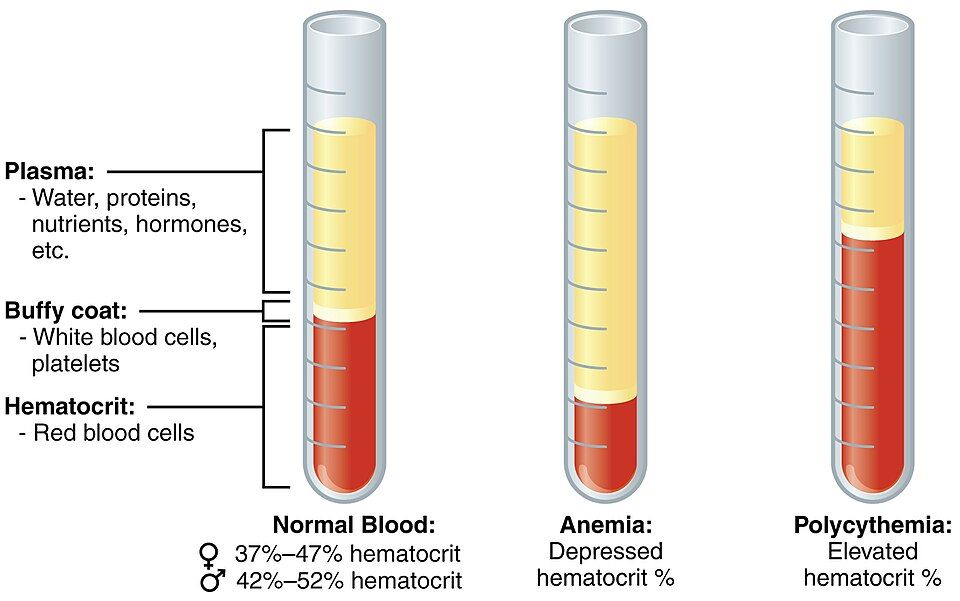
Put a tube of blood in a spinning machine called a centrifuge, and the heavier parts sink. You end up with three layers you can see. How big each layer is tells doctors a lot:
| Layer | What it holds | Share of blood |
|---|---|---|
| Plasma (top) | Water, plasma proteins, nutrients, gases, hormones, wastes, ions (salts) | About 55% |
| Buffy coat (middle) | Leukocytes (white cells) and platelets | Less than 1% |
| Erythrocytes (bottom) | Red blood cells. This share is the hematocrit. | About 45% |
That bottom layer has its own name. The hematocrit is the percent of whole blood that is red cells. Normal values are about 37–47 percent in women and 42–52 percent in men.
Plasma itself is about 92 percent water. Most of the rest is plasma proteins. Albumin is the most common one. It holds water inside the blood vessels and carries fatty molecules. Globulins include antibodies and proteins that carry things. Fibrinogen is the raw material for clots. One more term to learn now is serum. If you let blood clot and take the clot out, a pale fluid is left. That is serum, which is plasma without its clotting proteins.
Spin your own tube and pull apart the layers in the interactive below.
- Blood is a fluid connective tissue. It is cells (formed elements) floating in a liquid matrix (plasma). An adult has about 5 L, which is 8% of body weight.
- Its jobs fall into three groups: transport, regulation, and protection.
- Spun blood gives plasma (~55%), a thin buffy coat of white cells and platelets, and red cells (hematocrit, ~45%).
- Serum is plasma with the clotting proteins taken out.
Section 2The formed elements
Three kinds of passenger
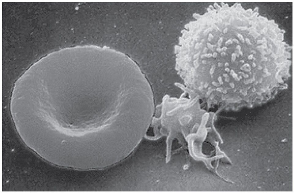
The living cargo of blood is called the formed elements. They come in three types. Erythrocytes are red blood cells. Leukocytes are white blood cells. Platelets are also called thrombocytes.
Their numbers are very uneven. Red cells rule, with about 5 million per microliter (a tiny drop) of blood. Platelets number a few hundred thousand. White cells are the rarest, with only about 5,000 to 10,000 per microliter. That is why the spun tube from Section 1 is almost all red on the bottom. The buffy coat is only a thin line.
Red cells: oxygen with everything stripped away
An erythrocyte is built to do one job well. It is a biconcave disc. That means it is a flat circle that is pinched in on both sides, like a donut with no hole. This shape gives it lots of surface for swapping gases. It also lets the cell fold so it can squeeze through capillaries, the tiny vessels narrower than itself.
The mature red cell makes room for as much hemoglobin as it can. To do this, it throws almost everything else out. It has no nucleus and no organelles, the small working parts of a cell. This has a cost. With no nucleus, the cell cannot repair itself. So it wears out in about 120 days. Then the spleen and liver recycle it. We will follow its oxygen work in Section 4.
White cells: the mobile defense force
Unlike red cells, every leukocyte keeps its nucleus. All of them are soldiers of the immune system. They split into two families. The split depends on whether they have tiny grains (granules) you can see.
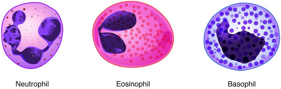
The granulocytes have grainy insides. They are neutrophils, eosinophils, and basophils. The agranulocytes have no visible grains. They are lymphocytes and monocytes. To learn them from most to least common, use this memory trick. Never Let Monkeys Eat Bananas stands for Neutrophils, Lymphocytes, Monocytes, Eosinophils, Basophils.
| White cell | Group | Rough share | Main job |
|---|---|---|---|
| Neutrophil | Granulocyte | 50–70% | First to arrive; eats bacteria (phagocytosis) |
| Lymphocyte | Agranulocyte | 20–30% | Targeted (adaptive) immunity: T, B, and NK cells |
| Monocyte | Agranulocyte | 2–8% | Turns into a macrophage, a big cell that eats germs |
| Eosinophil | Granulocyte | 1–4% | Attacks parasites; plays a part in allergy |
| Basophil | Granulocyte | <1% | Releases histamine and heparin; causes inflammation |
Platelets: fragments that plug leaks
Platelets are not whole cells at all. They are cell fragments, small pieces pinched off from giant marrow cells. These giant cells are called megakaryocytes. Platelets have no nucleus and live only about 10 days. They have one purpose. They rush to a wound, pile up, and start a clot. We will watch them do it in Section 5.
Meet all three formed elements, and the five white cells, in the interactive.
- The three formed elements are erythrocytes (oxygen), leukocytes (defense), and platelets (clotting). Red cells far outnumber the rest.
- The red cell is a biconcave disc with no nucleus. It is packed with hemoglobin and lasts about 120 days.
- The five white cells, from most to least: neutrophils, lymphocytes, monocytes, eosinophils, basophils (Never Let Monkeys Eat Bananas).
- Platelets are megakaryocyte fragments that start clotting.
Section 3Where blood cells are born
One stem cell, every blood cell
Red cells wear out in four months. Neutrophils wear out in just days. So your body must replace blood cells all the time. It makes hundreds of billions each day. The factory is the red bone marrow. The whole process of making blood cells is called hematopoiesis.
Amazingly, every one of these cells comes from a single kind of parent cell. It is the hematopoietic stem cell, also called a hemocytoblast. It is multipotent. This means it can become any blood cell, but only blood cells. It cannot become liver, nerve, or muscle. It also renews itself, so the supply never runs out. (Save the word pluripotent for embryonic stem cells and induced pluripotent stem cells. They can make tissues from all three germ layers, the first cell layers of an embryo.)
Two family lines: myeloid and lymphoid
Early on, the stem cell's offspring pick one of two branches. The myeloid line is the bigger one. It makes red cells and platelets (by way of megakaryocytes). It also makes most white cells: neutrophils, eosinophils, basophils, and monocytes. The lymphoid line makes only the lymphocytes. These are the T, B, and NK cells of targeted (adaptive) immunity.
Knowing which line a cell comes from is more than trivia. Blood cancers called leukemias are sorted as myeloid or lymphoid. The name depends on which branch of this tree went wrong.
| Line | Cells it makes | Notice |
|---|---|---|
| Myeloid | Erythrocytes, platelets, neutrophils, eosinophils, basophils, monocytes | The broad branch: almost everything |
| Lymphoid | Lymphocytes (T cells, B cells, NK cells) | The specialist branch: adaptive immunity |
Hormones set the pace
Blood cell making is not left to chance. Hormones and growth factors match it to what the body needs. The star is erythropoietin (EPO). The kidneys release it when they sense low oxygen. EPO tells the marrow to make more red cells. Thrombopoietin comes from the liver and drives platelet making. A group of colony-stimulating factors and interleukins boosts white cell making during an infection.
Building a red cell also needs raw materials. You need iron for hemoglobin. You also need vitamin B₁₂ and folate to make DNA. Run short on any of these and you become anemic. New red cells that are not quite finished are called reticulocytes. Doctors count them to see how hard the marrow is working.
Climb the family tree, one step at a time, in the interactive.
- Hematopoiesis happens in red bone marrow. Every blood cell comes from a multipotent hematopoietic stem cell.
- The stem cell branches into the myeloid line (red cells, platelets, most white cells) and the lymphoid line (lymphocytes).
- Erythropoietin from the kidney drives red cell making. Iron, B₁₂, and folate are the raw materials.
Section 4How blood carries oxygen
The molecule that does the carrying
Red cells matter because of one molecule, hemoglobin. Each hemoglobin is built from four protein chains. Two are alpha chains and two are beta chains, and they are called globins. Each chain holds a heme group. There is an atom of iron at the center of each heme. Oxygen binds to that iron.
One hemoglobin has four heme groups. So it can carry four O₂ molecules at once. A single red cell is stuffed with about 270 million hemoglobins. So one cell can carry over a billion oxygen molecules. Iron is the actual seat that oxygen sits on. That is why low iron so directly starves the tissues of oxygen.
Load in the lungs, unload in the tissues
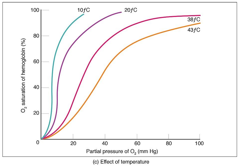
Hemoglobin's genius is that it grabs oxygen where there is plenty. Then it lets go where oxygen is scarce. In the lungs, the pressure of oxygen (partial pressure) is high. Here hemoglobin binds O₂ and becomes oxyhemoglobin. This oxygen-rich blood is a bright scarlet red.
Out in the tissues, cells have used up their oxygen. Here the oxygen pressure is low. Hemoglobin lets go of its load and becomes deoxyhemoglobin, a darker brick red. That color change is why blood in arteries looks so much brighter than blood in veins.
The binding is also cooperative. Once the first O₂ is on, the next ones bind more easily. So loading and unloading both happen fast. Some conditions push hemoglobin to release more oxygen. These are warmth, extra CO₂, and more acid. These are exactly the conditions in a hard-working muscle. So oxygen is dropped off right where it is needed.
And the return trip: carbon dioxide
Blood does not just bring oxygen. It also clears away the CO₂ that cells make. This waste rides back in three ways. The shares are worth knowing:
| Gas | How it travels | Share |
|---|---|---|
| O₂ | Bound to hemoglobin as oxyhemoglobin | ~98.5% |
| O₂ | Dissolved in plasma | ~1.5% |
| CO₂ | As bicarbonate (HCO₃⁻) in plasma | ~70% |
| CO₂ | Bound to hemoglobin (carbaminohemoglobin) | ~23% |
| CO₂ | Dissolved in plasma | ~7% |
Watch a red cell load up and drop off, over and over, in the interactive.
- Hemoglobin has four globin chains, each with an iron-holding heme. Each molecule carries up to four O₂.
- It forms oxyhemoglobin (bright red) in the lungs. It releases O₂ and becomes deoxyhemoglobin (darker) in the tissues.
- CO₂ travels back mostly as bicarbonate (~70%). Some rides on hemoglobin, and a little is dissolved.
- Carbon monoxide beats oxygen to hemoglobin and blocks delivery.
Section 5How bleeding stops
Three fast steps
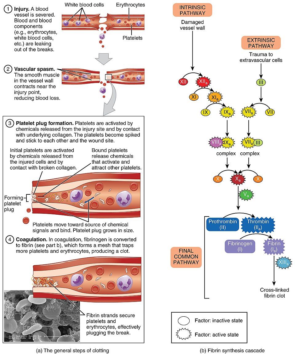
When a blood vessel is cut, the body must seal the leak before too much blood escapes. This is called hemostasis, which means "blood standing still." It happens in three steps that overlap. Each step buys time for the next. The whole thing can start within seconds.
| Step | What happens | Result |
|---|---|---|
| 1. Vascular spasm | Smooth muscle in the vessel wall tightens near the injury | The vessel narrows, which slows blood loss |
| 2. Platelet plug | Platelets stick to bare collagen, switch on, and pile up | A soft plug fills the hole |
| 3. Coagulation | A chain of clotting factors builds a fibrin mesh | A firm clot locks the plug in place |
The platelet plug snowballs
A vessel has a smooth inner lining. When it tears, the tough collagen under it is exposed. Platelets grab onto the collagen. A linking protein called von Willebrand factor helps them hold on. This contact changes platelets from smooth discs into spiky, sticky, active platelets.
Active platelets then release chemicals. These are ADP, thromboxane A₂, and serotonin. They call in and switch on even more platelets. This is a classic positive feedback loop. Each new platelet calls in others, so the plug grows fast until the hole is covered. This is also the step that aspirin weakens. Aspirin blocks thromboxane. That is why low-dose aspirin makes platelets less sticky.
Coagulation: turning liquid into gel
A platelet plug alone is weak. Coagulation, or clotting, makes it strong. A relay of plasma proteins called clotting factors switch each other on in a cascade, like falling dominoes. There are two starting paths, the intrinsic and the extrinsic pathway. They join into one common pathway.
The end has two moves. First, the enzyme thrombin is switched on. Then thrombin turns fibrinogen, a plasma protein that dissolves, into threads of fibrin that do not dissolve. These threads weave a net that traps red cells and platelets. That net is the actual clot.
Two helpers work behind the scenes. Calcium (clotting factor IV) is needed at several steps. The liver needs vitamin K to build several of the factors in the first place. Once the wound heals, an enzyme called plasmin breaks up the clot. This is called fibrinolysis, which means "fibrin breaking."
Step through how the leak is sealed and then made strong in the interactive.
- Hemostasis has three steps: vascular spasm → platelet plug → coagulation.
- The platelet plug grows by positive feedback, as active platelets call in more. Aspirin weakens this step.
- Coagulation ends with thrombin turning fibrinogen into a fibrin mesh. It needs calcium and vitamin K.
Section 6Blood types and transfusion
Antigens on the cell, antibodies in the plasma
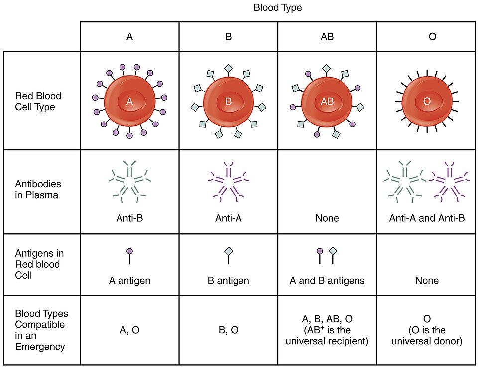
Your blood type depends on tiny flags on the surface of your red cells. These flags are called antigens (agglutinogens). In the ABO system there are two possible flags, A and B. Your genes decide which ones you have.
Here is the key rule. Your plasma has antibodies (agglutinins) against any antigen you lack. A type A person has A antigens and anti-B antibodies. A type B person has B antigens and anti-A antibodies. A type AB person has both antigens and no ABO antibodies. A type O person has neither antigen, but has both anti-A and anti-B.
Why a mismatch is dangerous
Say a patient gets red cells with an antigen that their plasma has antibodies against. Those antibodies grab the donor cells and clump them together. This is called agglutination. Then the cells burst, which is called hemolysis. The broken pieces can clog small vessels and overload the kidneys. A serious transfusion reaction can kill. That is why blood is always typed and cross-matched before it is given.
| Type | Antigen on red cells | Antibody in plasma | Can receive from |
|---|---|---|---|
| A | A | Anti-B | A, O |
| B | B | Anti-A | B, O |
| AB | A and B | None | A, B, AB, O: the universal recipient |
| O | None | Anti-A and Anti-B | O only. It is the universal donor of red cells. |
The Rh factor
On top of ABO is the Rh system. It is named for the rhesus monkey, which was used in the tests that found it. It has several antigens: C, c, D, E, and e. But one of them, D, triggers the immune system by far the most. It is the only one tested for as a routine. If you have D, you are Rh-positive. If not, you are Rh-negative.
ABO antibodies are there from early in life. Rh antibodies are different. An Rh-negative person makes anti-D antibodies only after being exposed to Rh-positive blood. This fact leads to a very important case in pregnancy care. It is called hemolytic disease of the newborn.
Say an Rh-negative mother carries an Rh-positive baby. Her body can become sensitized to the D antigen, which means it learns to attack it. In a later Rh-positive pregnancy, her antibodies may cross the placenta. There they attack the baby's red cells. The fix is simple and clever. A shot of Rh immune globulin (RhoGAM) clears the baby's cells from her blood before her immune system can react.
Pick a blood type in the interactive. See exactly who it can give to and take from.
- Your blood type is set by the antigens on your red cells. Your plasma holds antibodies against the antigens you lack.
- A mismatched transfusion causes agglutination and hemolysis. So blood is typed and cross-matched first.
- O-negative is the universal red cell donor. AB-positive is the universal recipient.
- Rh-negative mothers with Rh-positive babies risk hemolytic disease of the newborn. RhoGAM prevents it.
The chapter in one breath
- Blood is a fluid connective tissue, with formed elements floating in plasma. It handles transport, regulation, and protection.
- Spun blood splits into plasma (~55%), a thin buffy coat, and red cells (hematocrit, ~45%). Plasma is mostly water plus albumin, globulins, and fibrinogen.
- The formed elements are erythrocytes (oxygen), five kinds of leukocyte (defense), and platelets (clotting).
- Hematopoiesis in red marrow starts from one stem cell. It branches into the myeloid and lymphoid lines. The kidney's erythropoietin drives red cell making.
- Hemoglobin carries four O₂ on iron-heme seats. It loads in the lungs and unloads in the tissues. CO₂ rides back mostly as bicarbonate.
- Hemostasis seals a wound in three steps: vascular spasm, platelet plug, and coagulation. It ends in a fibrin mesh.
- Blood type is set by red cell antigens (A, B, Rh). Plasma holds antibodies against the ones you lack. This makes O-negative the universal donor and AB-positive the universal recipient.
ReferenceWords worth knowing
Here is a quick list of the terms in this chapter. Skim it now, or come back whenever a word trips you up.
- Agglutination
- The clumping of red blood cells when antibodies bind matching antigens. It happens in a mismatched transfusion.
- Albumin
- The most common plasma protein. It holds water in the blood vessels (osmotic pressure) and carries fatty molecules and drugs.
- Antibody (agglutinin)
- A plasma protein that finds and binds one certain antigen. ABO antibodies target the A or B antigen a person lacks.
- Antigen (agglutinogen)
- A marker on the surface of red cells (A, B, or Rh D). It sets your blood type and can trigger an immune response.
- Buffy coat
- The thin layer of white blood cells and platelets. It sits between the plasma and red cells in a spun sample.
- Coagulation
- The clotting chain, where factors switch each other on. It ends by turning fibrinogen into a fibrin mesh.
- Erythrocyte
- A red blood cell. It is a biconcave cell with no nucleus, packed with hemoglobin to carry oxygen.
- Erythropoietin (EPO)
- A hormone from the kidney. When oxygen is low, it tells the red bone marrow to make more red blood cells.
- Fibrin
- The protein thread that does not dissolve. Thrombin makes it from fibrinogen, and the threads form a mesh that traps cells in a clot.
- Formed elements
- The cell parts of blood: erythrocytes, leukocytes, and platelets.
- Hematocrit
- The percent of whole blood that is red blood cells. It is about 37–52%, and it differs between men and women.
- Hematopoiesis
- The making of blood cells in red bone marrow. All of them come from a multipotent hematopoietic stem cell.
- Hemoglobin
- The iron-holding protein in red cells, with four globin chains and four heme groups. It binds and carries oxygen.
- Hemostasis
- The stopping of bleeding. It happens through vascular spasm, a platelet plug, and coagulation.
- Leukocyte
- A white blood cell. These defenders have a nucleus and are split into granulocytes and agranulocytes.
- Plasma
- The liquid matrix of blood. It is about 92% water plus proteins, nutrients, gases, hormones, and wastes.
- Platelet (thrombocyte)
- A cell fragment from a megakaryocyte. It sticks to wounds and starts clotting.
- Rh factor
- The D antigen of the Rh blood group system. It is found in Rh-positive blood and missing in Rh-negative blood.
- Serum
- Plasma with its clotting proteins taken out. It is the fluid left after blood clots.
- Thrombin
- The enzyme that turns fibrinogen into fibrin. This is the last step in building a clot.