The Urinary System
Each minute, about one fifth of the blood your heart pumps takes a side trip. It flows through two fist-sized organs tucked against your back. It comes out cleaner and better balanced than it went in. This chapter follows that trip. You will meet the four organs of the urinary system, the body parts that make, store and get rid of urine. You will cut open a kidney to see how it is built. Then you will shrink down to the tiny nephron, where urine is made. You will watch three steps: filter, take back, and fine-tune. These steps turn 180 liters of filtered fluid a day into only about a liter and a half of urine.
Along the way, you will see how a few hormones, the body's chemical messengers, let the kidney set your blood volume and blood pressure. You will also see how a simple spinal reflex, an automatic action, empties the bladder. You learned to override it as a toddler. Keeping the inside of the body steady is the big theme of this whole book. The kidney is its master builder.
By the end of this chapter you can
- List the four organs of the urinary system and the job of each. Describe the many ways the kidneys keep the body steady, beyond making urine.
- Name the main gross structures of the kidney, the parts you can see without a microscope. These are the cortex, medulla, pyramids, calyces and renal pelvis. Trace the path urine takes to leave.
- Describe the nephron as the working unit of the kidney. Name its parts in order, from renal corpuscle to collecting duct.
- Explain the filtration membrane. Tell how the renal corpuscle makes a filtrate with no protein in it.
- Tell apart the three steps of making urine: filtration, reabsorption, and secretion. Say where each one happens.
- Explain how ADH, aldosterone, the renin–angiotensin–aldosterone system, and ANP change water and salt. Tell how this controls blood volume and pressure.
- Describe the micturition reflex. Explain how you control the outer sphincter to decide when to pee.
Section 1What the urinary system does
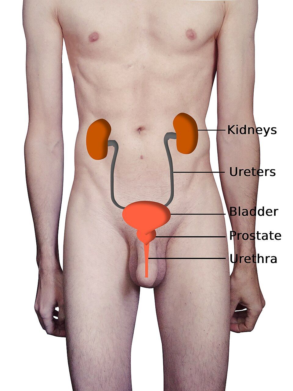
Not just a waste line
It is easy to think of the urinary system as just a way to get rid of waste. But that sells it short. Yes, the kidneys clear nitrogen wastes. One is urea, which comes from breaking down protein. Another is creatinine, which comes from muscle.
But their bigger job is homeostasis, the body's way of keeping things steady inside. Minute by minute, the kidneys decide how much water, sodium, potassium, calcium and acid to keep. They also decide how much to let go. This keeps your blood inside the narrow ranges your cells need to live. The kidney is where the idea of a steady inside turns into a real machine.
Drink a big glass of water, and the kidney gets rid of the extra within the hour. When you are low on water, it holds on tight. It makes the urine more concentrated, or stronger, to save every drop. The kidney also does jobs that have nothing to do with urine. It helps set blood pressure. It tells the bone marrow to make red blood cells. It also turns on vitamin D.
The four organs, in order
The plumbing is quite simple. Two kidneys make urine. Two ureters carry it down. One urinary bladder stores it. One urethra carries it out.
Everything up to the ureters is about chemistry, or changing the blood. Everything from the ureters down is about moving and storing urine. The kidneys sit high on the back wall of the belly, one on each side of the spine. They are retroperitoneal. This means they sit behind the lining of the belly, not inside it. The right kidney sits a bit lower than the left. That is because the large liver sits above it.
| Organ | Number | Main job |
|---|---|---|
| Kidneys | 2 | Filter blood and form urine. Balance water, salts and pH. Help set blood pressure and red blood cell count. |
| Ureters | 2 | Carry urine from kidney to bladder by peristalsis (waves of muscle squeezing) |
| Urinary bladder | 1 | Stores urine until it is a good time to let it out |
| Urethra | 1 | Carries urine from the bladder to the outside of the body |
A river of blood, a trickle of urine
The kidneys handle a huge amount of blood. Together they get about 20–25% of the entire cardiac output. That is the blood your heart pumps each minute. For their size, they get more blood than almost any other organ. From it, they filter about 180 liters of fluid every day.
If all of that left as urine, you would be in deep trouble. So the kidney takes back more than 99% of it. Only about 1–2 liters leave as urine. This plan is the secret to the kidney's control. It pushes a huge amount out. Then it picks exactly what to take back. That way, it can control dozens of substances one at a time.
Tap each of the four organs in the interactive. See where it sits and what it does.
- The urinary system has four organs. Two kidneys make urine. Two ureters move it. One bladder stores it. One urethra lets it out.
- The kidneys do much more than clear waste. They control water, electrolytes, and pH. They also help control blood pressure, red blood cell making, and vitamin D.
- The kidneys are retroperitoneal, behind the belly lining. They get about 20–25% of cardiac output. They filter about 180 L a day but pass only 1–2 L of urine.
Section 2Inside the kidney
The layers from outside in
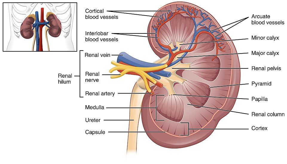
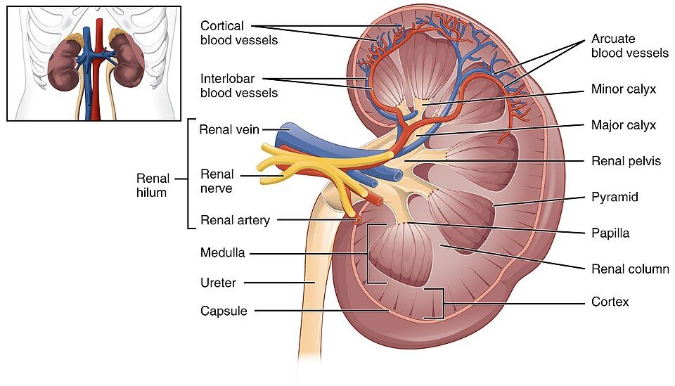
Slice a kidney down the middle like a bean. You will see neat layers. A tough, fiber-filled renal capsule wraps the outside. (Renal means "of the kidney.") Just under it is the renal cortex. This grainy outer layer is where filtering starts.
Deeper in is the renal medulla. It is made of 8–18 cone-shaped renal pyramids. The pyramids point inward. The tip of each cone is the renal papilla. It drips finished urine into spaces that collect it. Strips of cortex called renal columns dip down between the pyramids. They carry blood vessels.
The whole kidney curves around a notch on its inner edge, called the hilum. Here the renal artery comes in. The renal vein and the ureter go out.
The drainage route
Once urine drips off a papilla, the kidney does not change it again. It is just collected and drained. This route is worth learning. It names the parts a kidney stone passes through.
Urine leaves the papilla into a cup-shaped minor calyx. Several of these join to form a major calyx. The major calyces empty into a funnel called the renal pelvis. The renal pelvis narrows at the hilum and becomes the ureter. So the path is: papilla → minor calyx → major calyx → renal pelvis → ureter.
| Structure | Location | What happens there |
|---|---|---|
| Renal capsule | Outer surface | Tough coat that protects the kidney |
| Renal cortex | Outer working layer | Holds the renal corpuscles. Filtering starts here. |
| Renal medulla (pyramids) | Under the cortex | Holds the loops and collecting ducts that concentrate urine |
| Renal papilla | Tip of each pyramid | Drips finished urine into a minor calyx |
| Calyces & renal pelvis | Central collecting space | Funnel urine toward the ureter (no more changes are made) |
| Hilum | Notch on the inner edge | Where the renal artery, renal vein and ureter go in and out |
Two capillary beds in a row
The kidney's blood supply has one odd feature. It makes everything else work. Most organs have one bed of capillaries, the tiniest blood vessels. The kidney has two capillary beds in series, which means one after the other.
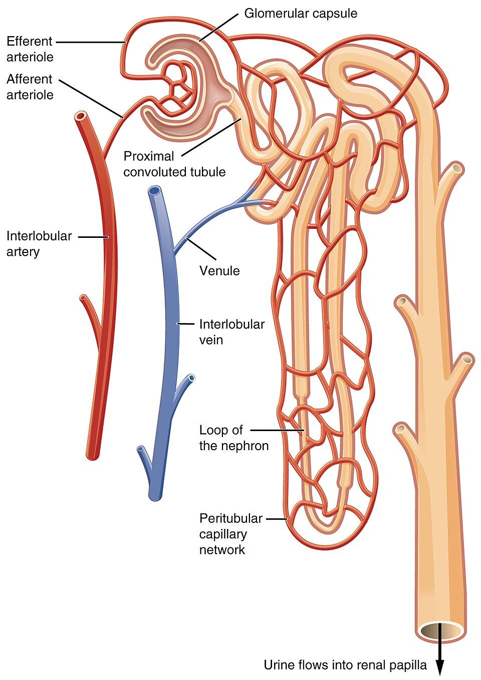
Blood comes in through the renal artery. It branches down to a tiny afferent arteriole, a small artery that carries blood in. Then it enters the first bed. This is a high-pressure knot of capillaries called the glomerulus. Filtering happens here.
Blood does not drain into a vein next. It leaves the glomerulus through an efferent arteriole, a small artery that carries blood out. Then it enters a second bed. These are the peritubular capillaries, which wrap around the tubules. With two beds, the kidney can filter under high pressure in the first bed. Then it can take things back under low pressure in the second. Two opposite jobs happen one after the other.
Slice the kidney in the interactive below and explore each zone.
- From the outside in: capsule → cortex → medulla (pyramids). Renal columns made of cortex sit between the pyramids.
- Urine drains: papilla → minor calyx → major calyx → renal pelvis → ureter. It is not changed after the papilla.
- The kidney has two capillary beds in series. The high-pressure glomerulus filters. The low-pressure peritubular capillaries take things back.
Section 3The nephron
A million tiny factories
The kidney does not filter like one big strainer. Its work is split among about one million tiny units in each kidney. These units are called nephrons. Each one is a whole little urine factory.
A nephron has two parts. The renal corpuscle makes the filtrate, the fluid filtered out of the blood. A long, winding renal tubule then turns that filtrate into urine. If you understand one nephron, you understand the whole kidney. The organ is just this same unit, repeated a million times.
The renal corpuscle and the filtration membrane
The renal corpuscle is where blood becomes filtrate. It has two parts. The glomerulus is that high-pressure ball of capillaries. It sits inside a cup called the glomerular capsule, or Bowman's capsule. Fluid squeezes out of the blood in the glomerulus. It crosses a three-layer filtration membrane and enters the capsule.
This membrane is very choosy. Its first layer is the fenestrated capillary wall. Fenestrated means "with windows," and this wall is full of pores. Its middle layer is a basement membrane. It has a negative charge that pushes proteins away. Its third layer is made of octopus-like cells called podocytes. Their "feet" lock together like fingers. They leave narrow gaps called filtration slits.
Together, these layers let water and small particles pass easily. They hold back blood cells and blood proteins. So normal filtrate is basically blood plasma with no protein. Protein or blood in the urine is a warning sign. It means this membrane may be damaged.
Following the tubule
From the capsule, filtrate flows through the tubule's parts in a set order. Each part has its own job. First comes the proximal convoluted tubule (PCT). Proximal means "near," and convoluted means "twisted." Next is the hairpin-shaped loop of Henle. It has a part going down and a part going up. Then comes the distal convoluted tubule (DCT). Distal means "far." Last is the collecting duct. It gathers filtrate from many nephrons and carries it toward the papilla.
Some nephrons sit almost fully in the cortex. These are cortical nephrons. A smaller group are the juxtamedullary nephrons. Juxta means "next to," so they sit next to the medulla. They have long loops that dive deep into the medulla. They are what make concentrated urine possible.
| Segment | Location | Main role |
|---|---|---|
| Renal corpuscle | Cortex | Filtration. Makes filtrate with no protein. |
| Proximal convoluted tubule (PCT) | Cortex | Bulk reabsorption, taking back most of the water, Na⁺, glucose and amino acids |
| Loop of Henle | Medulla | Makes the medulla salty, which lets the kidney concentrate urine |
| Distal convoluted tubule (DCT) | Cortex | Fine-tuning under hormone control (Na⁺, K⁺, Ca²⁺) |
| Collecting duct | Cortex & medulla | Last step of water reabsorption (controlled by ADH). Drains to the papilla. |
Trace the filtrate through every twist of the nephron in the interactive.
- The nephron is the working unit of the kidney. There are about 1 million in each kidney. Each has a renal corpuscle and a renal tubule.
- The filtration membrane has three layers: a capillary wall with pores, a basement membrane, and podocyte slits. It lets water and small particles through. It holds back cells and proteins.
- Tubule order: corpuscle → PCT → loop of Henle → DCT → collecting duct. Juxtamedullary nephrons have long loops. They make the urine concentrated.
Section 4How urine is made
Three steps: dump, reclaim, fine-tune
The kidney's plan for making urine seems wasteful. But it is actually very smart: dump almost everything out, then take back what you want. This happens in three steps, in order.
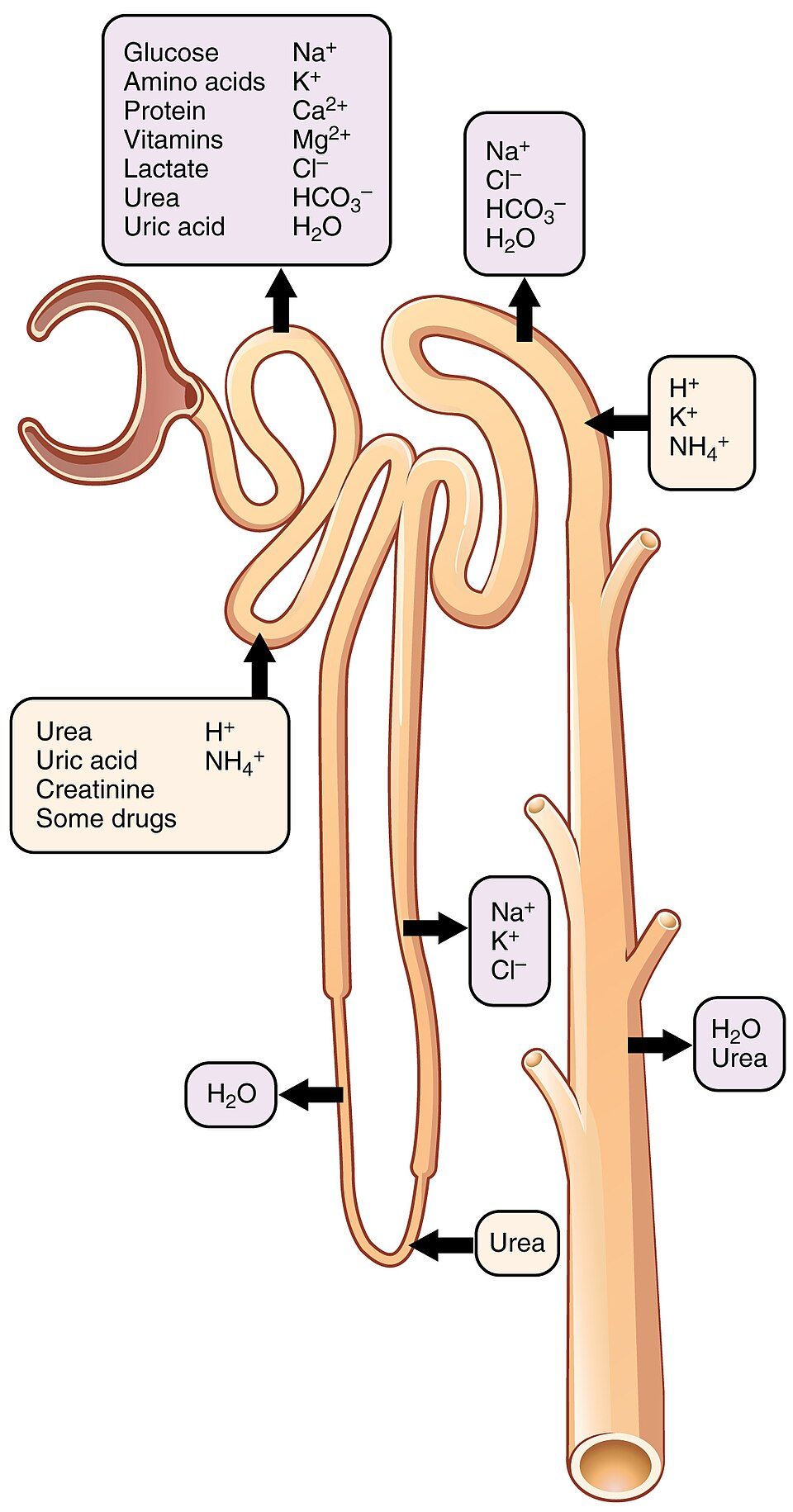
First, glomerular filtration pushes a large amount of fluid out of the blood and into the tubule. Second, tubular reabsorption takes back the water and useful substances. It returns them to the blood. Third, tubular secretion moves a few extra wastes and ions from the blood into the tubule to be thrown out. (Ions are tiny charged particles, like sodium.) Whatever is left in the tubule after all three steps is the urine. The smart part is control. The kidney filters a huge amount and then takes back only what it chooses. So it can adjust each substance on its own.
Step one: glomerular filtration
Filtering is driven by pressure alone. Blood enters the glomerulus under high pressure. That is because a wide afferent arteriole feeds in, and a narrow efferent one drains out. Think of a garden hose with your thumb over the end. The pressure forces fluid across the filtration membrane into the capsule.
All the glomeruli together make filtrate at a certain speed. This is called the glomerular filtration rate (GFR). It is normally about 125 mL per minute, which adds up to about 180 L a day. GFR is like a vital sign for the kidney. A falling GFR is the main measure of kidney failure. The kidney guards its GFR closely. It widens or narrows the afferent and efferent arterioles. This keeps filtering steady even when blood pressure goes up and down.
Steps two and three: reabsorption and secretion
If filtering were the whole story, you would pee 180 liters a day. You would dry out and die very quickly. Reabsorption, taking fluid back, saves you. The PCT alone takes back about 65% of the filtered water and sodium. It also takes back almost 100% of the glucose and amino acids. (Glucose is blood sugar. Amino acids are the building blocks of protein.) These nutrients are too valuable to waste.
The loop of Henle makes the medulla salty. Later, that salt helps pull water back by osmosis, the flow of water toward saltier places. By the time filtrate reaches the collecting duct, only small changes are left.
Meanwhile, secretion runs the other way. It moves substances from the blood into the tubule. These include hydrogen ions (H⁺) to control pH, extra potassium (K⁺), and many drugs and poisons. Secretion is how the kidney clears things that do not filter well.
| Process | Direction | Where | What moves |
|---|---|---|---|
| Filtration | Blood → tubule | Renal corpuscle | Water & small particles (pushed by pressure, about 125 mL/min) |
| Reabsorption | Tubule → blood | Whole tubule (mostly PCT) | Water, Na⁺, glucose, amino acids: the useful things |
| Secretion | Blood → tubule | PCT, DCT, collecting duct | H⁺, K⁺, drugs, poisons: extra waste |
Step through filtration, reabsorption and secretion in the interactive below.
- Making urine has three steps. Filtration goes from blood→tubule. Reabsorption goes from tubule→blood. Secretion goes from blood→tubule.
- GFR is about 125 mL/min. It is the key measure of how well the kidneys work. The kidney changes the width of its arterioles to keep GFR steady.
- The PCT takes back the most: about 65% of water and Na⁺, and almost all glucose. Secretion clears H⁺, K⁺ and drugs.
Section 5Hormonal control
Water: antidiuretic hormone
The kidney does not decide on its own how much water to keep. The body sends it signals with hormones. The most important water hormone is antidiuretic hormone (ADH), also called vasopressin. Antidiuretic means "against making lots of urine."
When you are low on water, your blood gets too concentrated. The hypothalamus in the brain senses this. Then the back part of the pituitary gland releases ADH. ADH acts on the collecting duct. It puts water channels called aquaporins into the duct's wall. Water then flows out through them into the salty medulla. The result is a small amount of concentrated urine, and your body saves water.
When you have plenty of water, ADH drops. The channels are removed, and water stays in the tubule. You make a large amount of dilute, or watery, urine. Alcohol blocks ADH. That is why a night of drinking leaves you dried out.
Salt and pressure: the renin–angiotensin–aldosterone system
To control blood pressure, the kidney manages sodium. Here is why. Where sodium goes, water follows. More water means more blood volume. More volume means higher pressure. The main system for this is the renin–angiotensin–aldosterone system (RAAS).
When blood pressure drops, the kidney's granular cells release renin. Renin changes a protein made by the liver into angiotensin I. Then an enzyme in the lungs, called ACE, changes it into angiotensin II. (An enzyme is a protein that speeds up a chemical change.)
Angiotensin II is powerful. First, it squeezes blood vessels narrower. This raises pressure right away. Second, it tells the adrenal gland to release aldosterone. Aldosterone tells the DCT and collecting duct to take back more sodium, and water follows it. At the same time, they get rid of potassium. More sodium taken back means more water kept. That means more blood volume and higher pressure. This boost is slower, but it lasts.
Letting go: atrial natriuretic peptide
Every control needs an opposite. Sometimes blood volume gets too high. Then the walls of the heart's upper chambers, the atria, get stretched. They release atrial natriuretic peptide (ANP). It does the reverse of RAAS. It makes you lose sodium and water in the urine. This lowers blood volume and pressure.
So ADH tunes water, RAAS raises pressure, and ANP lowers it. The kidney sits in the middle of this constant push and pull. It keeps your blood volume steady, and so it keeps your blood pressure steady too.
| Hormone | Trigger | Effect on the kidney | Net result |
|---|---|---|---|
| ADH (vasopressin) | Blood that is too concentrated or too low in volume | Adds aquaporins to the collecting duct → takes back water | Less urine, more concentrated. Water is saved. |
| Aldosterone | Angiotensin II; high K⁺ | Takes back Na⁺ (and water) and gets rid of K⁺ | ↑ blood volume & pressure |
| Renin / angiotensin II | Low blood pressure | Narrows blood vessels. Causes aldosterone release. | ↑ blood pressure (fast & slow) |
| ANP | High blood volume (atria stretched) | Makes you lose more Na⁺ and water in urine | ↓ blood volume & pressure |
Tap each hormone in the interactive. See how it shifts water and salt.
- ADH controls water. It puts aquaporins in the collecting duct. This makes urine concentrated and saves water.
- The RAAS controls sodium and pressure: low pressure → renin → angiotensin II → aldosterone → Na⁺ and water taken back → more blood volume and higher pressure.
- ANP works against RAAS. It gets rid of Na⁺ and water when blood volume is high. Together, these hormones keep blood pressure steady.
Section 6Storing and voiding urine
The storage plumbing
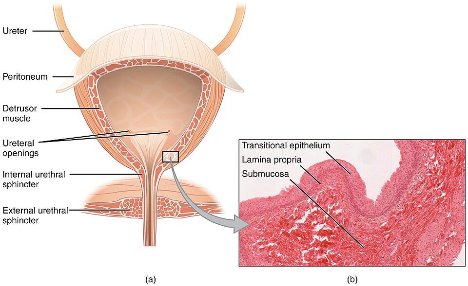
Once urine leaves the kidney, it is finished. The rest is just moving and storing it. The ureters squeeze urine down to the bladder with waves of muscle called peristalsis. So urine keeps moving even when you lie down.
The urinary bladder is a stretchy bag made of muscle. It is lined with transitional epithelium, a lining that can stretch flat as the bladder fills. When the bladder is empty, the lining falls into folds called rugae. The muscle in the bladder wall is the detrusor muscle. A smooth triangle on the bladder floor, the trigone, funnels urine toward the exit.
Two gates guard the way out. The internal urethral sphincter is smooth muscle. It works on its own, without your control. The external urethral sphincter is skeletal muscle. You control it on purpose. The urethra then carries urine out. It is short in females, about 4 cm. It is longer in males, about 20 cm, because it also runs through the penis.
The micturition reflex
Emptying the bladder starts as a simple spinal reflex. The bladder fills to about 200–400 mL. Then stretch receptors in the detrusor wall fire. These sensors send signals to the lowest (sacral) part of the spinal cord.
The cord answers with a parasympathetic command. This is part of the "rest and digest" nerve system. The command does two things at once. It contracts the detrusor muscle to squeeze the bladder. It also relaxes the internal sphincter to open the first gate. This is the micturition reflex. (Micturition means peeing.) A baby's nervous system has not yet learned control. So in a baby, the reflex just runs to the end, and the baby pees.
The voluntary override
The second gate is what makes you different from a baby. The external urethral sphincter is skeletal muscle that you can hold shut on purpose. Higher brain centers can allow peeing or put it off. These include a control area in the pons, a part of the brainstem, guided by the cortex.
Say the reflex fires at a bad time. Your brain keeps the external sphincter squeezed, and the urge passes for a while. When you decide it is time, the brain relaxes the external sphincter. The reflex finishes, and the bladder empties. Learning this override is what "potty training" teaches. Losing it can cause many kinds of incontinence, or leaking urine. This can happen through a spinal injury, a stroke or old age.
| Step | What happens |
|---|---|
| 1. Fill | Urine collects. The detrusor stretches as the amount nears 200–400 mL. |
| 2. Sense | Stretch receptors fire and send signals to the sacral spinal cord |
| 3. Reflex | Parasympathetic nerves contract the detrusor and relax the internal sphincter |
| 4. Decide | The brain (pons and cortex) holds or lets go of the external sphincter. This is the gate you control. |
| 5. Void | The external sphincter relaxes. The bladder empties through the urethra. |
Fill the bladder and empty it in the interactive. Watch the reflex play out.
- Ureters move urine by peristalsis. The bladder stores it behind two sphincters. Its key parts are the detrusor muscle, transitional epithelium and trigone.
- The micturition reflex is a reflex of the sacral spinal cord: stretch → parasympathetic nerves → the detrusor squeezes and the internal sphincter opens.
- The external sphincter is skeletal muscle you control on purpose, through the pons and cortex. This override lets you choose when to pee.
The chapter in one breath
- The urinary system has four organs: two kidneys, two ureters, one bladder and one urethra. They filter blood, balance water and salts, and drain urine. The kidneys also help set blood pressure, red blood cell count and vitamin D.
- A kidney has cortex on the outside and medulla (pyramids) inside. Urine drains papilla → calyces → renal pelvis → ureter. Blood passes through two capillary beds in series.
- The nephron is the working unit. There are about 1 million per kidney. Each has a renal corpuscle that filters. It also has a tubule that makes the urine (PCT → loop of Henle → DCT → collecting duct).
- Urine forms in three steps. Filtration goes from blood→tubule. Reabsorption goes from tubule→blood. Secretion goes from blood→tubule. GFR is about 125 mL/min.
- ADH saves water. The RAAS keeps sodium to raise blood pressure. ANP gets rid of sodium and water to lower it.
- The bladder stores urine. The micturition reflex lets it out. The external sphincter, which you control, lets you decide when to pee.
- Above all, the kidney is the body's great homeostatic organ. It turns the idea of a steady inside into a working machine.
ReferenceWords worth knowing
Here is a quick list of the terms in this chapter. Skim it now, or come back when a word trips you up.
- Aldosterone
- A hormone from the adrenal gland. It tells the distal tubule and collecting duct to take back sodium (and water with it) and get rid of potassium. This raises blood volume and pressure.
- Antidiuretic hormone (ADH)
- A hormone from the back part of the pituitary gland. It puts aquaporins into the collecting duct so water is taken back. This makes concentrated urine and saves body water.
- Calyx
- A cup-shaped space that catches urine dripping from a renal papilla. Minor calyces join into major calyces, which drain into the renal pelvis.
- Collecting duct
- The last part of the tubule, shared by many nephrons. Here ADH controls the last bit of water taken back before urine reaches the papilla.
- Detrusor muscle
- The smooth muscle of the bladder wall. It squeezes to push urine out during the micturition reflex.
- Glomerular filtration rate (GFR)
- The amount of filtrate all the glomeruli make each minute, normally about 125 mL/min. It is the main measure of how well the kidneys work.
- Glomerulus
- The high-pressure ball of capillaries inside the renal corpuscle. Here blood is filtered into the glomerular capsule.
- Juxtaglomerular apparatus
- The spot where a nephron's rising limb meets its afferent arteriole. Its cells sense salt and pressure and release renin.
- Loop of Henle
- The hairpin part of the tubule, with a part going down and a part going up. It makes the medulla salty, which is needed to concentrate urine.
- Micturition
- Peeing, or emptying the bladder. A spinal stretch reflex starts it, and you can allow it or put it off on purpose.
- Nephron
- The tiny working unit of the kidney, with about 1 million per kidney. It is made of a renal corpuscle and a renal tubule.
- Podocyte
- An octopus-shaped cell in the filtration membrane. Its "feet" form narrow filtration slits that help hold back proteins.
- Proximal convoluted tubule (PCT)
- The first part of the tubule after the corpuscle. It takes back most of the water, sodium, glucose and amino acids.
- Reabsorption (tubular)
- The movement of water and useful substances out of the tubule and back into the blood.
- Renal corpuscle
- The filtering part of the nephron. It is the glomerulus plus the glomerular (Bowman's) capsule around it.
- Renin
- An enzyme the kidney releases when blood pressure falls. It starts the chain of steps that makes angiotensin II and aldosterone.
- Retroperitoneal
- Sitting behind the peritoneum, the lining of the belly, against the back wall. The kidneys and ureters are found here.
- Secretion (tubular)
- The movement of wastes and ions (H⁺, K⁺, drugs) out of the blood and into the tubule to be thrown out.
- Trigone
- The smooth triangle on the bladder floor. Its corners are the two ureter openings and the opening to the urethra.
- Ureter
- The muscle tube that carries urine from a kidney to the bladder by peristalsis.
- Urethra
- The tube that carries urine from the bladder to the outside. It is short in females and longer in males.