The Reproductive System
Every other system in this book works to keep you alive. The reproductive system looks past you. Its job is to make a new person. To do that, your body does something no other tissue does. It builds a special cell called a gamete, a sex cell such as a sperm or an egg. A gamete carries exactly half of your genetic instructions. So when a sperm meets an egg, the full set comes back together.
In this chapter you will walk through both body plans. You will follow the male path from testis to exit, and the female path from ovary to uterus. You will see the two "production lines" that turn stem cells into gametes. They are spermatogenesis, the making of sperm, and oogenesis, the making of eggs. You will also track the rise and fall of hormones in the menstrual cycle, the monthly cycle of the female body. Under all of it sits one clever kind of cell division, meiosis. We save it for last. Once you see it, the rest of the chapter clicks into place.
By the end of this chapter you can
- Trace the path of sperm from the seminiferous tubules to the urethra. Name the glands that add fluid along the way.
- Describe spermatogenesis. Tell how one diploid stem cell becomes four haploid sperm, where it happens, and how long it takes.
- Name the main female reproductive organs. Give the job of the ovary, uterine tube, uterus, and the layers of the uterus.
- Explain oogenesis. Tell how its unequal divisions and its long, lifelong pause differ from sperm making.
- Put the phases of the ovarian and uterine cycles in order. Match each phase to its main hormone (FSH, LH, estrogen, progesterone).
- Tell meiosis apart from mitosis. Explain how crossing over and independent assortment make each gamete different.
- Define diploid and haploid. Explain why cutting the number of chromosomes in half is key to making a baby.
Section 1Male reproductive anatomy
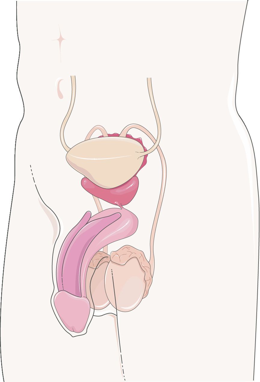
Two jobs: make sperm, deliver sperm
The male system has a simple task. It makes sperm, the male sex cells. Then it delivers them to the female body in a fluid that feeds them. Every part of the male anatomy helps with one of these two jobs.
Sperm are made in the two testes. The testes hang outside the body wall in a skin sac called the scrotum. That spot is no accident. Sperm making needs a temperature a few degrees cooler than the core of the body. A small muscle raises and lowers the testes to keep them at the right heat. So the scrotum works like a thermostat. Inside each testis are tightly coiled tubes called seminiferous tubules. Sperm are made along the walls of these tubes.
Following the road out
New sperm cannot swim yet, and they cannot fertilize an egg. First they move into the epididymis. This is a comma-shaped coil of tube that lies over the back of each testis. Sperm stay here for about two weeks. They grow up, learn to swim, and are stored.
Next they enter the ductus (vas) deferens. This muscular tube climbs out of the scrotum and loops over the bladder. It then joins the duct of a gland called the seminal vesicle. Together they form the ejaculatory duct. This short duct passes through the prostate. It empties into the urethra, the last tube, which carries semen out through the penis. In males the urethra is shared plumbing. It carries both urine and semen, but never at the same time.
| Structure | Role in the journey | Note |
|---|---|---|
| Seminiferous tubules | Where sperm are made | Coiled inside each testis |
| Epididymis | Sperm grow up, learn to swim, and are stored | About 6 m of coiled tube on the testis |
| Ductus (vas) deferens | Pushes sperm toward the urethra | Cut and tied in a vasectomy |
| Ejaculatory duct | Mixes sperm with fluid from the seminal vesicle | Passes through the prostate |
| Urethra | Carries semen (and urine) out | Shared tube in males |
The glands that make semen
Sperm are only a small part of semen, the fluid that leaves the body. Most of it is fluid from three kinds of helper glands. Each fluid has a job.
The two seminal vesicles add the most. Their fluid is thick and full of sugar. The sugar, called fructose, fuels the sperm's tails. The single prostate gland wraps around the urethra just below the bladder. It adds a milky fluid that is slightly alkaline, the opposite of acid. This helps sperm live through the acid in the female body. Last, the small bulbourethral glands give off a clear fluid before the rest. It makes the urethra slippery and cancels out any acid left from urine. Together these fluids wake up the sperm, feed them, and protect them for the trip.
Tap each part in the interactive. Trace the path sperm take from where they are made to the exit.
- Sperm are made in the seminiferous tubules of the testes. The testes sit in the scrotum to stay a bit cooler than the body.
- The road out runs testis → epididymis → ductus deferens → ejaculatory duct → urethra.
- Three glands add fluid. The seminal vesicles add fructose. The prostate adds alkaline fluid. The bulbourethral glands add a slippery fluid.
- In males the urethra is shared plumbing for both urine and semen.
Section 2Spermatogenesis
Turning a stem cell into a swimmer
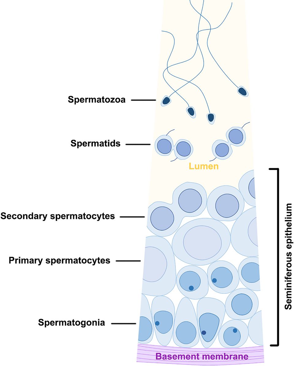
Spermatogenesis is the making of sperm. Think of it as an assembly line. It turns a diploid stem cell into grown-up sperm. It runs all the time along the walls of the seminiferous tubules, from puberty on.
It starts with a spermatogonium. This is a stem cell with the full 46 chromosomes. A full set like this is called diploid, or 2n. When a spermatogonium divides, one daughter cell stays behind as a stem cell. That is why a male never runs out. The other daughter becomes a primary spermatocyte. It is now set on the path to becoming sperm. It enters meiosis, the special division that cuts the number of chromosomes in half.
Two divisions, four cells
The math here is neat. The primary spermatocyte finishes meiosis I and becomes two secondary spermatocytes. Each one now has the haploid number, or half set (n = 23). Each of those then finishes meiosis II. That gives a total of four spermatids. In sperm making, the divisions are equal. Every cell gets a fair share of cytoplasm, the jelly inside the cell. So one starting cell makes four useful cells. Egg making is not like this.
Spermatids are round and cannot move. They still must be remodeled into sleek sperm. This last step is called spermiogenesis. The cell sheds extra cytoplasm. It packs its DNA into a small head. It puts a cap full of enzymes on the head, called the acrosome. And it grows a whip-like tail, the flagellum, to swim with. A middle piece packed with mitochondria, the cell's power plants, gives energy for the trip.
| Stage | Chromosomes | What happens |
|---|---|---|
| Spermatogonium | Diploid (2n) | Stem cell; one daughter stays a stem cell |
| Primary spermatocyte | Diploid (2n) | Enters and finishes meiosis I |
| Secondary spermatocyte | Haploid (n) | Two cells; finish meiosis II |
| Spermatid | Haploid (n) | Four cells; round and cannot swim |
| Sperm (spermatozoon) | Haploid (n) | Head + acrosome + midpiece + tail |
Helped along by nurse cells
The growing sperm cells do not look after themselves. Tall Sustentacular (Sertoli) cells sit among them. They are also called nurse cells. They feed the growing sperm. They also form the blood–testis barrier. This wall keeps the immune system from attacking sperm as if they were germs.
Between the tubules sit the interstitial (Leydig) cells. They make testosterone. This hormone drives sperm making. It also causes male traits such as a deep voice and facial hair. The whole process, from spermatogonium to finished sperm, takes about 64 to 74 days. A healthy male makes hundreds of millions of sperm every day.
Step through each division in the interactive. Watch a stem cell become four swimming sperm.
- A diploid spermatogonium makes four haploid sperm through two equal divisions of meiosis.
- Spermiogenesis reshapes round spermatids into sleek sperm with an acrosome, midpiece, and tail.
- Sertoli cells care for the sperm and form the blood–testis barrier. Leydig cells make testosterone.
- The full process takes about 64–74 days, and it never stops.
Section 3Female reproductive anatomy
A place to make an egg and a place to grow a baby
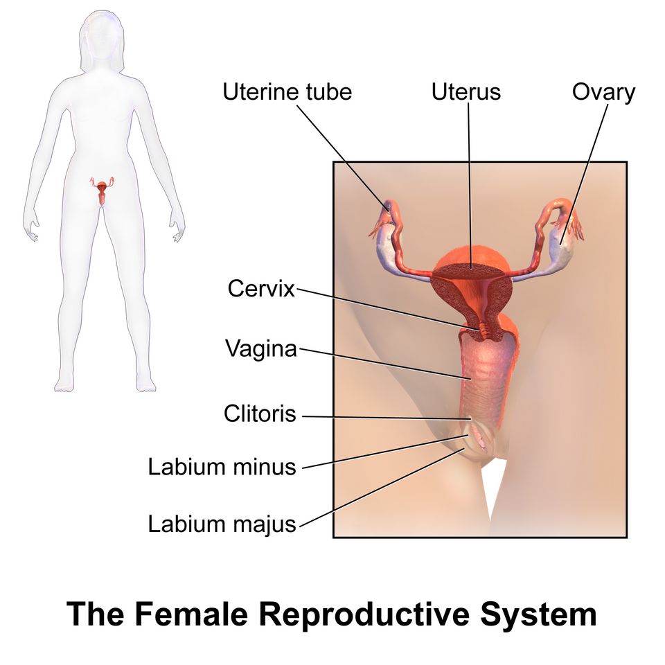
The female system does something the male system does not. It makes the gamete, the egg. But it also gives a place where a fertilized egg can attach and grow for nine months.
The two ovaries are each about the size of an almond. They sit low in the pelvis. They hold the whole lifetime supply of eggs. About once a month, an ovary lets go of a ripe egg. This is called ovulation. A uterine (fallopian) tube reaches toward each ovary. Its end has a fringe of finger-like parts called fimbriae. They sweep the released egg into the tube. If fertilization happens, it is usually here, in the outer third of the tube.
The uterus and its layers
The tube carries the egg toward the uterus, or womb. This is a muscular, pear-shaped organ. It is built to hold a growing baby. Its wall has three layers you should know.
The inner layer is the endometrium. This lining thickens each month to receive an embryo. If no embryo arrives, the lining is shed as the monthly period. The thick middle layer is the myometrium. It is smooth muscle that squeezes hard during labor. The thin outer layer is the perimetrium. It is a smooth, moist covering. The lower neck of the uterus is the cervix. It opens into the vagina. The vagina is a muscular canal. It is the birth canal, and it receives sperm during sex.
| Organ | Function | Note |
|---|---|---|
| Ovary | Makes eggs and the hormones estrogen & progesterone | Holds all eggs from birth |
| Uterine (fallopian) tube | Carries the egg toward the uterus; where fertilization usually happens | Fimbriae sweep in the egg |
| Uterus | Holds and feeds the growing baby | Endometrium / myometrium / perimetrium |
| Cervix | Neck of the uterus; opens into the vagina | Opens wide in labor |
| Vagina | Birth canal; receives semen | Muscular and able to stretch |
The ovary is also a gland
It is easy to think of the ovary as just a place to store eggs. But it is also an endocrine organ, a gland that makes hormones. Each month, a cluster of cells forms around the ripening egg. This cluster is the follicle. It pumps out estrogen. After ovulation, the leftover follicle turns into the corpus luteum. The corpus luteum makes progesterone.
These two hormones drive the changes in the lining of the uterus. Working with the brain, they run the whole monthly cycle. You will meet it in Section 5. So the ovary has two jobs. It releases the egg, and it controls the tissue that will receive it.
Tap each part in the interactive to explore the female organs from the front.
- The ovaries store eggs. They are also glands that make estrogen and progesterone.
- The uterine tube catches the released egg with its fimbriae. Fertilization usually happens in its outer third.
- The wall of the uterus has three layers. The endometrium is the lining that is shed. The myometrium is the muscle for labor. The perimetrium is the outer cover.
- The cervix opens into the vagina, the birth canal.
Section 4Oogenesis
The same tool, used very differently
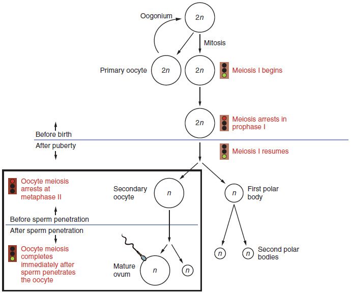
Egg making is called oogenesis. It uses the same meiosis as sperm making. But the female body runs it on a very different schedule, with a very different goal. The male makes four small sperm from one cell, and he does it without end. The female makes one large egg from one cell. She does it on a strict, once-a-month timetable.
Why the difference? It comes down to supplies. An egg must carry enough cytoplasm, food, and cell parts to start a new life. So the female body pours almost everything into a single egg.
Unequal divisions on purpose
Here is the twist. A primary oocyte is an early egg cell. When it finishes meiosis I, it does not split its cytoplasm evenly. It divides unequally. One daughter, the secondary oocyte, keeps nearly all the cytoplasm. The other becomes a tiny polar body. It gets almost nothing and later breaks down.
The same lopsided split happens again in meiosis II. But in humans, this second division only finishes if a sperm fertilizes the egg. In the end you get one working egg, called an ovum. You also get two or three polar bodies that will die off. One cell in, one useful egg out. It is the mirror image of the male's one in, four out.
| Feature | Oogenesis (egg) | Spermatogenesis (sperm) |
|---|---|---|
| Cells made from one starting cell | One egg (+ polar bodies) | Four sperm |
| How cytoplasm is split | Unequal; the egg keeps almost all of it | Equal |
| Timing | Starts before birth; one egg per cycle | Nonstop from puberty |
| Finishing meiosis II | Only if fertilized | Always finished |
| Lifetime total | About 400 eggs released | Trillions of sperm |
A process paused for decades
Oogenesis has the strangest timeline in the body. A female is born with all the eggs she will ever have. That is about one to two million primary oocytes. Before birth, they start meiosis I. Then they stop partway through and stay frozen. They wait like this for years.
Starting at puberty, one oocyte finishes meiosis I each month and is released. The rest keep waiting. So an egg released at age forty has been paused for four decades. This long wait helps explain why chromosome errors become more common as the mother gets older. Down syndrome is one example.
The supply shrinks over the years. By menopause, only about a thousand follicles are left. That is too few, and they respond too weakly, to keep the hormone cycle going. So the monthly cycles stop.
Step through the interactive. Watch the unequal divisions pack everything into one egg.
- Oogenesis makes one egg (plus polar bodies) from each starting cell. The cytoplasm is split unequally.
- All eggs start meiosis before birth and then pause. One finishes each cycle. Meiosis II finishes only at fertilization.
- A female is born with her lifetime egg supply. It drops to a very low level, about a thousand follicles, by menopause. Then the cycles stop.
- The long pause in meiosis helps explain why chromosome errors rise as the mother gets older.
Section 5The menstrual cycle
Two cycles running as one
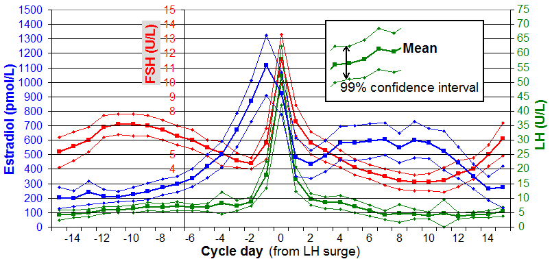
The menstrual cycle lasts about 28 days. It is really two cycles running in step. One happens in the ovary, and one happens in the uterus. Four hormones run the show.
Two come from the pituitary gland, a small gland under the brain. They are follicle-stimulating hormone (FSH) and luteinizing hormone (LH). The other two come from the ovary. They are estrogen and progesterone. The ovarian cycle tracks what happens to the egg and its follicle. The uterine cycle tracks the building and shedding of the lining. The two are timed so a fresh lining is ready just when an egg might arrive.
Follicular phase, ovulation, luteal phase
The cycle starts on day 1 with menstruation, the period. Last month's lining is shed. Next comes the follicular phase. FSH gets several follicles to grow. One takes the lead and pours out estrogen. Estrogen rebuilds the lining.
Rising estrogen then sets off a sudden jump in LH around day 14. This LH surge causes ovulation. The lead follicle bursts and lets go of its egg.
Next comes the luteal phase. The empty follicle becomes the corpus luteum. It makes progesterone. Progesterone keeps the lining thick and rich, ready in case an embryo arrives. If there is no pregnancy, the corpus luteum shrinks away. Progesterone and estrogen drop. With no hormones to hold it up, the lining is shed. Then the next cycle begins.
| Phase (days) | Main hormone | What is happening |
|---|---|---|
| Menstruation (1–5) | Estrogen and progesterone low; FSH rising | The old lining is shed while the next follicles start to grow |
| Follicular (1–13) | FSH, then estrogen | A follicle ripens; the lining rebuilds |
| Ovulation (~14) | LH surge | The follicle bursts; the egg is released |
| Luteal (15–28) | Progesterone | The corpus luteum makes the lining rich |
Feedback that flips
Here is a neat trick of this cycle. The same hormone can push in opposite ways, depending on timing. For most of the cycle, estrogen gives negative feedback. It quietly holds the pituitary back, like a brake.
But as the lead follicle ripens, estrogen climbs high and stays high. At that point the feedback flips to positive. Now high estrogen pushes the pituitary to release the LH surge. That surge causes ovulation. This is one of the few times the body uses positive feedback. As always, it is used to cause one big, one-time event. After ovulation, progesterone from the corpus luteum brings back negative feedback. Things calm down again.
Tap a phase in the interactive. See the hormones, the ovary, and the lining change together.
- Four hormones run the cycle. FSH and LH come from the pituitary. Estrogen and progesterone come from the ovary.
- The phases go menstruation → follicular → ovulation (LH surge, ~day 14) → luteal.
- Follicles make estrogen, which rebuilds the lining. The corpus luteum makes progesterone, which keeps the lining up.
- A high estrogen level flips to positive feedback. This sets off the LH surge that causes ovulation.
Section 6Meiosis
Why half is the whole point
We saved the engine for last. Every body cell you have is diploid. It has 46 chromosomes, the packages that hold your genes. They come in 23 pairs. One of each pair came from each of your parents.
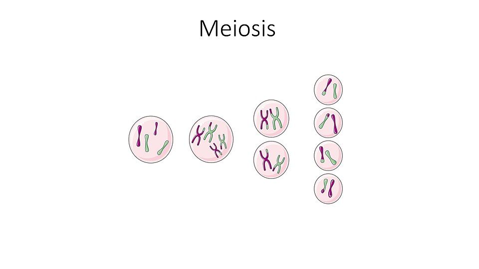
What if a sperm with 46 chromosomes joined an egg with 46? The child would have 92. The number would double every generation. The body's answer is meiosis. This special division makes haploid gametes, with just 23 chromosomes each. When a haploid sperm meets a haploid egg, the full diploid set of 46 comes back. So the count stays the same, generation after generation. Cutting the number in half is not a side effect of making gametes. It is the reason meiosis exists.
Two divisions, not one
Meiosis gets to the haploid number in two divisions in a row. The DNA is copied only once, before the first. In meiosis I, matching chromosomes line up in pairs. These are called homologous pairs. The pairs are pulled apart. This is the step that cuts the number in half, from diploid to haploid.
Meiosis II looks a lot like ordinary mitosis. The two copies of each chromosome finally split apart. The end result is four haploid cells. That means four sperm, or one egg plus polar bodies. Compare this with mitosis, the normal division that builds and repairs the body. Mitosis is one division. It makes two identical diploid cells.
| Feature | Mitosis | Meiosis |
|---|---|---|
| Purpose | Growth and repair | Make gametes |
| Divisions | One | Two |
| Daughter cells | 2 | 4 |
| Chromosome number | Diploid (2n), no change | Cut in half to haploid (n) |
| Genes | Exact copies | Each one different |
Shuffling the deck
Meiosis does more than cut the number in half. It also reshuffles your genes, so no two gametes are alike. It does this in two ways.
In crossing over, paired chromosomes swap matching pieces during meiosis I. This mixes the versions you got from each parent. In independent assortment, each pair lines up and splits at random. So which grandparent's chromosome ends up in a gamete is a coin flip. And that flip happens 23 times over. Together these make millions of different gametes from one person. That is why brothers and sisters differ. It is also why sexual reproduction makes the variety that evolution acts on.
Sometimes a pair fails to split apart. This is called nondisjunction. Then a gamete ends up with the wrong number of chromosomes. This causes conditions such as trisomy 21, also called Down syndrome.
Step through both divisions in the interactive. Watch the chromosomes drop by half and the genes shuffle.
- Meiosis cuts the chromosome number in half, from diploid (46) to haploid (23). Then fertilization can bring back the full set.
- It uses two divisions. Meiosis I pulls apart homologous pairs; this is the halving step. Meiosis II splits the sister copies.
- Crossing over and independent assortment make every gamete different.
- In nondisjunction, a pair fails to split. This gives a gamete the wrong number of chromosomes.
The chapter in one breath
- The reproductive system is here to make a new person. That takes haploid gametes that join to bring back the full set of chromosomes.
- In males, sperm are made in the seminiferous tubules. They travel epididymis → ductus deferens → ejaculatory duct → urethra. Along the way they mix with fluid from the seminal vesicles, prostate, and bulbourethral glands.
- Spermatogenesis makes four sperm from each starting cell through equal divisions. It never stops, and it takes about 64–74 days.
- In females, the ovary stores eggs and makes hormones. The uterine tube catches the egg. The uterus (endometrium, myometrium, perimetrium) holds a pregnancy.
- Oogenesis makes one egg plus polar bodies through unequal divisions. It starts before birth. Meiosis II finishes only at fertilization.
- The menstrual cycle is run by FSH, LH, estrogen, and progesterone. Follicles make estrogen. An LH surge causes ovulation. The corpus luteum makes progesterone.
- Meiosis cuts the chromosomes in half in two divisions. Meiosis I reduces, and meiosis II divides. Crossing over plus independent assortment make every gamete different.
ReferenceWords worth knowing
Here is a quick list of the terms in this chapter. Skim it now, or come back when a word trips you up.
- Acrosome
- The cap full of enzymes on the head of a sperm. It helps the sperm get through the outer coat of the egg.
- Corpus luteum
- What the empty follicle becomes after ovulation. It makes progesterone to keep up the lining of the uterus.
- Diploid (2n)
- Having the full set of chromosomes. In humans that is 46, in 23 pairs. Body cells are diploid.
- Ductus (vas) deferens
- The muscular tube that pushes sperm from the epididymis toward the urethra. It is cut in a vasectomy.
- Endometrium
- The inner lining of the uterus. It thickens each cycle to receive an embryo. If none attaches, it is shed as the period.
- Epididymis
- The coiled tube on the back of the testis. Sperm grow up, learn to swim, and are stored here.
- Estrogen
- A hormone made mainly by the growing follicle in the ovary. It rebuilds the lining of the uterus and sets off the LH surge.
- Follicle
- The cluster of cells in the ovary around a growing egg. It makes estrogen and releases the egg at ovulation.
- FSH (follicle-stimulating hormone)
- A hormone from the pituitary. It makes follicles grow in the ovary and helps sperm making in the testis.
- Gamete
- A haploid sex cell, either a sperm or an egg. It joins with another gamete at fertilization.
- Haploid (n)
- Having half the set of chromosomes. In humans that is 23. Gametes are haploid.
- LH (luteinizing hormone)
- A hormone from the pituitary. Its surge in the middle of the cycle causes ovulation and forms the corpus luteum.
- Meiosis
- The two-step cell division that cuts the chromosome number in half to make gametes. It also shuffles genes.
- Menopause
- The lasting end of monthly cycles. It happens when too few follicles are left, and they respond too weakly, to keep the hormone cycle going. The supply gets very low, but not to zero.
- Nondisjunction
- When a chromosome pair fails to split apart in meiosis. The gamete gets the wrong number of chromosomes.
- Oogenesis
- The making of eggs. Unequal divisions make one ovum and two or three polar bodies.
- Ovulation
- The release of a ripe egg from the ovary. It is caused by the LH surge in the middle of the cycle.
- Polar body
- A small cell made by the unequal divisions of oogenesis. It gets little cytoplasm and breaks down.
- Progesterone
- The hormone made by the corpus luteum. It keeps the lining of the uterus thick and rich after ovulation.
- Semen
- The fluid that leaves the male body at ejaculation. It is sperm plus fluids from the seminal vesicles, prostate, and bulbourethral glands.
- Seminiferous tubules
- The coiled tubes inside the testis where sperm are made.
- Spermatogenesis
- The making of sperm. Equal divisions make four haploid sperm from one starting cell.