Labour and Birth
Labour is the one event in nursing where two patients share a single circulation and a single clock. Everything you assess — the contraction, the cervix, the fetal heart, the colour of the fluid, the tone of the uterus after the placenta delivers — is a window onto whether oxygen is still crossing the placenta and whether the birthing parent is still holding onto their blood volume. This chapter builds labour the way it is practised: first the mechanics that decide whether a fetus can pass through a pelvis, then the stages and what normal progress actually looks like, then fetal surveillance and the interventions that restore oxygen delivery, then comfort and anaesthesia and the complications that follow the needle, and finally the third and fourth stages with the emergency sequences where the order of your actions is the intervention. Nothing here is intuitive by accident. Much of it is counter-intuitive on purpose, and that is exactly why it is memorised as a sequence.
By the end of this chapter you can
- Analyse the five Ps of labour and interpret fetal lie, presentation, attitude, position and station from a documented finding such as LOA at 0 station.
- Differentiate true labour from false labour and describe the first nursing action after spontaneous rupture of membranes.
- Sequence the cardinal movements and describe expected progress and nursing priorities in each of the four stages of labour.
- Interpret an electronic fetal monitoring strip using baseline, variability, accelerations and decelerations, assign a NICHD category, and carry out intrauterine resuscitation in priority order.
- Compare non-pharmacological comfort measures, systemic opioids and neuraxial analgesia, and state the first action for post-epidural hypotension.
- Execute the priority sequence for postpartum haemorrhage, magnesium sulfate toxicity, umbilical cord prolapse, shoulder dystocia and uterine rupture.
- Provide immediate newborn care at birth — warmth, skin-to-skin contact, delayed cord clamping and positive-pressure ventilation — and calculate weight-based delivery-room doses against the maximum recommended dose.
- Escalate an intrapartum red flag using SBAR and apply delegation limits that keep assessment, teaching, evaluation and the unstable patient with the registered nurse.
Section 1The Five Ps: What Decides Whether a Birth Can Happen
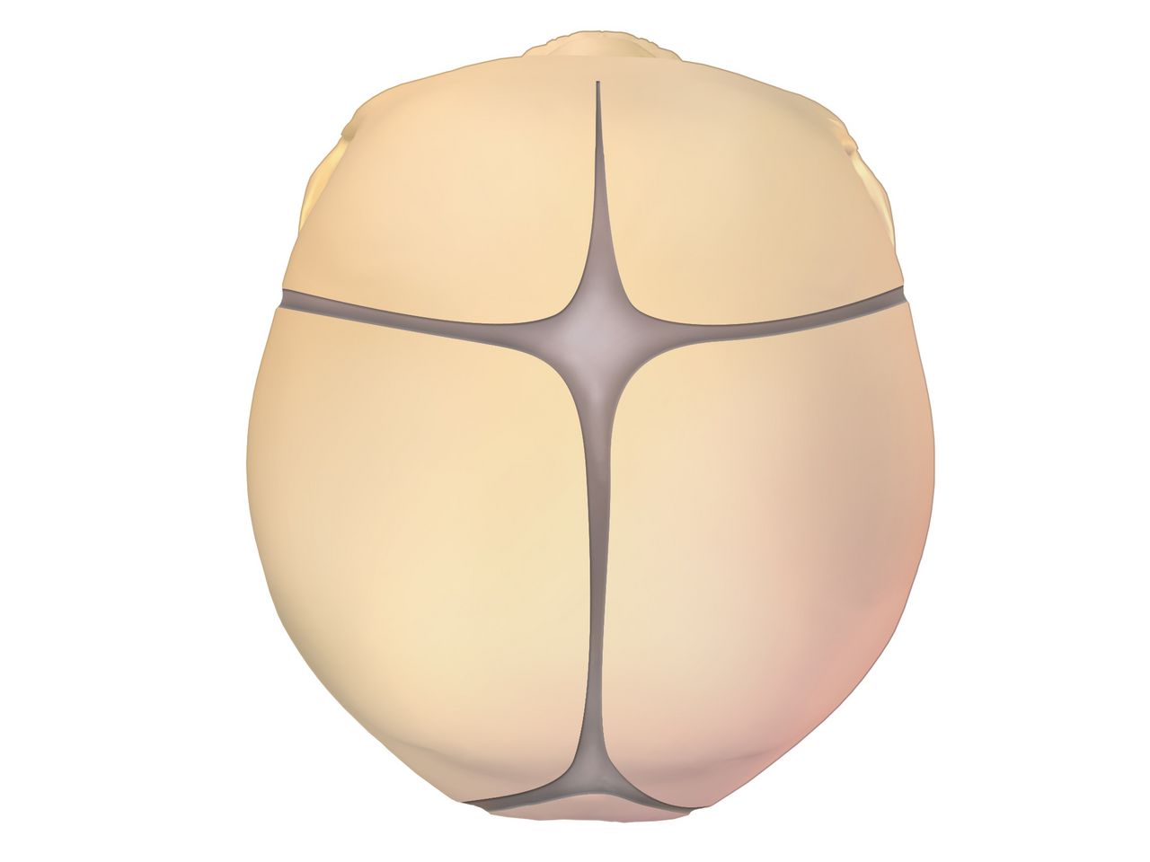
The passenger and the passage
The fetal skull is the widest part of the passenger, and it is deformable. The frontal, sagittal, coronal and lambdoid sutures are membranous seams that allow the parietal bones to overlap under pressure — moulding — which resolves over the first days of life. Where sutures meet, they form fontanelles: the diamond-shaped anterior fontanelle (roughly 2 to 3 cm across, closing at 12 to 18 months) and the small triangular posterior fontanelle (closing by 6 to 8 weeks). On vaginal examination the shape of the fontanelle your fingertip meets tells you the position of the head, which is why the examiner is palpating for a triangle rather than guessing.
Head size depends on attitude, the relationship of fetal parts to one another. With complete flexion — chin to chest — the presenting diameter is the suboccipitobregmatic, about 9.5 cm. Lose that flexion and the presenting diameter grows: a military attitude presents about 12 cm, a brow presentation about 13.5 cm, which is generally too large for the pelvis. Lie is the relationship of the fetal spine to the maternal spine and is longitudinal in about 99 per cent of term labours; transverse or oblique lie cannot be born vaginally. Presentation is the part entering the pelvis first: cephalic in roughly 96 per cent, breech in 3 to 4 per cent, shoulder in the remainder.
Position is recorded as three letters — the side of the maternal pelvis (L or R), the fetal reference point (O for occiput, S for sacrum in breech, M for mentum in face, A for acromion in shoulder), and the part of the pelvis it points toward (A anterior, P posterior, T transverse). LOA, left occiput anterior, is the most common and the most efficient. Occiput posterior positions produce intense back pain, a longer second stage and a greater chance of operative birth; hands-and-knees positioning, counterpressure and lunging help the fetus rotate. Station measures descent in centimetres above or below the ischial spines, from −5 to +5, with 0 station meaning the widest diameter of the head has passed the pelvic inlet — engagement.
The passage is the bony pelvis plus the soft tissues. The gynaecoid pelvis, present in roughly half of people, has a rounded inlet and wide outlet and is the most favourable. The android (heart-shaped inlet, narrow outlet), anthropoid (oval, favours occiput posterior) and platypelloid (flattened, the least favourable) types describe the rest. A diagonal conjugate of 11.5 cm or more on clinical pelvimetry suggests an adequate inlet. Pelvic capacity is not fixed: squatting and McRoberts positioning can increase the outlet diameter measurably, which is the physiological basis for the shoulder-dystocia manoeuvre you will meet in Section 5.
The powers, the position and the psyche
The primary power is the involuntary uterine contraction. Describe it in four terms. Frequency is measured from the beginning of one contraction to the beginning of the next, in minutes. Duration is the beginning to the end of the same contraction, in seconds. Intensity is the peak strength — by palpation graded mild (fundus indents like the tip of the nose), moderate (chin) or strong (forehead), or by intrauterine pressure catheter in mmHg. Resting tone is the pressure between contractions and should feel soft, under about 20 mmHg by catheter. Adequate labour by catheter is 200 or more Montevideo units in 10 minutes, calculated by adding the peak-minus-baseline amplitudes of every contraction in that window.
Contractions that are too frequent are as dangerous as contractions that are too weak, because the placenta refills only between them. Tachysystole is more than five contractions in 10 minutes averaged over a 30-minute window. Duration beyond 90 seconds or a resting tone that never softens compresses the spiral arteries and starves the intervillous space. Whenever tachysystole appears during an oxytocin infusion, the drug is the first suspect.
The secondary power is voluntary bearing down, which only helps once the cervix is completely dilated; pushing against a partly dilated cervix causes oedema and tearing. Maternal position is a power in its own right — upright and lateral positions use gravity, widen the outlet and reduce aortocaval compression, while flat supine positioning reduces venous return and can drop uterine perfusion. The psyche is not a soft extra. Fear and unrelieved pain raise circulating catecholamines, which reduce uterine blood flow and can slow a contraction pattern; continuous labour support measurably shortens labour and lowers caesarean rates.
Premonitory signs, true labour and rupture of membranes
In the weeks before labour the fetus settles into the pelvis (lightening), breathing eases and urinary frequency returns. Braxton Hicks contractions intensify, the cervix softens and begins to efface, the mucus plug is released as bloody show, weight may fall by half a kilogram to a kilogram and some people describe a burst of energy in the 24 to 48 hours before onset. None of these confirms labour. Only progressive cervical change does.
| Feature | True labour | False labour |
|---|---|---|
| Contractions | Regular, progressively closer, longer and stronger | Irregular, do not intensify |
| Effect of walking or a warm bath | Intensify or unchanged | Usually diminish or stop |
| Location of discomfort | Begins in the back and sweeps to the lower abdomen | Abdomen and groin only |
| Cervix | Progressive effacement and dilation — the defining finding | No change on serial examination |
| Show | Bloody show present and increasing | Absent or brownish discharge only |
Rupture of membranes is confirmed by pooling of fluid in the posterior fornix, a nitrazine strip turning blue-green (amniotic fluid pH about 6.5 to 7.5 against a vaginal pH of 4.5 to 5.5 — blood, semen and infection give false positives), ferning on a dried slide, or a commercial placental protein assay. The first nursing action after any rupture, spontaneous or artificial, is to assess the fetal heart rate for a full minute, because a gush of fluid can sweep the cord past the presenting part. Only then document the time, amount, colour and odour. Clear or with flecks of vernix is normal; green means meconium; port-wine suggests abruption; foul or cloudy fluid with maternal fever and fetal tachycardia suggests chorioamnionitis, and the risk of intra-amniotic infection climbs after about 18 to 24 hours of ruptured membranes.
When labour must be started, readiness is scored. The Bishop score rates dilation, effacement, station, cervical consistency and cervical position out of 13. A score of 8 or more in a nulliparous person predicts a vaginal birth about as likely as with spontaneous labour; 6 or less is unfavourable and calls for cervical ripening first — a dinoprostone vaginal insert (10 mg, removable, an advantage if tachysystole develops), misoprostol 25 micrograms vaginally every 3 to 6 hours, or a mechanical transcervical balloon inflated with 30 to 80 mL. Oxytocin is delayed 30 to 60 minutes after removing a dinoprostone insert and about 4 hours after a misoprostol dose.
Presentation, position and station describe whether a fetus can descend, but only progressive cervical change proves labour has begun — and the first action after membranes rupture is always a full minute of fetal heart rate.
Section 2The Mechanisms and Stages of Labour
The cardinal movements
The pelvis is not a straight tube. Its inlet is widest transversely and its outlet widest anteroposteriorly, so the fetus must turn as it travels. Engagement brings the biparietal diameter through the inlet. Descent continues throughout, driven by contractions, the pressure of amniotic fluid and, later, maternal pushing. Flexion is forced by resistance from the pelvic floor and reduces the presenting diameter to its smallest. Internal rotation turns the occiput towards anterior, and how far it travels depends on where it started: about 45 degrees from an oblique position such as LOA, about 90 degrees from a transverse position such as LOT, and about 135 degrees from occiput posterior. Extension occurs as the occiput passes under the symphysis and the head is born by extending over the perineum. Restitution and external rotation realign the head with the shoulders, and expulsion follows as the anterior shoulder slips under the symphysis, then the posterior shoulder, then the body.
Knowing this sequence is not academic. When the head is born but restitution does not occur and the chin retracts against the perineum — the turtle sign — you are watching a failed expulsion step, and the manoeuvres in Section 5 are all attempts to complete it.
First stage: latent, active and the honest truth about progress
The first stage runs from the onset of regular contractions to complete dilation. Contemporary practice divides it at 6 cm. The latent phase (0 to about 5 or 6 cm) can legitimately last many hours, particularly in a first labour or an induction, and its length alone is not a reason for caesarean birth. The active phase (6 cm to 10 cm) is when dilation accelerates. Historic Friedman curves demanding 1.2 cm per hour in a nullipara overestimate normal speed; modern data show a much wider range. Arrest of dilation is diagnosed only when the person is at least 6 cm with ruptured membranes and there has been no cervical change for 4 hours or more with adequate contractions, or 6 hours or more with inadequate contractions despite oxytocin.
Nursing care in the first stage is mostly surveillance and support. Vital signs every hour (every 30 minutes if membranes are ruptured or the temperature is rising), temperature every 4 hours intact and every 2 hours ruptured, contraction assessment with fetal heart rate at the interval dictated by risk, position change at least every 30 minutes, and bladder emptying every 2 hours. A full bladder is one of the commonest reversible causes of a stalled labour and of a fundus that will not stay firm afterwards. Vaginal examinations are kept to the minimum necessary once membranes are ruptured, because each one imports organisms.
Group B Streptococcus prophylaxis belongs here. A positive rectovaginal screen at 36 to 37 weeks, GBS bacteriuria in this pregnancy, a previous infant with GBS disease, or unknown status with fever, preterm labour or membranes ruptured 18 hours or more, all call for intrapartum penicillin G 5 million units IV loading then 2.5 to 3 million units every 4 hours. Adequate prophylaxis means at least 4 hours of antibiotic before birth. For a non-anaphylactic penicillin allergy use cefazolin; for severe allergy use clindamycin or vancomycin guided by susceptibility.
Second, third and fourth stages
The second stage runs from complete dilation to birth. It has a passive phase, when the fetus descends without active pushing (“labouring down”, especially useful with an epidural), and an active phase of bearing down. Open-glottis pushing with the urge, in an upright or side-lying position, is preferred to prolonged breath-holding, which raises intrathoracic pressure and reduces placental perfusion. Prolonged second stage is generally defined as more than 3 hours in a nullipara (4 with an epidural) or more than 2 hours in a multipara (3 with an epidural), and even then continued progress may justify continuing. Perineal support, warm compresses and controlled delivery of the head reduce severe lacerations; episiotomy is not performed routinely.
The third stage is birth of the infant to expulsion of the placenta, normally within 5 to 30 minutes. Signs of separation are a firm globular uterus, a rise of the fundus in the abdomen, lengthening of the visible cord and a sudden small gush of blood. A placenta delivering shiny fetal side first is Schultze; maternal side first is Duncan. Active management — a uterotonic (commonly oxytocin 10 units IM, or 10 to 40 units in 500 to 1000 mL of isotonic fluid IV), gentle controlled cord traction with counter-pressure above the symphysis, and fundal massage after delivery of the placenta — roughly halves the risk of haemorrhage. The placenta is inspected for completeness, because retained fragments are a classic delayed cause of bleeding.
The fourth stage is the first 1 to 4 hours after birth, the period of greatest haemorrhage risk. Assess fundus, lochia, bladder, perineum and vital signs every 15 minutes for the first hour, every 30 minutes for the next hour, then hourly. The fundus should be firm, midline and at or just below the umbilicus. A fundus that is boggy, above the umbilicus and displaced to the right almost always means a full bladder — have the patient void or catheterise, then reassess. Lochia rubra should be moderate; saturating a perineal pad in 15 minutes or less, or a steady trickle despite a firm fundus, is haemorrhage until proved otherwise.
| Stage | Boundaries | Typical duration | Nursing priority |
|---|---|---|---|
| First — latent | Onset to about 6 cm | Highly variable; hours | Support, hydration, ambulation, baseline monitoring |
| First — active | 6 cm to 10 cm | Faster, wide normal range | Fetal surveillance, comfort, position change, bladder |
| Second | Complete dilation to birth | Up to 3 h nullipara, 2 h multipara (add 1 h with epidural) | Pushing technique, perineal support, fetal heart after each contraction |
| Third | Birth to placenta | 5 to 30 minutes | Uterotonic, controlled cord traction, inspect placenta |
| Fourth | First 1 to 4 hours postpartum | 1 to 4 hours | Fundus, lochia, bladder, vitals; skin-to-skin and first feed |
Labour progress is judged by cervical and descent change over time against contemporary curves, and the fourth stage — fundus, lochia, bladder — is the hour when the greatest preventable death still happens.
Section 3Reading the Fetal Heart and Restoring Oxygen
What you are actually monitoring
Fetal oxygenation depends on a chain: maternal blood oxygen, maternal cardiac output, uterine artery flow, the intervillous space, the placenta itself, the umbilical cord and the fetal circulation. Every intrapartum intervention you will ever make targets one of those links. Before applying a monitor, perform Leopold manoeuvres with the bladder empty and a wedge under one hip: they identify the fetal part in the fundus, the location of the back (where you will hear the heart best), the presenting part, and its degree of descent.
For a low-risk labour, intermittent auscultation is an evidence-supported alternative to continuous monitoring: listen for 30 to 60 seconds immediately after a contraction, every 15 to 30 minutes in active first stage and every 5 to 15 minutes in second stage. Continuous electronic monitoring is used when risk factors exist — oxytocin, epidural, meconium, hypertensive disease, diabetes, growth restriction, prematurity, prior caesarean, an abnormal auscultated pattern. Internal monitoring with a fetal scalp electrode and an intrauterine pressure catheter requires ruptured membranes, adequate dilation and an engaged presenting part.
The five terms and the three categories
Baseline is the mean rate over a 10-minute window excluding accelerations, decelerations and marked variability, rounded to increments of 5, requiring at least 2 identifiable minutes. Normal is 110 to 160 bpm. Variability is the beat-to-beat fluctuation of the baseline and is the single most important indicator that the fetal autonomic nervous system is well oxygenated: absent (undetectable), minimal (5 bpm or less), moderate (6 to 25 bpm — the goal), or marked (more than 25 bpm). Accelerations at 32 weeks or more are a rise of at least 15 bpm lasting at least 15 seconds; before 32 weeks, 10 bpm for 10 seconds. They are reassuring.
Decelerations are classified by shape and timing, not depth. Early decelerations are gradual (onset to nadir 30 seconds or more) and mirror the contraction; they reflect head compression and require no intervention. Late decelerations are gradual with the nadir after the peak of the contraction and signal uteroplacental insufficiency — these are the ones that demand action. Variable decelerations are abrupt (onset to nadir under 30 seconds), at least 15 bpm below baseline for at least 15 seconds but under 2 minutes, and reflect cord compression. A prolonged deceleration lasts 2 minutes or more but under 10 minutes; 10 minutes or more is a baseline change.
| Pattern | Cause (VEAL CHOP) | First nursing action |
|---|---|---|
| Variable deceleration | Cord compression | Reposition — lateral, then knee-chest or Trendelenburg if persistent; check for prolapse |
| Early deceleration | Head compression | None; continue to observe and document |
| Acceleration | Oxygenation intact | None; reassuring |
| Late deceleration | Placental insufficiency | Full intrauterine resuscitation and notify the provider |
The NICHD three-tier system converts these findings into a decision. Category I requires all of: baseline 110 to 160, moderate variability, no late or variable decelerations; early decelerations and accelerations may be present or absent. It is normal and needs routine care. Category III is either absent variability accompanied by recurrent late decelerations, recurrent variable decelerations or bradycardia, or a sinusoidal pattern. It predicts abnormal fetal acid-base status and demands immediate intrauterine resuscitation and preparation for expedited birth. Category II is everything in between — the majority of tracings — and is indeterminate: it obliges surveillance, correction of what you can correct, and reassessment, not panic.
Intrauterine resuscitation, in order
When a tracing deteriorates, act in this sequence. First, reposition to a lateral position — it takes seconds, relieves aortocaval compression and often relieves cord compression at the same time. Second, stop or reduce the oxytocin, because the drug that is reducing placental refill is the one thing entirely under your control. Third, give an intravenous fluid bolus of 500 to 1000 mL of an isotonic non-dextrose solution to raise uterine perfusion. Fourth, perform a vaginal examination to check for cord prolapse and rapid descent. Fifth, notify the provider using SBAR. For tachysystole persisting after oxytocin is stopped, a tocolytic such as terbutaline 0.25 mg subcutaneously may be ordered. Amnioinfusion may be ordered for recurrent variable decelerations.
Supplemental oxygen at 10 L/min by non-rebreather mask has long appeared on this list, and many facilities still include it. Current evidence has not demonstrated improved neonatal outcomes from routine oxygen for a category II tracing, so follow your institution’s protocol but do not let reaching for a mask delay the repositioning, the oxytocin and the fluid that actually change uterine blood flow. Digital scalp stimulation producing an acceleration is reassuring and suggests a fetal pH above 7.20.
Moderate variability is the most reassuring finding on a strip, late decelerations mean placental insufficiency, and the first move for almost any worrying pattern is to reposition and stop the oxytocin — not to reach for oxygen.
Section 4Comfort, Analgesia and Anaesthesia
Where labour pain comes from, and what works without a drug
First-stage pain is visceral: cervical stretching and uterine ischaemia referred through T10 to L1, felt in the lower abdomen, back and thighs, and poorly localised. Second-stage pain is somatic: distension of the vagina and perineum carried by the pudendal nerve through S2 to S4, sharp and precisely located. This anatomy explains why a block adequate for the first stage may not cover the second, and why a pudendal block placed late covers perineal repair but not contractions.
Non-pharmacological measures are not a consolation prize. Continuous one-to-one labour support — a partner, a doula, a nurse who stays — reduces caesarean birth, shortens labour and improves satisfaction more reliably than most drugs. Hydrotherapy, warm packs, cold packs, counterpressure over the sacrum for occiput-posterior back pain, the double hip squeeze, position change every 20 to 30 minutes, birth balls, ambulation, rhythmic breathing, focal points, guided imagery, hypnobirthing, acupressure, TENS and intradermal sterile water papules for back pain all have supporting evidence and no fetal effect. Offer them first and keep offering them after an epidural is placed.
Systemic agents and nitrous oxide
Intravenous opioids give partial relief and cross the placenta. Fentanyl 50 to 100 micrograms IV has a short duration and less neonatal depression; butorphanol 1 to 2 mg and nalbuphine 10 mg are mixed agonist-antagonists that cause less nausea but must be avoided in an opioid-dependent patient because they can precipitate withdrawal in the parent and the fetus. The clinical rule is timing: an opioid given within roughly 1 to 4 hours of birth peaks in the newborn at the worst moment, producing respiratory depression and poor feeding. If birth is imminent, hold the dose and tell the provider why.
Two related safety points. A newborn depressed after maternal opioid is managed by effective positive-pressure ventilation, not naloxone; naloxone is no longer part of the initial delivery-room resuscitation sequence, and it must never be given to the newborn of an opioid-dependent parent because it can precipitate seizures. Nitrous oxide in a 50:50 blend with oxygen is self-administered by mask, started about 30 seconds before a contraction, and is safe because a sedated patient drops the mask; only the patient may hold it, and that includes not letting a well-meaning partner help.
Neuraxial analgesia and its complications
An epidural places a catheter in the epidural space for continuous local anaesthetic with an opioid; a spinal injects a single dose into cerebrospinal fluid for caesarean birth; a combined spinal-epidural gives rapid onset plus a catheter. Before placement: informed consent, IV access, a fluid preload or coload of 500 to 1000 mL of isotonic non-dextrose solution, baseline vital signs and fetal heart rate, an empty bladder, and positioning either sitting with the back arched or side-lying curled. Contraindications include patient refusal, coagulopathy or therapeutic anticoagulation, a platelet count below roughly 70 to 100 × 109/L, infection at the site or frank sepsis, uncorrected hypovolaemia and raised intracranial pressure.
Maternal hypotension is the most common complication — a systolic pressure below 100 mmHg or a fall of more than 20 per cent from baseline — and it reduces placental perfusion within seconds, so late decelerations often appear before the patient feels faint. Blood pressure is checked every 5 minutes for the first 15 to 20 minutes after dosing and after every top-up. The sequence when it drops: turn the patient to a lateral position or apply left uterine displacement first, then open the intravenous fluid, then apply oxygen, then notify anaesthesia and give an ordered vasopressor such as ephedrine or phenylephrine.
Other consequences follow from the physiology. Sensory block causes urinary retention — scan or catheterise every 2 hours. Motor block causes fall risk — the patient does not stand unassisted until motor strength and sensation return. Opioid in the mixture causes pruritus and occasionally delayed respiratory depression. A dural puncture produces a post-dural-puncture headache that is positional, worse sitting or standing and relieved lying flat; treat with hydration, analgesia and caffeine, and definitively with an epidural blood patch of 10 to 20 mL of the patient’s own blood. Maternal fever without infection is common and complicates newborn evaluation.
Continuous labour support is the best-evidenced comfort measure in obstetrics, and when an epidural drops the blood pressure the first action is to reposition for left uterine displacement — fluid, oxygen and vasopressors come after.
Section 5Birth, the Newborn’s First Minutes, and the Sequences That Save Lives
The first ten minutes of a newborn’s life
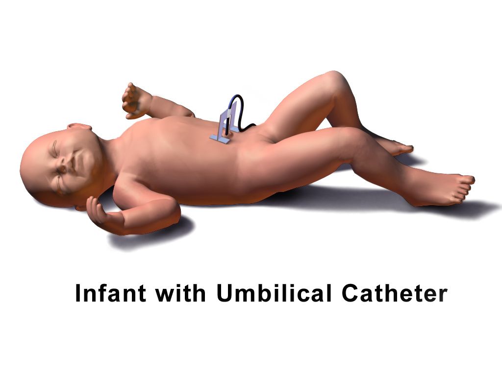
For a vigorous term newborn, the priorities are warmth, airway and contact. Dry thoroughly, remove the wet linen, place skin-to-skin with a cap, and delay cord clamping 30 to 60 seconds — a transfusion of roughly 20 to 30 mL/kg of placental blood that raises iron stores for months. Assessment at birth is continuous rather than scheduled: watch tone, respiratory effort and heart rate from the first seconds, because no recorded score directs resuscitation and you never wait for one before acting.
Normal newborn axillary temperature is 36.5 to 37.5 °C. If the newborn is not breathing or the heart rate is under 100, begin positive-pressure ventilation within the first minute of life — ventilation is the single most effective intervention. If the heart rate stays under 60 after 30 seconds of ventilation that visibly moves the chest, add compressions in a 3:1 ratio, delivering 90 compressions and 30 breaths each minute.
Postpartum haemorrhage: massage the fundus first
Haemorrhage is defined as cumulative blood loss of 1000 mL or more, or any blood loss accompanied by signs of hypovolaemia, within 24 hours of birth regardless of route. Visual estimation underestimates loss by 30 to 50 per cent, so measure it: weigh blood-soaked items, subtract the dry weight, and count 1 gram as 1 mL. Remember that a healthy pregnant person can lose 1500 mL before the blood pressure falls, because pregnancy adds 40 to 50 per cent to plasma volume — rising pulse, narrowing pulse pressure, restlessness and delayed capillary refill come first.
The causes are the four Ts: Tone (uterine atony, 70 to 80 per cent), Trauma (laceration, haematoma, inversion), Tissue (retained placenta) and Thrombin (coagulopathy). Because atony dominates, the first action for a boggy uterus with heavy bleeding is to massage the fundus, one hand cupping the fundus and the other braced above the symphysis to support the lower segment. Then empty the bladder, call for help, open the intravenous fluids, give uterotonics as ordered and quantify the loss. If the fundus is firm and the bleeding continues, the cause is not tone — look for a laceration or a haematoma, and remember that a steadily worsening perineal or rectal pain with a tense purple swelling is a haematoma.
| Uterotonic | Dose | Do not give if | Watch for |
|---|---|---|---|
| Oxytocin | 10 to 40 units in 500 to 1000 mL isotonic IV; or 10 units IM | Never as an undiluted IV push | Hypotension, tachycardia, water intoxication |
| Methylergonovine | 0.2 mg IM every 2 to 4 hours | Hypertension, preeclampsia, cardiac disease | Check BP before every dose; headache, chest pain |
| Carboprost | 250 micrograms IM every 15 to 90 min, max 8 doses (2 mg) | Asthma | Diarrhoea, vomiting, fever, bronchospasm |
| Misoprostol | 600 to 1000 micrograms oral, sublingual or rectal | Known prostaglandin allergy | Fever, shivering |
| Tranexamic acid | 1 g IV over 10 min, within 3 hours of birth | Active thromboembolic disease | Repeat once after 30 min if bleeding persists |
If pharmacology fails, escalation is mechanical and surgical: bimanual compression, an intrauterine balloon tamponade device, uterine artery embolisation, compression sutures, and hysterectomy as the last resort. Activate the massive transfusion protocol early rather than late.
Magnesium sulfate toxicity, cord prolapse, shoulder dystocia and uterine rupture
Pre-eclampsia is what puts most magnesium infusions in the room, so the criteria matter. It is new-onset blood pressure of 140/90 mmHg or higher on two readings at least 4 hours apart after 20 weeks of gestation, together with proteinuria of 300 mg or more in 24 hours, a protein-to-creatinine ratio of 0.3 or more, or 2+ on dipstick when no quantitative test is available. Proteinuria is not required if any severe feature is present: systolic 160 mmHg or higher or diastolic 110 mmHg or higher, platelets below 100 × 109/L, transaminases at twice the upper limit of normal, severe persistent right upper quadrant or epigastric pain, creatinine above 1.1 mg/dL or doubled from baseline, pulmonary oedema, or new cerebral or visual symptoms. HELLP — Haemolysis, Elevated Liver enzymes, Low Platelets — is the variant that catches people out, because the blood pressure may be only mildly raised or even normal and the patient presents complaining of right upper quadrant pain, nausea and feeling generally unwell. Its criteria are platelets below 100 × 109/L, AST or ALT at least twice normal, and LDH of 600 U/L or more with haemolysis on the blood film. Bedside care is the same for both: magnesium sulfate for seizure prophylaxis, antihypertensives for severe-range pressures, a low-stimulus room with side rails padded and suction and oxygen at hand, strict intake and output, and birth as the only definitive cure.
Magnesium sulfate is given for seizure prophylaxis in preeclampsia and for fetal neuroprotection, typically 4 to 6 g intravenously over 15 to 30 minutes then 1 to 2 g per hour by pump. The therapeutic range is about 4 to 7 mEq/L (4.8 to 8.4 mg/dL). Toxicity climbs in a fixed order: deep tendon reflexes disappear first, at roughly 8 to 12 mg/dL, then respiratory depression from about 12 mg/dL with respiratory arrest by 15 to 20 mg/dL, then cardiac conduction changes and cardiac arrest above roughly 25 mg/dL. That order is the whole reason you assess reflexes hourly — the reflex is the early warning, and by the time the respiratory rate falls you have already missed it. Hold the infusion and notify the provider for absent reflexes, a respiratory rate under 12 per minute, urine output under 30 mL/h (magnesium is cleared renally) or a falling level of consciousness. The first action for suspected toxicity is to stop the infusion, then give the antidote calcium gluconate 1 g (10 mL of a 10 per cent solution) intravenously over about 3 minutes, support ventilation, and draw a serum magnesium level. Expect the newborn to be hypotonic and possibly slow to breathe.
Umbilical cord prolapse follows rupture of membranes with an unengaged presenting part, and is more likely with polyhydramnios, malpresentation, prematurity, multiple gestation or a long cord. It shows as a sudden prolonged deceleration or severe recurrent variables immediately after membranes rupture, or as a pulsating cord felt or seen at the introitus. The first action is to insert a gloved hand and lift the presenting part off the cord, and to keep that hand in place until the infant is delivered — do not remove it, and do not attempt to push the cord back inside. Call for help without leaving the patient. Position knee-chest or Trendelenburg or exaggerated Sims with the hips elevated. Handle the cord as little as possible because touching it causes vasospasm; if it is outside the body, cover it with warm saline-moistened sterile gauze. Stop oxytocin, open fluids, give oxygen, and prepare for immediate caesarean birth.
Shoulder dystocia is the anterior shoulder impacted behind the symphysis after the head is born — the turtle sign. Risks include macrosomia, diabetes in pregnancy, previous dystocia and a prolonged second stage, but half of cases have no risk factor. Call for help and note the time, then work through HELPERR: McRoberts (two people hyperflex the thighs sharply onto the abdomen, flattening the sacral promontory and rotating the symphysis upward) plus suprapubic pressure resolves the majority. Fundal pressure is never used — it drives the shoulder harder into the symphysis and risks uterine rupture. Further steps are internal rotational manoeuvres, delivery of the posterior arm, and the all-fours (Gaskin) position. Complications include brachial plexus injury, clavicular or humeral fracture and hypoxic injury for the newborn, and haemorrhage and third or fourth degree laceration for the birthing parent.
Uterine rupture is rare and catastrophic. Suspect it with a previous classical incision or uterine surgery, a trial of labour after caesarean that is being induced or augmented, excessive oxytocin, grand multiparity or trauma. The most common presenting sign is an abnormal fetal heart rate — a prolonged deceleration or bradycardia, not pain. Other findings are sudden sharp tearing abdominal pain, loss of station as the presenting part retracts, cessation of contractions, palpable fetal parts through the abdominal wall and signs of hypovolaemic shock; external bleeding may be minimal because the haemorrhage is intra-abdominal. Stop the oxytocin, call the emergency team, give oxygen and fluids, and prepare for immediate laparotomy — the interval between rupture and birth is measured in minutes.
| Red flag | What it usually means | First action |
|---|---|---|
| Prolonged deceleration right after membranes rupture | Cord prolapse | Gloved hand elevates the presenting part and stays there; knee-chest |
| Boggy fundus, heavy lochia | Uterine atony | Massage the fundus, then empty the bladder and give uterotonics |
| Fundus firm but bleeding briskly | Laceration or haematoma | Inspect the perineum and cervix; call the provider |
| Absent deep tendon reflexes on magnesium | Impending respiratory depression | Stop the infusion, then calcium gluconate 1 g IV |
| Head born, chin retracts, no restitution | Shoulder dystocia | Call for help, note the time, McRoberts plus suprapubic pressure |
| Sudden bradycardia with tearing pain in a TOLAC | Uterine rupture | Stop oxytocin, activate emergency caesarean |
| Sudden dyspnoea, hypotension, then bleeding from every site | Amniotic fluid embolism | Call the code team, support airway and circulation, anticipate coagulopathy |
In obstetric emergencies the sequence is the treatment: massage the fundus first, stop the magnesium before you reach for calcium gluconate, lift the presenting part off a prolapsed cord and never push it back, and use McRoberts with suprapubic — never fundal — pressure.
The chapter in one breath
Labour is the interaction of five forces: a passenger whose skull moulds and whose position and station you can read from a three-letter abbreviation, a passage whose shape and capacity change with maternal positioning, powers described by frequency, duration, intensity and resting tone, and the position and psyche that quietly determine uterine blood flow. Only progressive cervical change proves labour has begun, and the first act after any rupture of membranes is a full minute of fetal heart rate. The four stages have expected patterns rather than deadlines, and the fourth stage — fundus, lochia, bladder, vital signs on a tightening schedule — carries the greatest preventable mortality. Fetal surveillance rests on five terms and three categories, with moderate variability the most reassuring finding and late decelerations the one that demands action; intrauterine resuscitation begins with repositioning and stopping the oxytocin. Comfort ranges from continuous labour support, the best-evidenced measure of all, to neuraxial analgesia whose commonest complication is hypotension corrected first by left uterine displacement. The newborn’s first minutes are warmth, skin-to-skin contact and delayed cord clamping, with positive-pressure ventilation begun inside the first minute if the infant is not breathing or the heart rate is under 100. And in every emergency — haemorrhage, magnesium toxicity, cord prolapse, shoulder dystocia, uterine rupture — the memorised order of actions is what saves the life.
Words worth knowing
| Term | What it means |
|---|---|
| Attitude | Relationship of fetal parts to one another; complete flexion gives the smallest presenting diameter, about 9.5 cm. |
| Lie | Relationship of the fetal spine to the maternal spine — longitudinal, transverse or oblique. |
| Presentation | The fetal part entering the pelvis first: cephalic, breech or shoulder. |
| Position | Three-letter notation of the fetal reference point within the pelvis, such as LOA (left occiput anterior). |
| Station | Descent in centimetres relative to the ischial spines, from −5 to +5; 0 indicates engagement. |
| Moulding | Overlapping of fetal skull bones at the unfused sutures during descent; resolves within days. |
| Effacement | Thinning and shortening of the cervix, expressed as a percentage. |
| Bishop score | Cervical readiness score out of 13; 8 or more is favourable for induction, 6 or less calls for ripening. |
| Tachysystole | More than five contractions in 10 minutes averaged over 30 minutes. |
| Montevideo units | Sum of contraction amplitudes above baseline over 10 minutes by intrauterine catheter; 200 or more is adequate. |
| Variability | Beat-to-beat fluctuation of the fetal heart baseline; moderate (6 to 25 bpm) indicates intact oxygenation. |
| Late deceleration | Gradual fall in fetal heart rate with the nadir after the contraction peak; uteroplacental insufficiency. |
| Category III tracing | Absent variability with recurrent late or variable decelerations or bradycardia, or a sinusoidal pattern; act immediately. |
| Intrauterine resuscitation | Reposition, stop oxytocin, fluid bolus, check for prolapse, notify — measures that restore fetal oxygen delivery. |
| Cardinal movements | Engagement, descent, flexion, internal rotation, extension, restitution and expulsion. |
| Quantitative blood loss | Measured rather than estimated loss; blood-soaked items are weighed with 1 gram counted as 1 mL. |
| Uterine atony | A soft, poorly contracted uterus; the cause of 70 to 80 per cent of postpartum haemorrhage. |
| McRoberts manoeuvre | Sharp hyperflexion of the thighs onto the abdomen to flatten the sacral promontory in shoulder dystocia. |
| Delayed cord clamping | Waiting 30 to 60 seconds before clamping, transfusing placental blood and improving newborn iron stores. |