HEPATOBILIARY AND PANCREATIC DISORDERS
The liver, gallbladder and pancreas sit quietly in the right upper quadrant and the epigastrium doing work no machine fully replaces — manufacturing albumin and clotting factors, clearing ammonia and bilirubin, metabolising nearly every drug you will ever hang, emulsifying fat and digesting protein. When they fail, they fail loudly and in patterns you can predict. The cirrhotic patient bleeds because the liver stopped making factor VII; he is confused because his gut is making ammonia his liver cannot clear; his belly is tight because portal pressure is pushing fluid into the peritoneum and his albumin is too low to pull it back. The patient with acute pancreatitis is hypotensive not because he is bleeding but because activated enzymes turned his retroperitoneum into a third space. Learn the mechanism once and the assessment findings, the laboratory values and the nursing priorities all fall out of it. This chapter teaches those mechanisms and then teaches you what to do first.
By the end of this chapter you can
- Explain the synthetic, metabolic and excretory functions of the liver and predict the clinical consequences of losing each one.
- Interpret a liver panel — ALT, AST, alkaline phosphatase, GGT, bilirubin, albumin and INR — and distinguish a hepatocellular from a cholestatic pattern.
- Compare hepatitis A through E by transmission, chronicity and prevention, and interpret basic hepatitis B serology.
- Describe the pathophysiology of portal hypertension and prioritise nursing care for ascites, oesophageal varices, hepatic encephalopathy, spontaneous bacterial peritonitis and hepatorenal syndrome.
- Differentiate biliary colic, acute cholecystitis and ascending cholangitis, and provide nursing care before and after laparoscopic cholecystectomy and ERCP.
- Explain the autodigestion mechanism of acute pancreatitis, cite the diagnostic lipase threshold, and manage fluids, pain, nutrition and the complications that kill.
- Teach a patient with chronic pancreatitis how to take pancreatic enzyme replacement therapy and recognise steatorrhoea.
- Identify the red flags in hepatobiliary and pancreatic disease that require an immediate SBAR call, and state what may be delegated to the LPN or UAP.
- Describe how hepatobiliary and pancreatic disease present atypically in the older adult, including delirium as a first sign of infection.
Section 1The Liver, the Biliary Tree and What the Numbers Mean
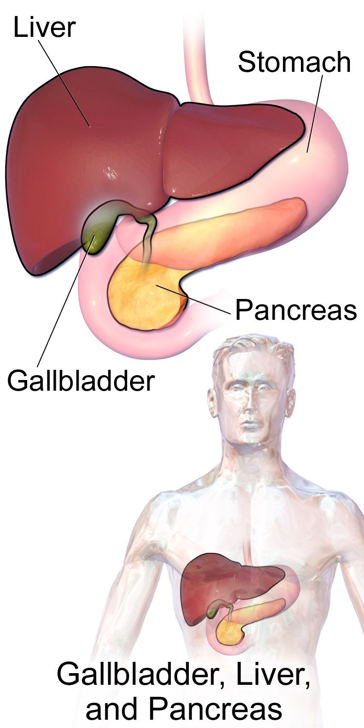
What the liver actually does — and what breaks when it stops
Group the liver’s jobs into four buckets and every finding in this chapter becomes predictable. Synthesis: the liver makes albumin, which holds fluid inside the vessel, and clotting factors I, II, V, VII, IX and X. Metabolism: it converts ammonia from gut protein breakdown into urea, stores and releases glucose, and biotransforms drugs through the cytochrome P450 system. Excretion: it conjugates bilirubin from senescent red cells and dumps it into bile. Storage and immune surveillance: it banks glycogen, iron, and the fat-soluble vitamins A, D, E and K, while Kupffer cells filter portal blood coming straight from the gut.
Now reverse each one. Lose synthesis and you get hypoalbuminaemia with oedema and ascites, plus a rising INR and a patient who bruises from a blood-pressure cuff. Lose metabolism and ammonia climbs, the patient becomes confused, blood glucose swings unpredictably, and every sedative you give lasts twice as long. Lose excretion and bilirubin backs up: yellow sclerae, dark tea-coloured urine, clay-coloured stools, and intractable pruritus from bile salts in the skin. Lose storage and vitamin K deficiency compounds the coagulopathy. Nothing in liver failure is arbitrary.
The liver has enormous reserve — it can lose a large fraction of functioning mass before laboratory values move — which is exactly why chronic liver disease is often silent until it is advanced. Your assessment must therefore be deliberate: skin and sclerae in natural light, palms for palmar erythema, chest and shoulders for spider angiomas, abdomen for distension, flank dullness and a fluid wave, hands held out for asterixis (the flapping, non-rhythmic loss of wrist extension), and the legs for pitting oedema. In darker-skinned patients look for jaundice in the sclerae, the hard palate and the palms, not in the general skin tone.
Jaundice: reading the colour
Bilirubin is the breakdown product of haem. Unconjugated (indirect) bilirubin is fat-soluble and travels bound to albumin; the hepatocyte conjugates it with glucuronic acid, making it water-soluble (direct) so it can be excreted in bile. Jaundice becomes visible when total bilirubin exceeds roughly 2 to 3 mg/dL, with a normal total of 0.3 to 1.2 mg/dL and a direct fraction of 0.1 to 0.3 mg/dL.
Where the problem sits tells you which fraction rises. Prehepatic jaundice — haemolysis, a transfusion reaction, a large resolving haematoma — overwhelms conjugation, so indirect bilirubin rises while stool and urine stay normal in colour. Intrahepatic jaundice — hepatitis, cirrhosis, drug injury — damages the cell that conjugates and excretes, so both fractions rise. Posthepatic or obstructive jaundice — a stone in the common bile duct, a pancreatic head tumour — blocks the exit, so conjugated bilirubin refluxes into blood. Because conjugated bilirubin is water-soluble it spills into urine (dark, tea-coloured, and it foams yellow) while none reaches the bowel (clay-coloured, pale stools). That triad — yellow skin, dark urine, pale stool — is obstruction until proved otherwise.
Pruritus in cholestasis is from bile salts deposited in skin, not from dryness. Antihistamines help little; cholestyramine, cool tepid baths, oatmeal or calamine, short clean fingernails, cotton clothing and keeping the room cool help more. Scratching an oedematous, coagulopathic patient buys skin tears and infection.
The liver panel is really two panels
Students memorise “LFTs” as a block and then cannot interpret them. Split them. The hepatocellular enzymes ALT (7 to 56 U/L) and AST (10 to 40 U/L) leak out of injured hepatocytes; ALT is more liver-specific because AST also lives in heart and skeletal muscle. Values in the thousands mean acute hepatocyte death — viral hepatitis, ischaemic hepatitis (shock liver) or paracetamol/acetaminophen toxicity. An AST-to-ALT ratio above 2:1 with only modest elevations classically points to alcohol-related liver disease.
The cholestatic enzymes alkaline phosphatase (30 to 120 U/L) and GGT (roughly 8 to 60 U/L) rise when bile flow is obstructed. Alkaline phosphatase also comes from bone, so a rise in both ALP and GGT localises the problem to the biliary tree; a rise in ALP with a normal GGT points to bone. Neither group of enzymes measures function — they measure damage. The true function tests are albumin (3.5 to 5.0 g/dL), which reflects synthesis over the preceding weeks, INR (0.8 to 1.1) and prothrombin time (11 to 12.5 seconds), which reflect synthesis over the preceding hours to days, and total bilirubin, which reflects excretion. A patient whose enzymes are falling while INR climbs is not improving — he is running out of hepatocytes.
| Test | Reference range | What a rise (or fall) tells you |
|---|---|---|
| ALT | 7–56 U/L | Hepatocellular injury; most liver-specific enzyme |
| AST | 10–40 U/L | Hepatocellular injury; also heart and muscle. AST:ALT above 2:1 suggests alcohol |
| Alkaline phosphatase | 30–120 U/L | Cholestasis or bone disease |
| GGT | 8–60 U/L | Confirms a high ALP is biliary; sensitive to alcohol |
| Total bilirubin | 0.3–1.2 mg/dL | Excretory failure or obstruction; jaundice visible above 2–3 mg/dL |
| Albumin | 3.5–5.0 g/dL | Synthetic function over weeks; low means oedema and ascites |
| INR / PT | 0.8–1.1 / 11–12.5 s | Synthetic function over hours to days; the fastest marker of decline |
| Ammonia | 15–45 mcg/dL | Impaired urea cycle; correlates poorly with mental status |
| Platelets | 150,000–400,000/mm3 | Low in cirrhosis from splenic sequestration and low thrombopoietin |
| Serum sodium | 135–145 mEq/L | Dilutional hyponatraemia marks advanced portal hypertension |
Imaging, biopsy and severity scoring
Right upper quadrant ultrasound is the first-line study for almost every hepatobiliary complaint: it is cheap, portable, radiation-free, and it detects gallstones, duct dilation, ascites and coarse hepatic echotexture. CT with contrast stages tumours, defines pancreatic necrosis and finds abscesses. MRCP images the ducts non-invasively; ERCP does the same but can also remove a stone or place a stent, at the cost of an invasive procedure with a real risk of post-procedure pancreatitis. Transient elastography (FibroScan) measures stiffness and increasingly replaces biopsy for staging fibrosis.
If a percutaneous liver biopsy is ordered, the nursing care is a small examination all by itself. Before: verify consent, check platelets and INR (many facilities require platelets above 50,000/mm3 and INR below 1.5), type and screen, hold anticoagulants as ordered, keep the patient NPO. During: the patient exhales and holds the breath at end-expiration so the diaphragm stops moving. After: position the patient on the right side with a rolled towel against the costal margin for two hours to tamponade the puncture site, bed rest for the ordered interval, and monitor vital signs frequently for the classic complications — haemorrhage (tachycardia, falling blood pressure, rising abdominal pain), bile peritonitis and pneumothorax (sudden dyspnoea, pleuritic pain).
Two scores drive real decisions. Child-Pugh classes A, B and C are built from bilirubin, albumin, INR, ascites and encephalopathy. MELD (bilirubin, INR, creatinine and sodium) is a pure number used to rank patients on the transplant list — sicker patients score higher and are transplanted sooner. Knowing that creatinine sits inside MELD explains why nephrotoxic drugs and careless diuresis in a cirrhotic patient are not minor errors.
Enzymes (ALT, AST, ALP, GGT) measure liver damage; albumin, INR and bilirubin measure liver function — and a rising INR with falling enzymes signals a liver that is dying, not healing.
Section 2Viral Hepatitis and Acute Liver Failure

The hepatitis alphabet, sorted by how it reaches the patient
Five viruses, two routes. A and E travel by the faecal-oral route — contaminated food, water and shellfish, poor hand hygiene, and in the case of E often contaminated drinking water in resource-limited settings. Neither becomes chronic in a patient with an intact immune system (hepatitis E can persist in the profoundly immunocompromised, such as a transplant recipient, but that is the exception). B, C and D travel in blood and body fluids — needles, sharps injuries, tattoos and piercings with unsterile equipment, sexual contact, and for B, perinatal transmission. All three can become chronic and all three can end in cirrhosis and hepatocellular carcinoma.
Hepatitis A has an incubation of roughly 15 to 50 days and is preventable with a two-dose vaccine six months apart; it is most contagious in the two weeks before jaundice appears, which is why outbreaks are traced backwards through food handlers. Hepatitis B has a longer incubation, 45 to 160 days, and becomes chronic in about 5 percent of infected adults but in up to 90 percent of infants infected perinatally — the reason the birth dose of vaccine matters so much. Hepatitis C rarely produces an acute illness anyone notices; roughly three-quarters of those infected develop chronic infection, and it remains a leading indication for liver transplant. Hepatitis D is a defective virus that can only infect a person who already carries hepatitis B surface antigen, so preventing B prevents D. Hepatitis E is usually self-limited but carries a mortality approaching 20 percent in the third trimester of pregnancy.
| Virus | Transmission | Chronic? | Prevention |
|---|---|---|---|
| Hepatitis A | Faecal-oral; food, water, shellfish | No | Vaccine (2 doses); hand hygiene; immune globulin post-exposure |
| Hepatitis B | Blood, body fluids, sexual, perinatal | Yes (about 5 percent of adults, up to 90 percent of infants) | Vaccine series; HBIG plus vaccine after exposure |
| Hepatitis C | Blood; injection drug use, unregulated tattooing | Yes (about 75 percent) | No vaccine; screen all adults once; direct-acting antivirals cure over 95 percent |
| Hepatitis D | Blood; requires coexisting hepatitis B | Yes | Hepatitis B vaccine |
| Hepatitis E | Faecal-oral; contaminated water | Rarely (immunocompromised) | Safe water; high mortality in pregnancy |
Hepatitis B serology without tears
Three results answer almost every question. HBsAg (surface antigen) means the virus is present — the patient is infected and infectious right now; persistence beyond six months defines chronic infection. Anti-HBs (surface antibody) means immunity, either from resolved infection or from vaccination. Anti-HBc (core antibody) means the patient has actually encountered the wild virus at some point; the IgM subtype indicates recent or acute infection. Vaccination produces anti-HBs alone, so a nurse with anti-HBs positive and anti-HBc negative is vaccinated, while anti-HBs plus anti-HBc positive means natural resolved infection. HBeAg is a marker of high viral replication and high infectivity.
Clinically, hepatitis moves through three phases. The prodromal (preicteric) phase brings malaise, anorexia, nausea, low-grade fever, myalgia, right upper quadrant discomfort and often a distaste for cigarettes or food — nonspecific enough to be mistaken for influenza. The icteric phase brings jaundice, dark urine, pale stools, pruritus and tender hepatomegaly. The convalescent phase can drag on for weeks to months with fatigue that patients find demoralising because they “look fine.”
Nursing care, prevention and exposure management
Care in acute viral hepatitis is largely supportive: rest balanced against activity as tolerated, small frequent high-calorie meals with the largest meal in the morning when nausea is least, adequate fluids, and absolute avoidance of alcohol and of hepatotoxic drugs. Antiemetics are given cautiously and phenothiazines are avoided where possible because of cholestatic potential. Teach the patient with hepatitis A about scrupulous hand hygiene, not preparing food for others, and not sharing utensils; teach the patient with hepatitis B or C not to share razors, toothbrushes or needles, to use barrier protection, and never to donate blood, organs or semen.
Occupational exposure is a chapter you may live rather than read. After a needlestick, wash the site with soap and water immediately, report it at once, and let occupational health test the source and the exposed worker. For hepatitis B, an unvaccinated or non-immune worker receives HBIG plus the vaccine series, ideally within 24 hours and no later than seven days after percutaneous exposure. There is no post-exposure prophylaxis for hepatitis C — the protocol is baseline and serial testing, with early direct-acting antiviral treatment if seroconversion occurs. Standard precautions handle all of the blood-borne types; contact precautions are added for hepatitis A only when the patient is incontinent or nappy-dependent.
Chronic hepatitis C is now curable in more than 95 percent of patients with 8 to 12 weeks of oral direct-acting antivirals. The nursing work has shifted from managing interferon misery to adherence and access: one missed week can compromise the cure, and many patients need help navigating prior authorisation.
Acute liver failure and the paracetamol problem
Acute (fulminant) liver failure is the rapid loss of hepatic function with coagulopathy (INR of 1.5 or greater) and encephalopathy in a patient with no prior liver disease. In North America the single most common cause is acetaminophen (paracetamol) toxicity, often unintentional — a patient taking a prescription opioid-acetaminophen combination plus an over-the-counter cold preparation plus a sleep aid, none of which he recognises as containing the same drug. The maximum for a healthy adult is 4 g in 24 hours; in chronic liver disease, malnutrition or regular alcohol use, 2 g or less per day is safer.
The mechanism is worth knowing because it explains the antidote. Most acetaminophen is glucuronidated safely; a minority is oxidised to the toxic metabolite NAPQI, which glutathione neutralises. In overdose, glutathione stores are exhausted and NAPQI destroys centrilobular hepatocytes. N-acetylcysteine replenishes glutathione and is dramatically effective when started within 8 hours of ingestion, though it is still given later. It is dosed by weight, typically starting with a 150 mg/kg intravenous loading dose per protocol; the oral form smells of rotten eggs and is far better tolerated chilled, in juice, through a straw.
Nursing priorities in acute liver failure are ABC first and neurological status second. Grade III to IV encephalopathy threatens the airway — a patient who cannot protect it needs intubation, not another dose of lactulose. Monitor glucose hourly or per protocol because a failing liver cannot mobilise glycogen and profound hypoglycaemia is common. Watch for cerebral oedema, the leading cause of death: elevate the head of bed 30 degrees, keep the neck midline, cluster care, and minimise suctioning and other stimulation. Avoid sedatives if humanly possible; they are metabolised by the organ that has stopped working. Transplant evaluation is not a last resort here — it is an early call.
Hepatitis A and E come from what the patient swallows and never become chronic; B, C and D come from blood and body fluids and can end in cirrhosis, cancer or transplant — and in acute liver failure the deteriorating mental status, not the bilirubin, is the finding that changes your plan.
Section 3Cirrhosis, Portal Hypertension and Their Complications
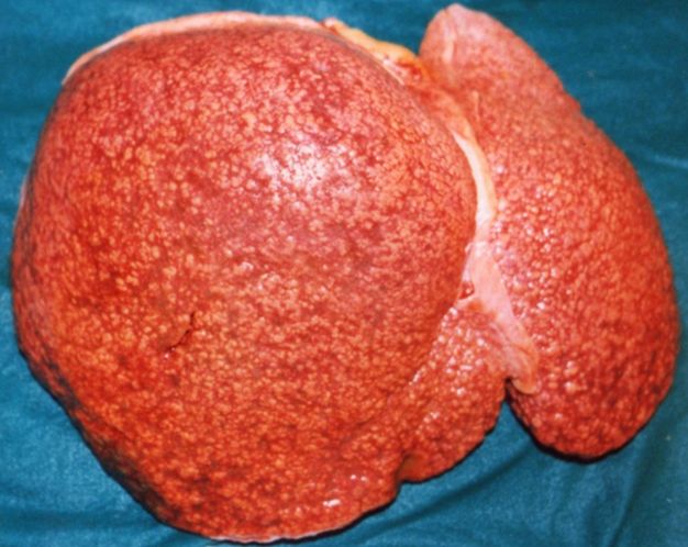
From injury to fibrosis to portal hypertension
Repeated hepatocyte injury — from alcohol, chronic hepatitis B or C, metabolic dysfunction-associated steatotic liver disease (formerly NAFLD), autoimmune disease, haemochromatosis or biliary obstruction — activates hepatic stellate cells, which lay down collagen. Over years the architecture is replaced by regenerative nodules separated by fibrous bands. Two things follow. First, functioning hepatocyte mass falls, producing the synthetic and metabolic failures of Section 1. Second, and more mechanically, blood coming from the entire gut through the portal vein can no longer flow easily through the stiff liver. Portal pressure rises.
Trace that pressure and you have the whole clinical picture. Blood backs up into the spleen, which enlarges and sequesters platelets, producing thrombocytopenia. It backs up into collateral veins that were never designed for the volume — the submucosal veins of the oesophagus and stomach become varices, the periumbilical veins become caput medusae, the rectal veins become haemorrhoids. Hydrostatic pressure in the splanchnic bed, combined with low oncotic pressure from hypoalbuminaemia, pushes fluid into the peritoneum as ascites. Splanchnic vasodilation lowers effective arterial volume, activating the renin-angiotensin-aldosterone system, which retains sodium and water — making the ascites worse and diluting serum sodium. And portal blood carrying gut-derived ammonia now shunts around the liver instead of through it, producing hepatic encephalopathy.
Assessment reflects all of it: jaundice, spider angiomas, palmar erythema, gynaecomastia and testicular atrophy in men (the failing liver cannot metabolise oestrogen), muscle wasting with a protuberant abdomen, ecchymoses, Dupuytren contracture, foetor hepaticus, asterixis and, in men and women alike, a striking loss of temporal muscle mass. Do not be reassured by a blood pressure that looks acceptable on paper; cirrhotic patients often run baseline systolics in the 90s because of splanchnic vasodilation, so a reading of 100/60 that you would call normal in anyone else may already represent significant volume loss in this patient. Compare every pressure with that patient’s own baseline and follow the trend — a falling systolic with a climbing heart rate and a narrowing pulse pressure warns you of bleeding well before the absolute number looks alarming.
Ascites and spontaneous bacterial peritonitis
Ascites is managed in a fixed order. First, sodium restriction to about 2,000 mg per day — this is the intervention patients most often sabotage, usually unknowingly, through canned soup, deli meat, condiments and restaurant food. Fluid restriction is added only when serum sodium falls below roughly 125 mEq/L. Second, diuretics: spironolactone (an aldosterone antagonist, which addresses the actual mechanism) paired with furosemide, classically in a 100 mg to 40 mg ratio to keep potassium balanced. Monitor potassium closely — spironolactone raises it, furosemide lowers it — along with sodium and creatinine. Target weight loss is about 0.5 kg per day without peripheral oedema and up to 1 kg per day with oedema; faster diuresis precipitates renal injury and encephalopathy.
Third, paracentesis for tense or refractory ascites. Have the patient void immediately beforehand to reduce bladder puncture risk, position upright or in high Fowler, measure abdominal girth at a marked point and obtain a weight before and after, and monitor for hypotension during and after drainage. When more than 5 litres are removed, intravenous albumin of roughly 6 to 8 g per litre removed is given to prevent post-paracentesis circulatory dysfunction. TIPS (transjugular intrahepatic portosystemic shunt) is reserved for refractory cases; it reliably lowers portal pressure and just as reliably worsens encephalopathy, because it shunts ammonia-laden blood past the liver by design.
Spontaneous bacterial peritonitis is the infection of ascitic fluid without a perforation, and it is the complication students miss. The classic presentation — fever, diffuse abdominal pain, rebound tenderness — is frequently absent. In many patients the only sign is new or worsening confusion, a rising creatinine, or unexplained hypotension. Diagnosis is by diagnostic paracentesis showing an ascitic fluid polymorphonuclear count of 250 cells/mm3 or more. Treatment is a third-generation cephalosporin such as cefotaxime or ceftriaxone plus intravenous albumin (1.5 g/kg on day 1 and 1 g/kg on day 3) to protect the kidneys, followed by lifelong prophylaxis. A SAAG (serum-ascites albumin gradient) of 1.1 g/dL or greater confirms that the ascites is from portal hypertension rather than from malignancy or infection of the peritoneum itself.
Any cirrhotic patient with new confusion gets three questions answered before you reach for lactulose: is he bleeding, is he infected, is he dry? Gastrointestinal haemorrhage, spontaneous bacterial peritonitis, over-diuresis and constipation precipitate far more encephalopathy than dietary protein ever does.
Oesophageal varices: the bleed that kills
Varices are thin-walled submucosal veins under high pressure. When one ruptures the patient can lose litres in minutes, and the coagulopathy and thrombocytopenia of cirrhosis mean the bleeding does not stop on its own. Presentation is haematemesis (bright red or coffee-ground), melaena, tachycardia, hypotension and altered mental status.
Priorities are pure ABC. Airway first — a patient vomiting blood with encephalopathy will aspirate; suction is at the bedside and intubation is often required before endoscopy. Then two large-bore intravenous lines (18 gauge or larger), type and crossmatch, volume resuscitation and blood products with a restrictive transfusion target (generally haemoglobin around 7 g/dL, because over-transfusion raises portal pressure and re-triggers bleeding). Pharmacologically, octreotide reduces splanchnic blood flow — a 50 mcg bolus followed by a 50 mcg/hr infusion for 3 to 5 days — and prophylactic ceftriaxone is given because bacterial infection follows variceal bleeding with depressing regularity and worsens outcomes. Definitive control is endoscopic variceal band ligation within 12 hours. Balloon tamponade with a Sengstaken-Blakemore or Minnesota tube is a rescue bridge only, with scissors taped at the head of the bed for airway obstruction if the gastric balloon migrates.
Prevention matters more than rescue. Patients with known varices take a nonselective beta blocker — propranolol, nadolol or carvedilol — titrated to a resting heart rate of about 55 to 60, which lowers portal pressure. Cardioselective agents such as metoprolol do not work for this. Teach the patient to avoid NSAIDs and aspirin, to avoid straining and heavy lifting (Valsalva raises portal pressure), to treat constipation, to chew thoroughly and avoid coarse or sharp foods, and to seek care immediately for vomiting blood or black tarry stools.
Hepatic encephalopathy and hepatorenal syndrome
Hepatic encephalopathy runs a spectrum from a subtle reversal of sleep-wake cycle and slowed handwriting (grade I), through lethargy and disorientation with asterixis (grade II), to marked confusion and somnolence but rousable (grade III), to coma (grade IV). Serial mental-status assessment is the single most important nursing observation on a cirrhotic patient, and the change from grade II to grade III is an airway decision.
Lactulose is first-line. It is a nonabsorbable disaccharide that acidifies the colon, converting ammonia (NH3) to ammonium (NH4+), which cannot cross back into blood, and it purges it out as an osmotic laxative. Titrate to two to three soft stools per day — too few and the patient stays encephalopathic, too many and he becomes dehydrated and hypokalaemic, which paradoxically worsens encephalopathy. It can be given orally, by nasogastric tube or as a retention enema. Rifaximin 550 mg twice daily is a minimally absorbed antibiotic that reduces ammonia-producing gut flora and is added to prevent recurrence. Note that serum ammonia correlates poorly with clinical severity: treat the patient, not the number. And modern practice does not restrict protein — these patients are catabolic and need 1.2 to 1.5 g/kg/day, ideally with a late evening snack to shorten the overnight fast.
Hepatorenal syndrome is functional kidney failure in advanced liver disease: intense renal vasoconstriction in response to splanchnic vasodilation, with structurally normal kidneys. It is marked by a rising creatinine, oliguria, and a very low urine sodium (below 10 mEq/L) that does not correct with volume. It is precipitated by over-diuresis, large-volume paracentesis without albumin, NSAIDs, aminoglycosides and contrast. Treatment is albumin plus vasoconstrictors such as terlipressin or midodrine with octreotide, but the definitive treatment is liver transplant. Prevention is nursing work: protect these kidneys.
SBAR — call now. “S: Mr Reyes in 412 with alcohol-related cirrhosis just vomited about 400 mL of bright red blood. B: Known grade II oesophageal varices, admitted three days ago for ascites, on spironolactone and furosemide. A: Heart rate 128, blood pressure 84/50, he is confused and cool to touch; two 18-gauge lines are in, he is on oxygen, suction is at the bedside and I have sent a type and crossmatch and a stat CBC. R: I need you at the bedside now, and I need orders for blood products, octreotide, ceftriaxone and urgent endoscopy.”
| Complication | Mechanism | Key finding | First-line management |
|---|---|---|---|
| Ascites | Portal hypertension plus low albumin plus sodium retention | Distension, flank dullness, fluid wave, weight gain | 2 g sodium diet, spironolactone plus furosemide, paracentesis |
| Spontaneous bacterial peritonitis | Bacterial translocation into ascitic fluid | New confusion or rising creatinine; ascitic PMN 250/mm3 or more | Cefotaxime or ceftriaxone plus albumin, then prophylaxis |
| Oesophageal varices | Collateral flow through submucosal veins | Haematemesis, melaena, shock | Airway, volume, octreotide, ceftriaxone, band ligation |
| Hepatic encephalopathy | Gut ammonia bypasses the liver | Asterixis, sleep reversal, confusion | Lactulose to 2–3 soft stools daily, rifaximin, treat the trigger |
| Hepatorenal syndrome | Renal vasoconstriction with normal kidneys | Rising creatinine, oliguria, urine sodium below 10 mEq/L | Albumin plus vasoconstrictors; transplant is definitive |
Every major complication of cirrhosis traces back to one of two failures — the liver cannot synthesise, or blood cannot get through it — and in a bleeding varix the first nursing action is protecting the airway, not hanging the octreotide.
Section 4Gallbladder and Biliary Disorders
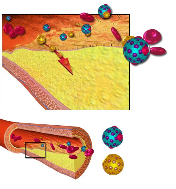
Stones form, and then stones move
The gallbladder stores and concentrates bile and contracts in response to cholecystokinin when fat reaches the duodenum. Cholelithiasis develops when bile becomes supersaturated with cholesterol, when the gallbladder empties sluggishly, or when excess bilirubin forms pigment stones (as in haemolysis or cirrhosis). The classic risk profile is remembered as the four Fs — female, fair, fat, forty — and it is worth extending: pregnancy and oestrogen therapy, rapid weight loss or bariatric surgery, prolonged fasting or total parenteral nutrition, diabetes, and Native American or Hispanic ancestry all raise risk.
Whether the patient has symptoms depends entirely on whether a stone moves. Stones sitting in the gallbladder are often silent. A stone that lodges in the cystic duct during contraction produces biliary colic: severe, steady right upper quadrant or epigastric pain radiating to the right shoulder or scapula, typically 30 minutes to a few hours after a fatty meal, often with nausea and vomiting, resolving when the stone falls back. There is no fever and no leucocytosis, because nothing is inflamed yet. A stone that lodges in the common bile duct is choledocholithiasis and now obstructs bile drainage for the liver and, because the pancreatic duct joins at the ampulla, potentially for the pancreas as well — jaundice, dark urine, clay stools, and gallstone pancreatitis.
Acute cholecystitis and ascending cholangitis
Acute cholecystitis is what happens when the stone does not fall back: the obstructed gallbladder distends, its wall becomes inflamed and oedematous, and bacteria colonise the stagnant bile. Now the pain is persistent rather than colicky, there is fever, leucocytosis with a white count typically 12,000 to 20,000/mm3, guarding, and a positive Murphy sign — the patient stops inspiration abruptly when you palpate under the right costal margin. Ultrasound shows a thickened gallbladder wall, pericholecystic fluid and stones. Untreated it progresses to gangrene and perforation. Acalculous cholecystitis, seen in critically ill, ventilated or TPN-dependent patients without stones, is easy to miss and carries a higher mortality.
Ascending cholangitis is infection of the obstructed biliary tree and it is a surgical and infectious emergency. Charcot triad — fever with rigors, jaundice and right upper quadrant pain — identifies it. When hypotension and altered mental status are added, you have Reynolds pentad, meaning septic shock, and the patient needs blood cultures, broad-spectrum antibiotics, aggressive fluid resuscitation and urgent biliary decompression by ERCP. Do not wait for the fever to declare itself in an older adult: in a patient over 75 the first and sometimes only sign of cholangitis is new confusion or a fall, with a normal or even low temperature and a white count that has not budged.
| Condition | Pain pattern | Fever / WBC | Bilirubin and enzymes | Urgency |
|---|---|---|---|---|
| Biliary colic | Episodic, post-fatty meal, resolves | Absent / normal | Normal | Elective |
| Acute cholecystitis | Constant RUQ, positive Murphy sign | Present / 12,000–20,000/mm3 | Mildly elevated | Same admission surgery |
| Choledocholithiasis | RUQ with jaundice | Variable | Bilirubin and ALP clearly elevated | ERCP |
| Ascending cholangitis | RUQ with rigors | High fever / marked leucocytosis | Bilirubin and ALP markedly elevated | Emergency — sepsis |
| Gallstone pancreatitis | Epigastric boring to the back | Present | Lipase 3 times upper limit or more | Emergency |
Procedures and the nursing care around them
Laparoscopic cholecystectomy is the standard, usually same-day or one-night stay. Post-operatively, the finding students find surprising is referred right shoulder pain from residual carbon dioxide irritating the diaphragm. Treat it with early ambulation, position changes, a left side-lying knee-to-chest position and analgesia, and reassure the patient it resolves in 24 to 48 hours — but do not dismiss shoulder pain accompanied by fever, rising abdominal pain, tachycardia or bile-stained drainage, which suggests a bile leak or duct injury. Encourage incentive spirometry: the incisions sit right under the diaphragm, splinting is instinctive, and atelectasis is the commonest complication.
An open cholecystectomy may leave a T-tube in the common bile duct. Keep the collection bag below the level of the gallbladder to prevent reflux, never irrigate or clamp it without an order, expect 300 to 500 mL of bile-coloured drainage in the first 24 hours declining thereafter, and report drainage exceeding 500 mL per day or an abrupt stop. When the tube is clamped before removal (often before and after meals), watch for pain, nausea and chills, which mean bile is not draining into the duodenum. Return of brown stool colour confirms bile is reaching the bowel.
ERCP deserves its own teaching. Keep the patient NPO beforehand, verify consent and allergies, and afterwards keep the patient NPO until the gag reflex returns. The complication to watch for is post-ERCP pancreatitis: new or worsening epigastric pain radiating to the back, nausea and a rising lipase in the hours after the procedure. Perforation and bleeding are rarer but real. Report new severe abdominal pain, fever or vomiting immediately.
Delegation. UAP may obtain vital signs on a stable post-cholecystectomy patient, assist with early ambulation, record intake and output and empty a measured drainage bag, and remind the patient to use the incentive spirometer at the ordered interval. The LPN may give scheduled oral analgesia, perform routine dressing changes and reinforce teaching. The RN keeps the initial and ongoing assessment, T-tube management decisions, the first evaluation of new shoulder or abdominal pain, blood products, intravenous push analgesia in many facilities, and all discharge teaching.
Teaching the patient going home
After cholecystectomy the patient no longer stores bile — bile drips continuously into the duodenum instead of arriving in a bolus. Most people tolerate a normal diet within weeks, but for the first four to six weeks advise smaller, lower-fat meals and gradual reintroduction of fatty foods; loose stools are common early and usually settle. Teach incision care, the signs of infection, and the specific reasons to call: fever above 38.3 degrees Celsius (101 degrees Fahrenheit), worsening rather than improving pain, persistent vomiting, jaundice, dark urine or clay-coloured stools. No heavy lifting for the interval the surgeon specifies. For patients managed without surgery, teach the low-fat diet and the pattern of pain that means the stone has moved into the duct.
Biliary disease is graded by whether the stone is stuck and whether the bile is infected — and Charcot triad in any patient, or new confusion in an older one, means cholangitis and sepsis until proved otherwise.
Section 5Acute and Chronic Pancreatitis and Pancreatic Cancer
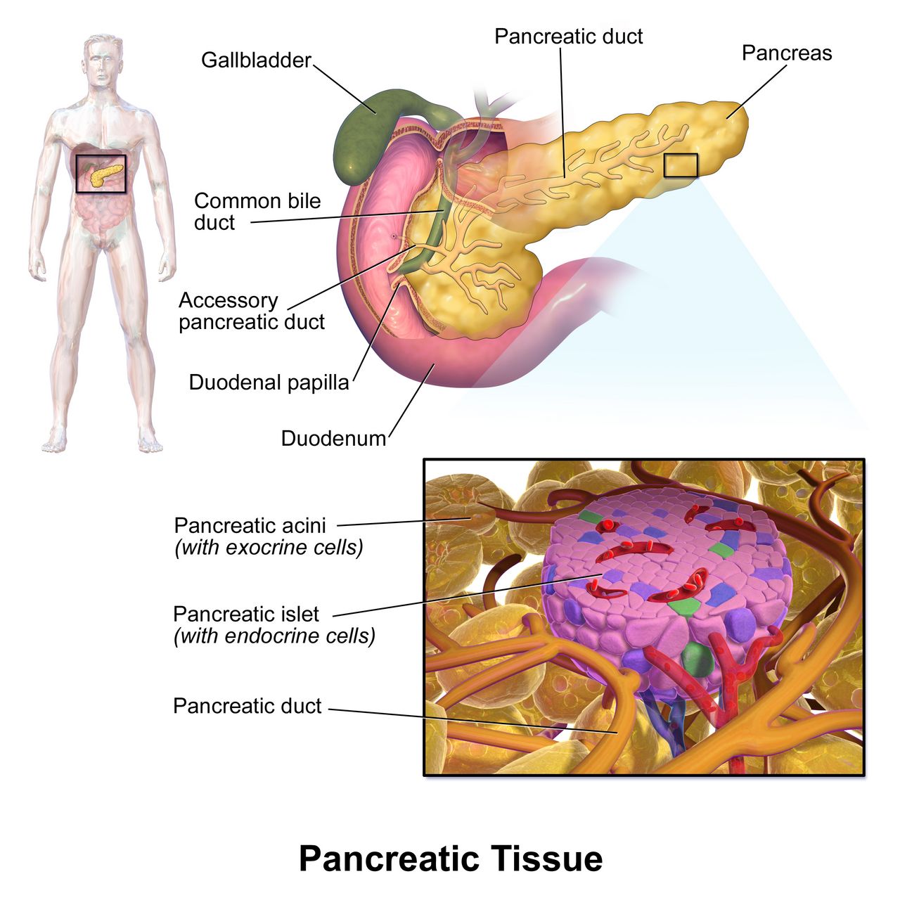
Acute pancreatitis: the gland digests itself
The pancreas secretes amylase and lipase in fully active form, but it manufactures its proteases as inactive precursors — zymogens such as trypsinogen, chymotrypsinogen and proelastase — which are normally activated only after they reach the duodenum. In acute pancreatitis, trypsinogen is activated inside the gland, and that premature activation triggers a cascade that converts the remaining zymogens and begins autodigestion. Lipase digests fat, proteases digest protein and vessel walls, and the resulting inflammation causes massive capillary leak. Two causes account for most cases: gallstones obstructing the ampulla, and alcohol. Others include hypertriglyceridaemia (usually above 1,000 mg/dL), hypercalcaemia, ERCP, abdominal trauma, and drugs such as azathioprine, valproic acid, thiazides, sulfonamides and some HIV medications.
Every classic manifestation follows from the mechanism. Pain is severe, boring, epigastric or left upper quadrant, radiating straight through to the back — the pancreas is retroperitoneal — and is characteristically worse supine and relieved by leaning forward or curling into a knee-chest position. Nausea and vomiting that does not relieve the pain, abdominal distension and diminished bowel sounds from a reactive ileus, and low-grade fever are usual. Hypotension and tachycardia reflect third spacing: litres of plasma sequestered in the retroperitoneum and peritoneum, producing hypovolaemic shock with no visible blood loss. Enzymatic fat necrosis binds calcium (saponification), causing hypocalcaemia — look for Chvostek and Trousseau signs, perioral tingling and, in severe cases, tetany or dysrhythmias. Haemorrhagic pancreatitis may produce Cullen sign (periumbilical ecchymosis) or Grey Turner sign (flank ecchymosis), late and ominous findings.
Diagnosis requires two of three: characteristic pain, lipase or amylase at least three times the upper limit of normal, and imaging findings. Lipase (normal roughly 0 to 160 U/L) is preferred because it is more pancreas-specific, rises within 4 to 8 hours and stays elevated 8 to 14 days; amylase (normal roughly 30 to 110 U/L) rises and falls faster and is elevated in many non-pancreatic conditions. Note firmly that the height of the enzyme does not indicate severity — severity is judged by organ failure, by scoring systems such as BISAP or Ranson criteria, and by CT findings of necrosis. Supporting labs include a rising haematocrit from haemoconcentration, rising BUN, leucocytosis, hyperglycaemia (islet cell injury), low calcium (normal 8.6 to 10.2 mg/dL) and elevated ALT above roughly 150 U/L, which strongly suggests a gallstone cause.
Managing the acute attack
There is no drug that stops pancreatitis. Management is supportive and the nursing contribution is enormous. Fluid resuscitation is the single most important early intervention: lactated Ringer’s at roughly 5 to 10 mL/kg/hr initially, titrated to a urine output of at least 0.5 mL/kg/hr, a falling BUN and a normalising haematocrit. More is not better — over-resuscitation causes pulmonary oedema and abdominal compartment syndrome — so this is goal-directed, not maximal, and the nurse’s hourly output and respiratory assessment drive the titration.
Pain control is aggressive and usually opioid-based, frequently by patient-controlled analgesia. Older texts warned that morphine causes sphincter of Oddi spasm; the evidence does not show worse outcomes, and untreated pain has real consequences including splinting and atelectasis. Many programmes and facilities still prefer hydromorphone or fentanyl — follow your protocol, and know why the question is asked. Position the patient side-lying with knees flexed and the head of bed elevated, which relieves pain and improves ventilation.
Nutrition has changed and students are often taught the old answer. Prolonged NPO with total parenteral nutrition is no longer standard. In mild pancreatitis, early oral feeding with a low-fat diet within 24 hours as tolerated improves outcomes; in severe disease, enteral feeding by nasogastric or nasojejunal tube within 24 to 72 hours is preferred over parenteral because it preserves gut mucosal integrity and lowers infection rates. A nasogastric tube to suction is used for ileus, intractable vomiting or distension — not routinely. Alcohol and smoking cessation counselling begins during the admission, not at discharge.
Complications define the mortality. Local: pancreatic pseudocyst (a fluid collection that may present as a palpable epigastric mass with persistent pain and a persistently elevated amylase, and may rupture or bleed), infected necrosis (fever and clinical deterioration at 7 to 14 days, requiring CT-guided aspiration and antibiotics), abscess, and fistula. Systemic: hypovolaemic shock, acute respiratory distress syndrome (the reason a rising respiratory rate and falling oxygen saturation in a pancreatitis patient is never “just pain”), acute kidney injury, disseminated intravascular coagulation, hypocalcaemia and hyperglycaemia.
Red flags — call now in pancreatitis: respiratory rate above 24 with falling saturation (ARDS), urine output under 0.5 mL/kg/hr despite fluids, new tetany or a positive Chvostek sign, systolic blood pressure falling with a rising heart rate, new fever and deterioration after day 5 to 7 (infected necrosis), and any new abdominal rigidity or Grey Turner sign.
Chronic pancreatitis and enzyme replacement
Repeated or continuous injury — most often alcohol, and increasingly genetic and autoimmune causes — replaces functional acini with fibrosis and calcification. The pancreas is a gland with both exocrine and endocrine roles, so chronic destruction produces two separate failures. Exocrine insufficiency means fat and protein are not digested: steatorrhoea (bulky, greasy, foul-smelling stools that float and are hard to flush), weight loss despite adequate intake, and deficiency of the fat-soluble vitamins A, D, E and K. Endocrine insufficiency means islet cell loss and diabetes, which is brittle because glucagon-producing cells are lost alongside beta cells, making hypoglycaemia both more likely and harder to counter-regulate. Chronic pain is the third pillar and is often the hardest problem, with a genuine risk of opioid dependence.
Diagnosis rests on imaging — CT or MRCP showing calcifications, a dilated or beaded duct and atrophy — because enzymes may be normal or even low in a burned-out gland. A faecal elastase-1 test confirms exocrine insufficiency. Management is enzyme replacement, glucose control, pain management, nutritional support with fat-soluble vitamin supplementation, and absolute abstinence from alcohol and tobacco.
Pancreatic enzyme replacement therapy teaching is precise and highly testable. Take it with every meal and every snack, not before or after — the enzymes must mix with food to work. Swallow enteric-coated capsules whole; do not crush or chew them, because gastric acid destroys the enzyme. If the patient cannot swallow capsules, the microspheres may be sprinkled on a small amount of an acidic soft food such as applesauce and swallowed immediately without chewing. Do not mix with milk or other alkaline foods. Wipe the lips and rinse the mouth after dosing, since residual enzyme irritates skin and mucosa. Acid suppression with a proton pump inhibitor is often added to protect the enzyme. Evaluate effectiveness by weight gain and by the disappearance of steatorrhoea — the stool is the outcome measure.
Pancreatic cancer
Adenocarcinoma of the pancreatic head is the classic presentation and it announces itself, when it announces itself at all, with painless progressive jaundice as the tumour compresses the common bile duct: yellow sclerae, dark urine, clay stools and relentless pruritus, accompanied by anorexia and marked weight loss. A palpable, non-tender distended gallbladder in a jaundiced patient (Courvoisier sign) suggests malignant obstruction rather than stones. New-onset diabetes in an older adult with weight loss deserves suspicion. Migratory thrombophlebitis and unexplained venous thromboembolism are recognised paraneoplastic clues. CA 19-9 is used to follow treatment response, not to screen. Risk factors include smoking, chronic pancreatitis, diabetes, obesity and family history.
Surgical resection by pancreaticoduodenectomy (the Whipple procedure) — removal of the pancreatic head, duodenum, gallbladder, distal common bile duct and often part of the stomach, with reconstruction — is the only curative option and is possible in a minority of patients. Post-operative nursing is high acuity: multiple drains and often a biliary stent, monitoring for anastomotic leak (fever, rising drain amylase, increasing pain), delayed gastric emptying, haemorrhage, glucose instability and infection. For unresectable disease, care is palliative — biliary stenting to relieve jaundice and pruritus, enzyme replacement, aggressive pain management including coeliac plexus block, nutritional support and early involvement of palliative care.
Older-adult variation. Present the classic picture back to yourself and then subtract it. Cholecystitis and cholangitis in a patient over 75 may bring no fever, no leucocytosis and no localised tenderness — only confusion, anorexia or a fall. Pancreatitis may present with hypotension attributed to “dehydration.” Reduced hepatic mass and blood flow slow drug metabolism, so standard doses of benzodiazepines, opioids and paracetamol behave like larger ones. Delirium is a vital sign in the older adult.
Acute pancreatitis kills through third-space fluid loss and organ failure rather than through the enzyme number, so goal-directed fluid resuscitation, pain control, early enteral nutrition and vigilance for ARDS and hypocalcaemia are the nursing priorities.
The chapter in one breath
The liver synthesises albumin and clotting factors, metabolises ammonia and drugs, and excretes bilirubin, so its failure produces oedema and ascites, a rising INR, encephalopathy and jaundice in a sequence you can predict. Enzymes measure damage; albumin, INR and bilirubin measure function. Viral hepatitis splits cleanly into the faecal-oral, never-chronic types A and E and the blood-borne, potentially chronic types B, C and D, and hepatitis B serology reduces to three markers: surface antigen means infection, surface antibody means immunity, core antibody means true exposure. Acetaminophen is the commonest cause of acute liver failure, and N-acetylcysteine works best within eight hours. In cirrhosis, portal hypertension drives ascites, spontaneous bacterial peritonitis, varices, encephalopathy and hepatorenal syndrome — and new confusion means look first for bleeding, infection or dehydration. Biliary disease is graded by whether the stone is stuck and whether the bile is infected; Charcot triad is cholangitis and cholangitis is sepsis. Acute pancreatitis is autodigestion with massive third spacing: fluids, pain control, early enteral nutrition and watching for ARDS and hypocalcaemia. Throughout, priorities follow ABC, and in older adults delirium may be the only warning you get.
Words worth knowing
| Term | What it means |
|---|---|
| Asterixis | Non-rhythmic flapping loss of wrist extension when the arms are extended; a hallmark of hepatic encephalopathy |
| Ascites | Accumulation of fluid in the peritoneal cavity from portal hypertension plus hypoalbuminaemia |
| SAAG | Serum-ascites albumin gradient; a value of 1.1 g/dL or greater indicates portal hypertension as the cause |
| Portal hypertension | Elevated pressure in the portal venous system from resistance to flow through a fibrotic liver |
| Cholestasis | Impaired bile flow; produces a rise in alkaline phosphatase, GGT and conjugated bilirubin with pruritus |
| Spontaneous bacterial peritonitis | Infection of ascitic fluid without perforation; diagnosed at an ascitic PMN count of 250 cells/mm3 or more |
| Hepatorenal syndrome | Functional renal failure in advanced liver disease with structurally normal kidneys and urine sodium below 10 mEq/L |
| MELD score | Bilirubin, INR, creatinine and sodium combined to rank transplant priority |
| Child-Pugh class | Severity grading of cirrhosis using bilirubin, albumin, INR, ascites and encephalopathy |
| TIPS | Transjugular intrahepatic portosystemic shunt; lowers portal pressure but worsens encephalopathy |
| Variceal band ligation | Endoscopic banding of bleeding oesophageal varices; the definitive haemostatic treatment |
| HBsAg | Hepatitis B surface antigen; its presence means active, infectious hepatitis B |
| Anti-HBs | Hepatitis B surface antibody; indicates immunity from vaccination or resolved infection |
| Murphy sign | Abrupt cessation of inspiration on right subcostal palpation; suggests acute cholecystitis |
| Charcot triad | Fever, jaundice and right upper quadrant pain; indicates ascending cholangitis |
| Reynolds pentad | Charcot triad plus hypotension and altered mental status; indicates septic shock from cholangitis |
| Choledocholithiasis | A gallstone lodged in the common bile duct, causing obstructive jaundice or gallstone pancreatitis |
| Cullen and Grey Turner signs | Periumbilical and flank ecchymoses respectively; late signs of haemorrhagic pancreatitis |
| Steatorrhoea | Bulky, greasy, foul-smelling floating stools from undigested fat in exocrine pancreatic insufficiency |
| PERT | Pancreatic enzyme replacement therapy; taken with every meal and snack, capsules swallowed whole |