Renal and Urinary Disorders
The kidneys are the quietest organs on your assignment and the least forgiving. They hold sodium and water within a few percent of target, keep potassium inside a window barely one and a half milliequivalents wide, excrete the daily acid load, activate vitamin D, and signal the marrow to make red cells — and they do all of it without producing a single symptom you can see from the doorway. When they fail, the failure announces itself somewhere else: in a peaked T wave on the monitor, in crackles at the bases, in a confused eighty‑two‑year‑old who was oriented yesterday, in a creatinine that crept from 0.9 to 1.4 while everyone was watching the surgical site. This chapter teaches you to read those distant signals, to separate the kidney that is under‑perfused from the kidney that is injured from the kidney that is merely obstructed, and to know which of your four patients needs you first.
By the end of this chapter you can
- Explain how glomerular filtration, tubular reabsorption and the renin‑angiotensin‑aldosterone system generate the findings seen in renal disease.
- Interpret creatinine, BUN, eGFR, urinalysis, urine casts and FeNa against real reference ranges.
- Differentiate prerenal, intrarenal and postrenal acute kidney injury and describe the four phases of AKI.
- Prioritise nursing care for hyperkalemia, fluid overload and uremia, including the correct order of emergency potassium management.
- Stage chronic kidney disease and manage its systemic complications: anemia, CKD mineral and bone disorder, metabolic acidosis and cardiovascular risk.
- Compare hemodialysis, peritoneal dialysis, CRRT and transplant, and protect an arteriovenous fistula.
- Manage urinary tract infection, pyelonephritis, urolithiasis, urinary retention and incontinence, and prevent CAUTI.
- Identify renal red flags requiring immediate provider notification and communicate them using SBAR.
- Apply delegation rules and describe how renal and urinary disease presents differently in the older adult.
Section 1How the Kidney Works and How You Assess It
The nephron in one page
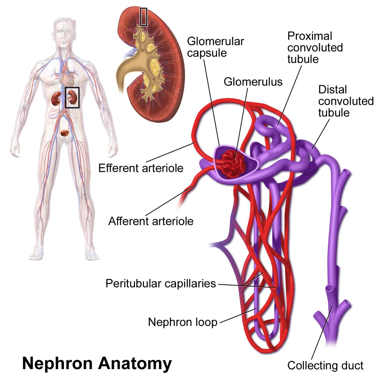
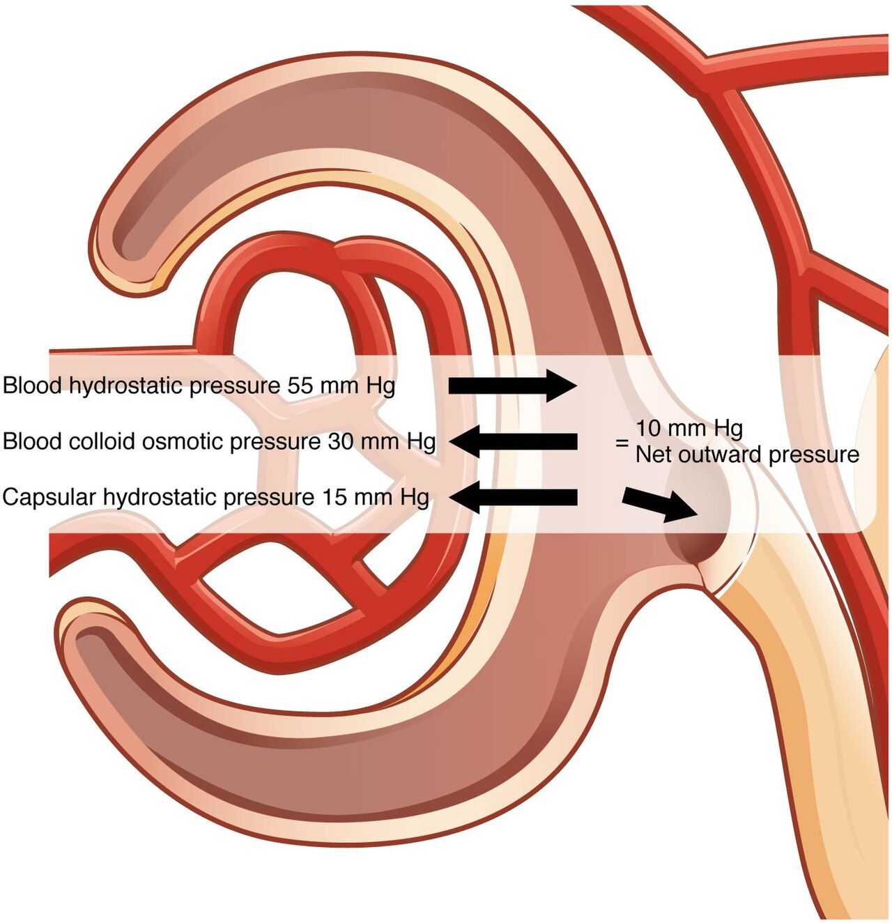
Each kidney holds roughly one million nephrons, and each nephron begins with a tuft of capillaries — the glomerulus — sitting inside a cup called Bowman capsule. Blood arrives under pressure through the afferent arteriole and leaves through the narrower efferent arteriole. That deliberate bottleneck is what drives filtration: about 180 litres of plasma are pushed across the glomerular membrane every day. The membrane is a size and charge filter. Water, electrolytes, glucose, urea and creatinine cross freely; albumin and red cells should not. When you see protein or blood in the urine, the filter itself has been damaged.
What follows is reclamation. The proximal tubule takes back roughly two‑thirds of the filtered sodium and water plus essentially all of the glucose, amino acids and bicarbonate. The loop of Henle builds the medullary concentration gradient that makes concentrated urine possible — and it is the site loop diuretics block. The distal tubule and collecting duct are where fine control happens: aldosterone drives sodium reabsorption in exchange for potassium and hydrogen ion, and antidiuretic hormone opens water channels so water follows. Of the 180 litres filtered, only about 1 to 2 litres leave as urine.
Three hormonal jobs matter enormously in disease. First, renin: when renal perfusion falls, the juxtaglomerular cells release renin, which starts the cascade ending in angiotensin II (vasoconstriction, efferent arteriole constriction to preserve filtration) and aldosterone (sodium and water retention). Second, erythropoietin: peritubular cells sense oxygen delivery and tell the marrow to make red cells. Third, activation of vitamin D: the kidney performs the final hydroxylation that produces calcitriol, without which the gut cannot absorb calcium. Remember those three and the anemia, hypertension and bone disease of chronic kidney disease stop being a list to memorise and become consequences you can predict.
Reading the numbers: creatinine, BUN and eGFR
Serum creatinine (roughly 0.6 to 1.2 mg/dL; about 0.7 to 1.3 in men and 0.6 to 1.1 in women) is the workhorse. Creatinine is a waste product of muscle metabolism produced at a fairly steady rate and cleared almost entirely by filtration, so it rises when filtration falls. Two cautions. It is insensitive early — roughly half of function can be lost before creatinine leaves the normal range — and it depends on muscle mass. A frail 80‑year‑old woman with a creatinine of 1.1 mg/dL may have lost more than half her filtration. Never call a creatinine “normal” in an older or cachectic patient without looking at the estimated GFR.
Blood urea nitrogen (10 to 20 mg/dL) reflects filtration too, but it is contaminated by everything else that affects urea: dehydration, high protein intake, gastrointestinal bleeding, steroids and catabolic states all raise it, while liver failure and malnutrition lower it. That contamination is actually useful. A BUN‑to‑creatinine ratio above 20 to 1 points toward a prerenal cause, because a dry kidney reabsorbs urea avidly along with sodium and water while creatinine keeps moving. A ratio near 10 or 15 to 1 with both values rising suggests intrinsic damage.
Estimated GFR is calculated from creatinine, age and sex, and it is the number that actually drives clinical decisions: staging, drug dosing, contrast decisions and referral for dialysis planning. Normal is above 90 mL/min/1.73 m². Before you give any renally cleared drug — enoxaparin, gabapentin, metformin, vancomycin, many antibiotics — look at the calculated clearance, not the creatinine. One caveat matters at the bedside: eGFR is normalised to body surface area, so for weight‑sensitive drugs such as enoxaparin pharmacy dose‑adjusts from the Cockcroft‑Gault creatinine clearance, which falls far faster than eGFR in a small, low‑weight older adult.
Urine as data
Urine output is the cheapest renal monitor in the hospital, and it is often delegated to unlicensed personnel and then never looked at. The adult threshold to know is 0.5 mL/kg/hr. For a 70 kg patient that is 35 mL per hour, or roughly 210 mL over a six‑hour shift segment. Oliguria is less than 400 mL in 24 hours (or output under 0.5 mL/kg/hr for six hours); anuria is less than 100 mL in 24 hours and should make you think obstruction or catastrophic vascular event until proven otherwise. Pair intake and output with a daily weight taken at the same time, on the same scale, in the same clothing: one kilogram equals approximately one litre of fluid, and weight is far more reliable than an intake and output record that nobody charted at lunch.
The urinalysis tells you where the injury lives. Specific gravity normally ranges 1.005 to 1.030; a high value with concentrated dark urine suggests a dry patient whose tubules still work, while a fixed value near 1.010 (the specific gravity of plasma) suggests tubules that have lost the ability to concentrate at all. Protein should be absent to trace; persistent proteinuria means glomerular disease. The urine albumin‑to‑creatinine ratio categorises albuminuria as A1 (under 30 mg/g), A2 (30 to 300 mg/g) or A3 (over 300 mg/g), and A2 or higher predicts progression.
Casts are cylindrical moulds of the tubule and they are close to diagnostic. Muddy brown granular casts mean acute tubular necrosis. Red blood cell casts mean glomerulonephritis — blood is coming through the filter, not from a stone or a catheter. White blood cell casts mean infection or inflammation inside the kidney, that is pyelonephritis or interstitial nephritis, rather than a bladder infection. Hyaline casts are usually benign and appear with dehydration or exercise.
| Test | Reference range | What a change tells you |
|---|---|---|
| Creatinine | 0.6–1.2 mg/dL | Rise of 0.3 mg/dL in 48 h meets AKI criteria; falsely reassuring in low muscle mass |
| BUN | 10–20 mg/dL | Disproportionate rise suggests prerenal state, GI bleed or catabolism |
| eGFR | Above 90 mL/min/1.73 m² | Stages CKD and drives all renal drug dosing |
| Potassium | 3.5–5.0 mEq/L | Above 5.5 is urgent in oliguria; above 6.0 with ECG change is an emergency |
| Bicarbonate | 22–26 mEq/L | Falls in renal failure — the kidney cannot excrete the daily acid load |
| Phosphorus | 2.5–4.5 mg/dL | Rises as GFR falls; drives secondary hyperparathyroidism |
| Calcium (total) | 8.5–10.5 mg/dL | Falls in CKD from loss of calcitriol and phosphate binding |
| Hemoglobin | 12–16 g/dL (F), 14–18 g/dL (M) | Falls with erythropoietin deficiency; CKD target on therapy is 10–11 g/dL |
| Albumin | 3.5–5.0 g/dL | Under 3.0 with heavy proteinuria suggests nephrotic syndrome |
Imaging, biopsy and the aging kidney
Renal ultrasound is first‑line and answers the single most urgent structural question: is there hydronephrosis, meaning obstruction? It uses no contrast and no radiation, needs no preparation, and can be done at the bedside. Non‑contrast helical CT is the gold standard for stones. CT with iodinated contrast and coronary angiography carry a risk of contrast‑associated AKI, which typically shows a creatinine rise within 48 to 72 hours; prevention is isotonic intravenous fluid before and after, holding other nephrotoxins, and using the smallest contrast volume possible. Metformin is held at the time of contrast when eGFR is under 30, and restarted after 48 hours once renal function is confirmed stable.
Percutaneous kidney biopsy is how glomerular disease is diagnosed. It is a bleeding procedure into a highly vascular organ. Before: check coagulation studies, hemoglobin, type and screen, and confirm consent and held anticoagulants. After: bed rest lying supine with pressure over the site for four to six hours, vital signs on a tapering schedule, serial hemoglobin, and inspection of every urine specimen. Gross hematuria that persists beyond the first few voids, flank pain, hypotension or a falling hemoglobin means bleeding — notify immediately. Teach the patient to avoid heavy lifting and strenuous activity for about two weeks.
Age changes everything you have just read. Nephron number falls steadily from about age 40, renal blood flow declines, the concentrating and diluting ability narrows, and the thirst response blunts. The practical consequences on your floor: an older adult becomes prerenal after one day of poor intake and a dose of furosemide, tolerates a nephrotoxin far worse than a 30‑year‑old, and carries a “normal” creatinine that hides real impairment. Add polypharmacy and the older adult is the patient in whom AKI is both most likely and most likely to be missed.
Creatinine is a late and muscle‑dependent signal, so judge kidney function by the trend, the eGFR and the urine output together — never by a single value in isolation.
Section 2Acute Kidney Injury
Three doors into the same room
Acute kidney injury is an abrupt decline in filtration, defined by a creatinine rise of at least 0.3 mg/dL within 48 hours, a creatinine at least 1.5 times baseline within seven days, or urine output under 0.5 mL/kg/hr for six hours. It complicates a large fraction of hospital admissions and carries real mortality. The classification is not academic: prerenal injury is fixed with volume, postrenal injury is fixed with drainage, and intrarenal injury is mostly supported while the tubules heal.
Prerenal AKI is a perfusion problem in a structurally normal kidney — roughly two‑thirds of cases. Causes are anything that lowers effective renal blood flow: hemorrhage, vomiting and diarrhea, aggressive diuresis, burns, sepsis with vasodilation, heart failure with poor forward flow, and cirrhosis. Drugs contribute directly. NSAIDs constrict the afferent arteriole and shut the tap; ACE inhibitors and ARBs dilate the efferent arteriole and drop filtration pressure. Either is tolerated in a well‑perfused patient and dangerous in a dry one. The classic dangerous combination on a med‑surg floor is an ACE inhibitor plus a diuretic plus an NSAID in a patient who has been nothing by mouth — sometimes called the triple whammy.
Intrarenal (intrinsic) AKI is damage to the tissue itself. The most common form is acute tubular necrosis, produced either by prolonged ischemia — prerenal injury that was not corrected — or by nephrotoxins: aminoglycosides, vancomycin, amphotericin B, cisplatin, iodinated contrast, and pigment loads from rhabdomyolysis (myoglobin, tea‑coloured urine, creatine kinase often in the thousands to tens of thousands) or hemolysis. Acute interstitial nephritis is usually an allergic reaction to a drug — proton pump inhibitors, beta‑lactams, sulfonamides — and may come with rash, fever and eosinophilia. Acute glomerulonephritis is inflammation of the filter: post‑streptococcal disease appears one to three weeks after pharyngitis or three to six weeks after skin infection, with tea‑coloured urine, red cell casts, hypertension, periorbital edema, an elevated ASO titre and a low complement C3. Nephrotic syndrome is the other glomerular pattern — proteinuria over 3.5 g per 24 hours, albumin under 3.0 g/dL, massive edema, hyperlipidemia and a hypercoagulable state from urinary loss of antithrombin.
Postrenal AKI is obstruction: benign prostatic hyperplasia, bilateral stones, tumour, retroperitoneal fibrosis, a clotted or kinked catheter. It is the least common cause and the most rewarding to find, because relieving the obstruction often restores function completely if it is caught early. Any patient who abruptly stops making urine deserves a bladder scan and a look at the catheter before anyone reaches for fluids.
| Finding | Prerenal | Intrarenal (ATN) | Postrenal |
|---|---|---|---|
| BUN‑to‑creatinine ratio | Above 20:1 | Near 10–15:1 | Variable |
| Urine sodium | Under 20 mEq/L | Above 40 mEq/L | Variable |
| Fractional excretion of sodium (FeNa) — valid only off diuretics | Under 1% | Above 2% | Variable |
| Fractional excretion of urea — use instead when a diuretic has been given | Under about 35% | Above about 50% | Variable |
| Urine osmolality | Above 500 mOsm/kg | Under 350 mOsm/kg | Variable |
| Urine sediment | Bland, hyaline casts | Muddy brown granular casts | Bland, may show crystals |
| Bedside clue | Dry mucosa, tachycardia, orthostasis, low CVP | Recent contrast, nephrotoxin, hypotension, sepsis | Distended bladder, no catheter output, hydronephrosis |
| First move | Restore volume and perfusion | Stop the toxin, support, treat cause | Drain: irrigate or replace catheter, relieve obstruction |
One caveat governs that whole table. FeNa and urine sodium are interpretable only in a patient who has not had a diuretic. A loop or thiazide diuretic forces natriuresis, so a volume‑depleted prerenal patient who received furosemide within roughly the previous 24 hours characteristically shows a urine sodium above 40 mEq/L and a FeNa above 2% — the intrarenal pattern — and filing that patient under acute tubular necrosis means withholding the volume they actually need. Since “aggressive diuresis” and “a diuretic in a patient who was already dry” are among the commonest prerenal causes on a med‑surg floor, this is not a rare exception. When a diuretic has been given, use the fractional excretion of urea instead: under about 35% suggests a prerenal state, because urea reabsorption is not blocked by these drugs. Note too that FeNa can read under 1% despite genuine intrinsic injury in contrast nephropathy, pigment nephropathy from rhabdomyolysis, hepatorenal syndrome and acute glomerulonephritis. No single index outranks the bedside picture, the medication record and the response to a fluid challenge.
The four phases and what each one costs you
Onset (initiation) runs from the insult to the first fall in urine output — hours to days — and it is the only window in which the injury can often be reversed. This is when your assessment matters most: the falling output, the rising creatinine, the hypotensive episode nobody escalated.
Oliguric phase typically lasts one to three weeks, and urine output falls below 400 mL per day. This is the dangerous phase. Everything the kidney was supposed to excrete accumulates: potassium climbs, phosphorus climbs, hydrogen ion climbs (metabolic acidosis with a bicarbonate under 22 mEq/L), water accumulates producing hypertension, edema, jugular venous distension, crackles and dilutional hyponatremia, and urea climbs toward uremia. The single most lethal problem here is hyperkalemia. Nursing care is fluid restriction, strict intake and output, daily weights, dietary potassium and phosphorus restriction, and vigilant medication review.
Diuretic phase begins when tubules start recovering filtration before they recover concentrating ability. Output can reach 3 to 5 litres a day and occasionally 10. Now the risk inverts: hypovolemia, hypotension, hypokalemia and hyponatremia. Patients who survived the oliguric phase are lost here when nobody notices that a patient putting out 400 mL an hour is being under‑replaced. Monitor electrolytes at least daily and replace fluid and potassium as ordered — and note that replacement potassium is never given as an intravenous push or bolus under any circumstance: it is always diluted, infused on a pump at a controlled rate, and given with the patient monitored.
Recovery may take up to twelve months, and function may not return fully. A patient who has had AKI carries a permanently higher risk of chronic kidney disease, which is a teaching point at discharge: avoid NSAIDs, stay hydrated, and get renal function rechecked.
Management and the hyperkalemia emergency
Medical management is cause‑directed. Prerenal injury gets isotonic crystalloid and, in shock, vasopressors to restore a mean arterial pressure that perfuses the kidney. Postrenal injury gets drainage. Intrinsic injury gets removal of the offending agent, careful fluid balance, and dose adjustment of every renally cleared drug. Loop diuretics do not treat AKI and do not improve survival — they manage volume in a patient who still makes urine. Renal replacement therapy is started for the indications summarised by AEIOU: refractory Acidosis, Electrolyte disturbance (chiefly hyperkalemia), Intoxications, fluid Overload unresponsive to diuretics, and Uremic complications such as pericarditis, encephalopathy or bleeding.
Hyperkalemia is the emergency you will actually face. Potassium above 5.5 mEq/L in an oliguric patient is urgent; above 6.0 with ECG changes is a code waiting to happen. ECG evolution is orderly: tall peaked T waves first, then a flattened or absent P wave and a prolonged PR interval, then a widening QRS that blends into a sine wave and then asystole or ventricular fibrillation. The nursing sequence is: recognise, place the patient on continuous cardiac monitoring, obtain a 12‑lead ECG, notify the provider, hold all potassium sources (intravenous fluids with potassium, potassium‑sparing diuretics, ACE inhibitors and ARBs, salt substitutes, dietary potassium), and prepare the drugs below in the correct order.
| Order | Agent | What it does | Nursing implication |
|---|---|---|---|
| 1 | Calcium gluconate 1 g IV over 2–3 min | Stabilises the myocardial membrane; does not lower potassium | Give first when ECG changes are present; onset 1–3 min, lasts only 30–60 min; monitor continuously; incompatible with bicarbonate in the same line |
| 2 | Regular insulin 10 units IV with 25 g dextrose | Drives potassium into cells | Onset 15–30 min, lasts 4–6 h; check glucose before, at 30 min, and hourly for several hours — hypoglycemia is common and delayed |
| 3 | Albuterol 10–20 mg nebulised | Beta‑2 shift into cells | Four to eight times the bronchodilator dose; expect tachycardia and tremor |
| 4 | Sodium bicarbonate | Shifts potassium only if the patient is acidotic | Adjunct only; watch for volume and sodium load, and hypocalcemia |
| 5 | Loop diuretic, patiromer or sodium zirconium cyclosilicate | Actually removes potassium from the body | Diuretic works only if urine is being made; binders act over hours and are not emergency therapy; sodium polystyrene sulfonate has largely fallen out of favour because of bowel necrosis |
| 6 | Hemodialysis | Definitive removal | For refractory or severe hyperkalemia; ensure access, consent and nephrology involvement |
Remember the concept that separates a passing student from a safe nurse: step 1 protects the myocardium without lowering the potassium by a single point, and steps 2 through 4 only buy time by hiding potassium inside cells. The potassium is still in the body and it will come back out. Nothing is finished until step 5 or 6 has actually removed it, and the potassium is rechecked.
In acute kidney injury, first decide whether the problem is perfusion, parenchyma or plumbing — then treat hyperkalemia before anything else, and remember that stabilising and shifting potassium is not the same as removing it.
Section 3Chronic Kidney Disease and End‑Stage Renal Disease
Staging and the two diseases that cause most of it
Chronic kidney disease is abnormality of kidney structure or function present for more than three months. Diabetes causes roughly 40% of cases in the United States and hypertension another 25% or so; glomerulonephritis, polycystic kidney disease and repeated AKI account for much of the rest. The mechanism of progression is common to all of them: as nephrons are lost, the survivors hyperfilter to compensate, that hyperfiltration scars them, and the disease becomes self‑sustaining. This is exactly why treatment aims at lowering intraglomerular pressure rather than at any specific original insult.
| Stage | eGFR (mL/min/1.73 m²) | What you will see and do |
|---|---|---|
| G1 | 90 or above, with kidney damage such as albuminuria | Asymptomatic; screen, control blood pressure and glucose |
| G2 | 60–89 with damage | Still asymptomatic; slow progression, avoid nephrotoxins |
| G3a / G3b | 45–59 / 30–44 | Complications appear: anemia, rising phosphorus and PTH, acidosis, hypertension; adjust drug doses; nephrology referral |
| G4 | 15–29 | Symptomatic; prepare for renal replacement — access placement, modality education, transplant evaluation |
| G5 | Under 15 | End‑stage; dialysis or transplant, or conservative comfort‑focused management |
Albuminuria is staged alongside GFR because it independently predicts progression and cardiovascular death: A1 is under 30 mg/g, A2 is 30 to 300 mg/g, A3 is above 300 mg/g. A patient at G2 with A3 is at higher risk than a patient at G3a with A1. When a student asks why we test urine on a patient whose creatinine is normal, this is the answer.
What uremia does to every system
Cardiovascular disease is what actually kills most patients with CKD, long before they reach dialysis. Volume overload plus renin activation plus vascular calcification produces hypertension, left ventricular hypertrophy and accelerated atherosclerosis. Late uremia adds pericarditis: a friction rub, pleuritic chest pain relieved by leaning forward, and a risk of tamponade — muffled heart tones, hypotension, jugular venous distension. That is an urgent dialysis indication, not a wait‑until‑morning finding.
Hematologic. Anemia comes from erythropoietin deficiency, shortened red cell survival, iron deficiency and blood loss in the dialyser. It is normocytic and normochromic. Uremia also causes platelet dysfunction, so these patients bruise, ooze from puncture sites and bleed from the gut with a normal platelet count. Treatment is iron repletion first — check transferrin saturation and ferritin — then an erythropoiesis‑stimulating agent titrated to a hemoglobin of about 10 to 11 g/dL. Do not chase a normal hemoglobin: higher targets increase stroke, thrombosis and death.
CKD mineral and bone disorder follows a predictable chain. The failing kidney cannot excrete phosphorus, so phosphorus rises; phosphorus binds calcium and the kidney can no longer activate vitamin D, so calcium falls; low calcium and high phosphorus drive the parathyroids to secrete PTH continuously; PTH pulls calcium out of bone. The result is renal osteodystrophy — weak bones, fractures, bone pain — plus soft‑tissue and vascular calcification and intractable itching. Treatment is dietary phosphorus restriction, phosphate binders given with meals (calcium acetate, sevelamer, lanthanum — a binder given between meals binds nothing), active vitamin D, and calcimimetics such as cinacalcet, which lower PTH but can cause hypocalcemia.
Metabolic and neurologic. Bicarbonate falls below 22 mEq/L because the kidney cannot excrete acid or regenerate bicarbonate; chronic acidosis worsens bone disease and muscle wasting and is treated with oral sodium bicarbonate. Potassium retention worsens as GFR falls under about 20. Uremic encephalopathy brings fatigue, poor concentration, sleep reversal, asterixis, and in older adults it commonly presents as delirium. Peripheral neuropathy causes restless legs and burning feet. Gastrointestinal effects include anorexia, metallic taste, nausea, uremic fetor and gastritis with bleeding risk. Integumentary findings are a sallow grey‑yellow tone, severe pruritus and uremic frost in extreme cases. Sexual dysfunction, infertility and immune suppression round out a genuinely miserable syndrome.
Slowing it down: drugs, diet and teaching
Four drug classes carry the evidence. ACE inhibitors and ARBs dilate the efferent arteriole, lowering intraglomerular pressure and albuminuria. Expect and tolerate a creatinine rise of up to about 30% after starting; a larger rise, or a potassium above 5.5 mEq/L, means hold and call. Never combine an ACE inhibitor with an ARB. SGLT2 inhibitors (dapagliflozin, empagliflozin) now slow progression in both diabetic and non‑diabetic CKD with albuminuria; a small acute dip in eGFR after starting is expected and is not injury; teach genital hygiene because of mycotic infections, watch for volume depletion, and know that euglycemic diabetic ketoacidosis is a rare but real adverse effect. Finerenone, a non‑steroidal mineralocorticoid receptor antagonist, adds benefit in diabetic CKD — monitor potassium closely. Statins address the cardiovascular risk that is the true cause of death. Blood pressure control — usually a target near 130/80 mmHg or lower, individualised — and glycemic control remain foundational.
Nutrition is a nursing responsibility as much as a dietitian’s. Before dialysis, protein is moderately restricted (roughly 0.6 to 0.8 g/kg/day) to reduce the nitrogenous load; once on hemodialysis, protein needs increase to about 1.0 to 1.2 g/kg/day because amino acids are lost into the dialysate. This reversal is a favourite examination item. Sodium is limited to under 2 g/day, potassium to roughly 2 to 3 g/day, phosphorus to 800 to 1000 mg/day — which means teaching patients to avoid dark colas, processed cheese, and anything with “phos” on the ingredient list, since additive phosphorus is almost completely absorbed. Fluid is commonly restricted to about 1000 mL plus the previous day’s urine output. Teach patients to spread fluid across the day, use ice chips (counted as half their volume), sugar‑free hard candy and frequent mouth care for thirst.
Discharge teaching priorities: take blood pressure and record it, weigh daily and report a gain of more than 2 kg (about 4 to 5 pounds) in a day or 2.5 kg between dialysis treatments, take phosphate binders with the first bite of every meal and snack, never take NSAIDs or magnesium‑ or aluminium‑containing antacids without asking, check with the pharmacist before any new drug or herbal product, get the influenza, COVID‑19, pneumococcal and hepatitis B vaccines, and protect the non‑dominant arm for future access even before dialysis is needed.
Chronic kidney disease is staged by eGFR and albuminuria, and almost every complication traces back to four lost functions — excreting water and potassium, excreting acid and phosphorus, activating vitamin D, and making erythropoietin.
Section 4Dialysis, Access and Transplantation
Hemodialysis and protecting the access
In hemodialysis, blood is pumped from the patient through a dialyser containing thousands of semipermeable fibres while dialysate flows in the opposite direction on the other side of the membrane. Solutes move by diffusion down their concentration gradients — urea, creatinine and potassium out of the blood; bicarbonate and calcium in as prescribed. Water is removed separately by ultrafiltration, a pressure gradient set by the amount of fluid the patient needs to lose to reach the prescribed “dry weight.” A typical outpatient schedule is three to four hours, three times a week. Anticoagulation, usually heparin, keeps the circuit from clotting and is a bleeding consideration afterward.
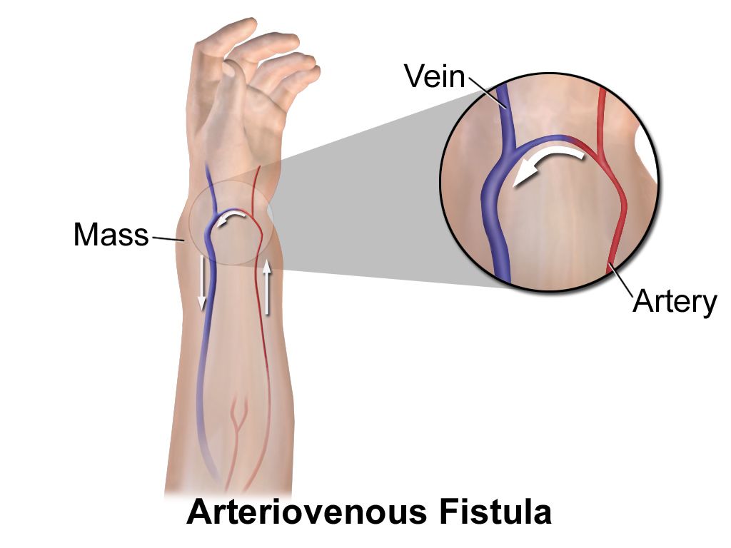
Access determines survival. An arteriovenous fistula is a surgical anastomosis of the patient’s own artery and vein, usually in the non‑dominant forearm. It has the lowest infection rate and longest patency but needs to mature — typically at least six weeks and often two to three months — before it can be cannulated. An AV graft uses synthetic tubing, is usable in about two to three weeks, and clots and infects more often. A tunnelled central venous catheter works immediately but carries the highest infection and central stenosis risk and is the least desirable long‑term choice.
Assessing and protecting a fistula is a nursing skill you must be able to perform without prompting. Palpate for a thrill — a buzzing vibration — and auscultate for a bruit — a swishing sound — at least every shift and any time the patient reports a change. Absence of both means the access has clotted, and that is an urgent call: a fistula can sometimes be salvaged within hours. On the access extremity: no blood pressure cuffs, no venipuncture, no intravenous lines, no restraints, no tight clothing or jewellery, and no sleeping on that arm. Post a sign at the bedside. Also assess distal circulation for steal syndrome — a cool, pale, painful, numb hand distal to the access means the fistula is diverting blood from the extremity.
Around a treatment: weigh before and after (the difference is fluid removed), check whether antihypertensives should be held to prevent intradialytic hypotension, and know which of the patient’s medications are dialysable and therefore scheduled after the run — many antibiotics, water‑soluble vitamins and some antiepileptics. Afterward, assess for hypotension, dizziness, cramps, bleeding from needle sites and the post‑dialysis weight. Dialysis disequilibrium syndrome occurs mainly during the first treatments in a very uremic patient: urea is cleared from blood faster than from brain, water follows the osmotic gradient into brain tissue, and the patient develops headache, nausea, restlessness, confusion and, at worst, seizures. Slow or stop the treatment and notify immediately.
Peritoneal dialysis
Peritoneal dialysis uses the patient’s own peritoneum as the membrane. Warmed dextrose‑based dialysate is instilled through a permanent abdominal catheter (fill), left in place for a prescribed dwell during which solutes diffuse into it and water follows the osmotic pull of the dextrose, and then drained out by gravity (drain). It can be done manually four or five times a day or by a cycler overnight. Its advantages are autonomy, no needles, gentler fluid shifts and a more liberal diet; its disadvantages are the daily discipline it demands, protein loss, hyperglycemia and weight gain from absorbed dextrose, and hernia formation.
The nursing essentials: use strict aseptic technique at every connection, warm the dialysate with a dry warmer or heating pad and never in a microwave, record fill and drain volumes precisely, and weigh the patient after draining. Effluent should be clear and pale yellow. Cloudy effluent is peritonitis until proven otherwise — often with abdominal pain, rebound tenderness and fever — and requires a specimen for cell count, Gram stain and culture, followed by intraperitoneal antibiotics. If outflow is less than inflow, check for a kinked or clamped line, constipation (the most common and most overlooked cause), fibrin in the catheter, and reposition the patient before assuming catheter failure. Peritoneal dialysis is contraindicated with extensive abdominal adhesions, recent abdominal surgery or an unrepaired hernia.
Continuous therapies and transplantation
Continuous renal replacement therapy removes solute and fluid slowly and continuously over 24 hours in the intensive care unit. Because volume is removed gradually, it is far better tolerated by the hemodynamically unstable septic or post‑operative patient than intermittent hemodialysis. It usually requires anticoagulation of the circuit — regional citrate is now preferred over systemic heparin and demands monitoring of ionised calcium — along with one‑to‑one nursing and hourly balance calculation; electrolytes and phosphorus fall and need replacement.
Kidney transplantation offers the best survival and quality of life. The graft is placed in the iliac fossa — palpable and tender if rejecting — and the native kidneys are usually left in place. In the immediate post‑operative period, monitor urine output hourly; a well‑functioning living‑donor graft may produce enormous volumes, and replacement is usually ordered as a percentage of output. Falling urine output, a rising creatinine, fever, weight gain, hypertension and tenderness over the graft suggest rejection, and every one of those is a call to the transplant team.
Immunosuppression is lifelong and non‑negotiable. Calcineurin inhibitors (tacrolimus, cyclosporine) require trough monitoring and are themselves nephrotoxic; antimetabolites (mycophenolate) cause gastrointestinal upset and marrow suppression and are teratogenic; corticosteroids bring hyperglycemia, bone loss and mood change. The composite risk is infection and malignancy. Teach hand hygiene, avoidance of sick contacts, no live vaccines, sun protection and annual skin examination, and above all that a missed dose is never trivial — the graft can be lost in days. A low‑grade fever in a transplant patient is a serious finding, not a nuisance.
Dialysis buys time, not cure — and the nurse’s highest‑value contributions are protecting the access, catching peritonitis or a clotted fistula early, and knowing which drugs to hold or to time around the treatment.
Section 5Infection, Obstruction and the Lower Urinary Tract
Urinary tract infection, pyelonephritis and CAUTI
Bacteria — most often Escherichia coli from the perineum — ascend the short female urethra into the bladder. Cystitis produces dysuria, urgency, frequency, suprapubic pain and cloudy, malodorous urine, sometimes with hematuria; fever is usually absent. Urinalysis shows positive leukocyte esterase and nitrites, more than 10 white cells per high‑power field and bacteria; a culture with 100,000 colony‑forming units per millilitre confirms it. Always obtain the culture before the first antibiotic dose. Uncomplicated cystitis is treated with nitrofurantoin (avoid when creatinine clearance is under about 30 mL/min, because it will not concentrate in the urine), trimethoprim‑sulfamethoxazole or fosfomycin. Phenazopyridine relieves burning, turns urine and tears bright orange, will permanently stain contact lenses, is limited to about two days, and is not an antibiotic — patients who stop the antibiotic because the burning stopped are a recurring problem.
Pyelonephritis is infection of the kidney itself: fever above 38.5°C, chills, flank pain, costovertebral angle tenderness, nausea and vomiting on top of lower tract symptoms, with white cell casts in the urine. It requires systemic antibiotics — ceftriaxone or a fluoroquinolone, intravenously if the patient is vomiting or unstable — along with hydration and analgesia. The danger is urosepsis: hypotension, tachycardia, tachypnea, altered mental status, rising lactate. Infection behind an obstruction is a urologic emergency; antibiotics alone will not fix it and the tract must be drained, often with a ureteral stent or a percutaneous nephrostomy.
Catheter‑associated urinary tract infection is the most common preventable hospital infection and it is largely nursing‑controlled. Prevent it by inserting a catheter only when genuinely indicated, using sterile technique, maintaining a closed drainage system, keeping the bag below the level of the bladder and off the floor, securing the tubing to prevent traction and dependent loops, performing daily perineal hygiene with soap and water, and — the intervention with the largest effect — reviewing daily whether the catheter can come out and removing it as soon as possible. Never irrigate routinely, never disconnect the system to obtain a specimen (use the sampling port after disinfecting it), and never raise the bag above the bladder during transfers.
Older adults deserve a paragraph of their own. Classic dysuria may be absent; instead you see new confusion, a fall, incontinence in a previously continent patient, anorexia or simply “not herself.” Fever may be blunted, so a temperature of 37.8°C in an 85‑year‑old can be significant. At the same time, asymptomatic bacteriuria is common in the elderly and should not be treated — a positive culture in a patient with no symptoms does not explain a delirium, and treating it drives resistance and Clostridioides difficile. The safe nursing stance is to work up the confused older adult fully rather than blaming the urine reflexively. Teach prevention: adequate fluids, front‑to‑back wiping, voiding after intercourse, cotton underwear, avoiding douches and prolonged retention.
Urolithiasis
Stones form when solutes supersaturate the urine, most often because of low fluid intake. About 75 to 80% are calcium oxalate or calcium phosphate. Uric acid stones form in acidic urine and are radiolucent. Struvite stones form only in the presence of urease‑producing organisms such as Proteus, grow into large staghorn calculi and are always infection‑associated. Cystine stones are rare and genetic. The pain — renal colic — is sudden, severe, wave‑like flank pain radiating to the groin or testicle or labia, with a patient who cannot lie still, plus nausea, vomiting, hematuria and often urinary frequency once the stone reaches the ureterovesical junction. Non‑contrast CT confirms it.
Management for a stone under about 5 mm is expulsive: aggressive fluids, analgesia with an NSAID such as ketorolac (used cautiously and briefly, and avoided if renal function is impaired) with opioids as needed, an antiemetic, and tamsulosin to relax ureteral smooth muscle. Strain every urine and send any recovered fragment for analysis — that analysis determines the whole prevention plan. Larger or obstructing stones need extracorporeal shock wave lithotripsy, ureteroscopy with laser fragmentation, or percutaneous nephrolithotomy. After lithotripsy, teach that bruising over the flank and hematuria for several days are expected, that fragments may pass for weeks, and that fluids and straining continue.
| Stone type | Urine pH | Prevention teaching |
|---|---|---|
| Calcium oxalate | Any | 2.5–3 L fluid daily; limit sodium and animal protein; limit high‑oxalate foods (spinach, rhubarb, nuts, beets, chocolate, tea); do not restrict dietary calcium — low calcium intake increases oxalate absorption and makes stones worse |
| Uric acid | Acidic | Limit purines (organ meats, sardines, red meat, beer); alkalinise urine with potassium citrate; allopurinol if indicated |
| Struvite | Alkaline | Eradicate the infection and remove the stone completely — it will regrow around residual fragments |
| Cystine | Acidic | High fluid intake around the clock including overnight; urinary alkalinisation |
Retention, obstruction, incontinence and hematuria
Benign prostatic hyperplasia is near‑universal with age and produces obstructive symptoms (hesitancy, weak stream, straining, incomplete emptying, dribbling) and irritative ones (frequency, urgency, nocturia). Alpha‑1 blockers such as tamsulosin relax the bladder neck and work within days — teach to take at bedtime and rise slowly because of orthostatic hypotension, and to tell any eye surgeon about the drug because of intraoperative floppy iris syndrome. 5‑alpha reductase inhibitors such as finasteride shrink the gland but take up to six months to work, lower PSA by roughly half (so the laboratory must know), and must not be handled by anyone who is or may become pregnant because of teratogenicity. After transurethral resection of the prostate, expect continuous bladder irrigation; keep the outflow pink to light red, increase the irrigation rate if it darkens or clots appear, and remember to subtract irrigant from output when charting.
Acute urinary retention presents as suprapubic pain and distension with an inability to void, and in the older adult sometimes as agitation alone. It is also produced by drugs: anticholinergics, antihistamines, opioids, tricyclics and some decongestants. Confirm with a bladder scanner before catheterising — that is the assessment step students most often skip. A post‑void residual above roughly 150 to 200 mL is abnormal. Older teaching directed clamping the catheter after 500 to 1000 mL; current evidence supports complete drainage in most adults, so follow your facility policy, and in either case monitor for post‑obstructive diuresis, hypotension and electrolyte loss afterward.
Incontinence is not a normal part of aging and is worth naming precisely, because the treatment differs. Stress incontinence leaks with cough, laugh or lift and responds to pelvic floor (Kegel) exercises and weight loss. Urge incontinence is a sudden overwhelming need from detrusor overactivity, treated with bladder training and timed voiding, then a beta‑3 agonist (mirabegron — monitor blood pressure) or an antimuscarinic such as oxybutynin, which is best avoided in older adults because of dry mouth, constipation, retention and confusion. Overflow incontinence is constant dribbling from a chronically full bladder, usually obstruction or a neurogenic bladder, and is managed by relieving the obstruction or by intermittent catheterisation. Functional incontinence means the urinary tract works but the patient cannot reach the toilet — the intervention is a bedside commode, a call light within reach, accessible clothing and scheduled toileting, not a catheter.
Finally, one symptom deserves its own alarm. Painless gross hematuria is bladder cancer until proven otherwise, particularly in a smoker over 50. It is often intermittent, so patients dismiss it when it stops. Diagnosis is by cystoscopy with biopsy. Superficial disease is treated by transurethral resection with intravesical therapy such as BCG; teach patients to sit to void for six hours after instillation, to add bleach to the toilet and let it stand, and to wash hands thoroughly. Patients with a urinary diversion or ileal conduit need stoma teaching, expectation that mucus in the urine is normal, and lifelong follow‑up.
Lower tract disease is mostly infection or obstruction — scan the bladder before you catheterise, culture before you treat, remove the catheter as early as possible, and treat painless gross hematuria as cancer until a urologist says otherwise.
The chapter in one breath
The kidney filters 180 litres a day and returns nearly all of it, so renal disease is silent until the compensations fail. Assess it with the trend in creatinine, the eGFR, the urine output against 0.5 mL/kg/hr, a daily weight and a urinalysis whose casts tell you where the injury lives. Acute kidney injury comes through three doors — perfusion, parenchyma, plumbing — and the distinction dictates whether you give volume, remove a toxin or drain an obstruction; through its oliguric phase the killer is hyperkalemia, where calcium stabilises the myocardium, insulin with dextrose and albuterol shift potassium into cells, and only diuresis, binders or dialysis actually remove it. Chronic kidney disease is staged by eGFR and albuminuria, and its anemia, bone disease, acidosis, hypertension and uremia all follow from four lost kidney functions. Dialysis replaces clearance, not the kidney: protect the fistula, palpate the thrill, treat cloudy peritoneal effluent as peritonitis. Downstream, most disease is infection or obstruction — culture first, scan the bladder before catheterising, remove catheters early, strain every urine, and never dismiss painless gross hematuria.
Words worth knowing
| Term | What it means |
|---|---|
| Glomerular filtration rate | Volume of plasma filtered per minute; the single best measure of kidney function, normally above 90 mL/min/1.73 m² |
| Azotemia | Accumulation of nitrogenous wastes (urea, creatinine) in the blood, detected by laboratory testing |
| Uremia | The clinical syndrome produced by azotemia — nausea, pruritus, encephalopathy, pericarditis, bleeding |
| Oliguria / anuria | Urine output under 400 mL per 24 hours / under 100 mL per 24 hours |
| Fractional excretion of sodium (FeNa) | Percentage of filtered sodium excreted; under 1% suggests prerenal, above 2% suggests tubular injury — valid only in a patient who has not had a diuretic |
| Fractional excretion of urea | Percentage of filtered urea excreted; used in place of FeNa after a loop or thiazide diuretic, under about 35% suggests prerenal |
| Acute tubular necrosis | The most common intrinsic AKI, from ischemia or nephrotoxin, marked by muddy brown granular casts |
| Nephrotoxin | An agent that damages the kidney — NSAIDs, aminoglycosides, vancomycin, contrast, amphotericin B, cisplatin |
| Nephrotic syndrome | Proteinuria above 3.5 g per 24 hours with hypoalbuminemia, edema, hyperlipidemia and thrombosis risk |
| Glomerulonephritis | Inflammation of the glomeruli producing hematuria with red cell casts, hypertension and edema |
| Albuminuria | Albumin in the urine, staged A1 to A3 by albumin‑to‑creatinine ratio; predicts CKD progression |
| CKD mineral and bone disorder | High phosphorus, low calcitriol, low calcium and secondary hyperparathyroidism causing renal osteodystrophy |
| Erythropoietin | Kidney hormone stimulating red cell production; its loss causes the normocytic anemia of CKD |
| Ultrafiltration | Pressure‑driven removal of water during dialysis, set by the difference between actual and dry weight |
| Dry weight | The target post‑dialysis weight at which the patient is neither fluid overloaded nor depleted |
| Arteriovenous fistula | Surgically created artery‑to‑vein connection for hemodialysis access; assessed by thrill and bruit |
| Dialysis disequilibrium syndrome | Cerebral edema from rapid urea clearance, seen in early treatments; headache, confusion, seizures |
| Peritonitis | Infection of the peritoneal cavity in peritoneal dialysis, signalled first by cloudy effluent |
| Renal colic | Severe wave‑like flank pain radiating to the groin caused by a stone in the ureter |
| Costovertebral angle tenderness | Pain on percussion over the kidney, a hallmark of pyelonephritis |
| Post‑void residual | Urine remaining after voiding, measured by bladder scan; above roughly 150–200 mL is abnormal |