Maternal-Newborn and Paediatric Nursing
Family-Centred Care, Reproductive Health, Equity and Accountability
Maternity and paediatric nursing is the only branch of practice in which the patient arrives attached to another patient, and in which the person who will deliver most of the care after discharge is standing in the room the whole time. That fact reorganises everything: assessment, teaching, consent, escalation and the way you speak. This opening chapter builds the floor the rest of the book stands on. You will meet the four pillars of family-centred care and the language that carries them; you will re-learn reproductive anatomy and the hormonal cycle well enough to explain contraception, infertility and pregnancy dating to a patient; and you will take on the two foundations that decide whether any of that ever reaches the patient in front of you — cultural humility and the social conditions that shape who survives childbearing and who does not, and the law and ethics of consent, confidentiality and the nurse’s own accountability. Everything the later chapters teach about labour, birth, the newborn and the sick child is built on this floor.
By the end of this chapter you can
- Explain the four core concepts of family-centred care — dignity and respect, information sharing, participation and collaboration — and apply atraumatic care and cultural humility to a maternity or paediatric encounter.
- Rewrite blaming clinical language (“failure to progress”, “poor feeder”) into accurate, non-blaming description, and use inclusive terms such as birthing parent and chestfeeding where they fit the situation.
- Apply the five rights of delegation and state which tasks stay with the RN — assessment, teaching, evaluation and any unstable patient.
- Describe the ovarian and endometrial cycles, the FSH/LH/oestrogen/progesterone sequence and the timing of ovulation, outline spermatogenesis and its 64-to-74-day interval, and use Nägele’s rule to date a pregnancy.
- Tier contraceptive methods by typical-use effectiveness, identify absolute contraindications to combined hormonal contraception, and counsel on emergency contraception within its time windows.
- Recognise primary and secondary dysmenorrhoea, endometriosis and polycystic ovary syndrome, classify abnormal uterine bleeding with PALM-COEIN, outline the infertility work-up beginning with a semen analysis, and describe routine sexually transmitted infection and cervical screening and preconception care including folic acid dosing.
- Distinguish cultural competence from cultural humility, trace how the social determinants of health reach a maternity or paediatric bedside, and explain disparities in maternal mortality, preterm birth and infant mortality as consequences of structural racism and unequal access rather than of race itself.
- Work with a trained interpreter correctly, apply trauma-informed principles, recognise how implicit bias turns a dismissed report of pain into a preventable death, and escalate on the patient’s own concern using SBAR with a named red flag and a specific request.
- State what makes consent informed, describe decision-making capacity as understanding, appreciation, reasoning and the communication of a consistent choice and distinguish it from agreement, separate the nurse’s role as witness from the provider’s duty to obtain it, uphold the right to refuse — including in pregnancy — and apply confidentiality, documentation, scope-of-practice and mandatory reporting duties to a maternity or paediatric encounter.
Section 1The Family as the Unit of Care
The four pillars, and what each one looks like at 3 a.m.
Family-centred care rests on four concepts that are easy to recite and hard to practise. Dignity and respect means you build the plan around what this family values, not around what the last family valued. Information sharing means you give complete, unfiltered, timely information in language the family can actually use — a parent who is told the potassium is 2.9 mmol/L and nothing else has been informed of nothing. Participation means the family chooses the level of involvement they want, which may be more than you expected or less. Collaboration means families are consulted about unit policy, discharge design and their own child’s plan, not merely about which arm you take blood from.
At the bedside these translate into concrete behaviours. Sit down. Use the child’s name and the parents’ names, and ask what the child is called at home. Ask the parent what they are noticing — parents detect deterioration before monitors do, and a parent saying “he is not himself” is a clinical finding you document and act on, not reassurance you dispense. Offer family presence during procedures and, where unit policy permits, during resuscitation; the evidence is that presence does not impair the team and reduces long-term parental distress, provided a designated staff member stays with the family to narrate what is happening.
Family structure is whatever the family says it is: two parents, one parent, grandparents raising grandchildren, blended households, same-sex parents, foster and kinship carers, a birthing person with a doula and no partner. Document the actual caregivers and who holds legal decision-making authority, because those two are not always the same person. Ask, do not assume, who may receive clinical information.
Atraumatic care and the language we use
Atraumatic care means minimising physical and psychological distress from the care itself. Practically: cluster painful procedures, do them in a treatment room rather than in the child’s bed so the bed stays a safe place, use topical anaesthetic for planned venepuncture, offer sucrose and non-nutritive sucking for neonatal procedural pain, allow a parent to hold the infant skin-to-skin during heel sticks, and let a preschooler make real choices about things that are genuinely optional (which arm, which plaster) while never offering a false choice about something that must happen.
Language is a clinical intervention. The phrase failure to progress assigns blame to a person for the behaviour of a uterus; describe the labour pattern instead — “cervical change from 5 to 6 cm over four hours with adequate contractions”. A newborn is not a “poor feeder”; the observation is “latch is shallow, transfers 5 mL in 20 minutes, tires at the breast”. A parent is not “non-compliant”; the finding is “has missed two appointments, reports no transport and no childcare”, which is a problem a nurse can solve. Use birthing parent and chestfeeding where they fit and mother and breastfeeding where they fit; ask the patient which words they use, record it, and use it consistently.
Trauma-informed practice belongs in the same paragraph. A substantial proportion of birthing patients have a history of sexual trauma, and the intimate examinations of maternity care can re-trigger it, so care is delivered on the assumption that trauma may be present. Section 4 sets out the principles and the practical rules that follow from it.
Consent, assent and who may sign
A minor generally cannot give legal consent; a parent or guardian gives permission and the child gives assent, which is expected from about seven years of age and involves developmentally appropriate explanation and a genuine willingness to proceed. Emancipated minors (married, in military service, living independently, or so declared by a court) consent for themselves. A mature minor is one whom a clinician or a court judges to have sufficient maturity to consent to a particular treatment, and mature-minor provisions and category-specific statutes extend independent consent further. Every United States jurisdiction allows a minor to consent to diagnosis and treatment of sexually transmitted infections, though the ages and conditions differ; consent for contraception, pregnancy care and mental health or substance use care varies far more widely by jurisdiction, so you must know your own. In a true emergency, consent for life-saving treatment is implied.
Delegation and what stays with the RN
| Task | RN responsibility | May be delegated to LPN/LVN | May be delegated to UAP |
|---|---|---|---|
| Initial and ongoing assessment; interpreting findings | Yes (non-delegable) | No (may collect data and report) | No |
| Patient and family teaching; evaluating understanding | Yes (non-delegable) | May reinforce teaching the RN has already given; the RN evaluates understanding | No |
| Care of the unstable patient or the patient whose status is changing | Yes (non-delegable) | No | No |
| Evaluating response to an intervention | Yes (non-delegable) | No | No |
| Fundal assessment and evaluation of lochia (the vaginal discharge that follows birth) in the first 24 hours | Yes (non-delegable) | No (may report what is observed while giving care) | No |
| Interpreting fetal heart rate patterns (the continuous tracing of the baby’s heart rate in labour); titrating oxytocin or magnesium (infusions adjusted against the patient’s minute-to-minute response) — both taken up in the labour and birth chapter | Yes (non-delegable) | No | No |
| Vital signs, intake and output, daily weights on a stable patient | — | Yes | Yes |
| Bathing, positioning, feeding an established stable feeder, ambulation | — | Yes | Yes |
| Oral and most routine medications | — | Yes (route rules vary by state) | No |
| IV push medication; blood product administration | Yes (non-delegable in most jurisdictions) | No, unless the state nurse practice act expressly permits it | No |
Apply the five rights of delegation every time: right task, right circumstance, right person, right direction and communication, right supervision and evaluation. Delegating a vital sign set is safe on a stable postpartum patient at hour 20; the identical task is not safe at hour two on a patient with a boggy fundus — a uterus that feels soft rather than firm, meaning it is not clamping its own vessels shut — because the value needs interpreting in real time by the person accountable for the outcome. Note also that assessment, teaching, evaluation and the unstable patient are the memorable core rather than the whole list: your jurisdiction’s medication and monitoring rules decide which further tasks are non-delegable, and in most jurisdictions titrating oxytocin or magnesium, and giving IV push medication or a blood product, are among them.
In maternity and paediatrics the family is the unit of care and the permanent care team, so the RN’s non-delegable work — assessment, teaching, evaluation and the unstable patient — is also the work that determines what happens after discharge.
Section 2Reproductive Anatomy and the Menstrual Cycle
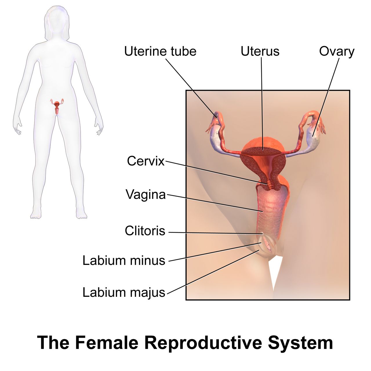
Structures, and why each one matters clinically
The uterus has three layers. The endometrium is the functional lining that thickens, secretes and sheds each cycle and is the bed the blastocyst implants into. The myometrium is three interlacing layers of smooth muscle; the middle layer wraps figure-of-eight fibres around the radial and arcuate vessels that run through the muscle wall, so when it contracts after birth it clamps those vessels shut. The muscle is in that sense a living ligature, and a uterus that stays soft is a uterus that is not compressing its own vessels — what is done about that when bleeding follows a birth belongs to the postpartum and obstetric-emergency chapters. The perimetrium is the outer serosa.
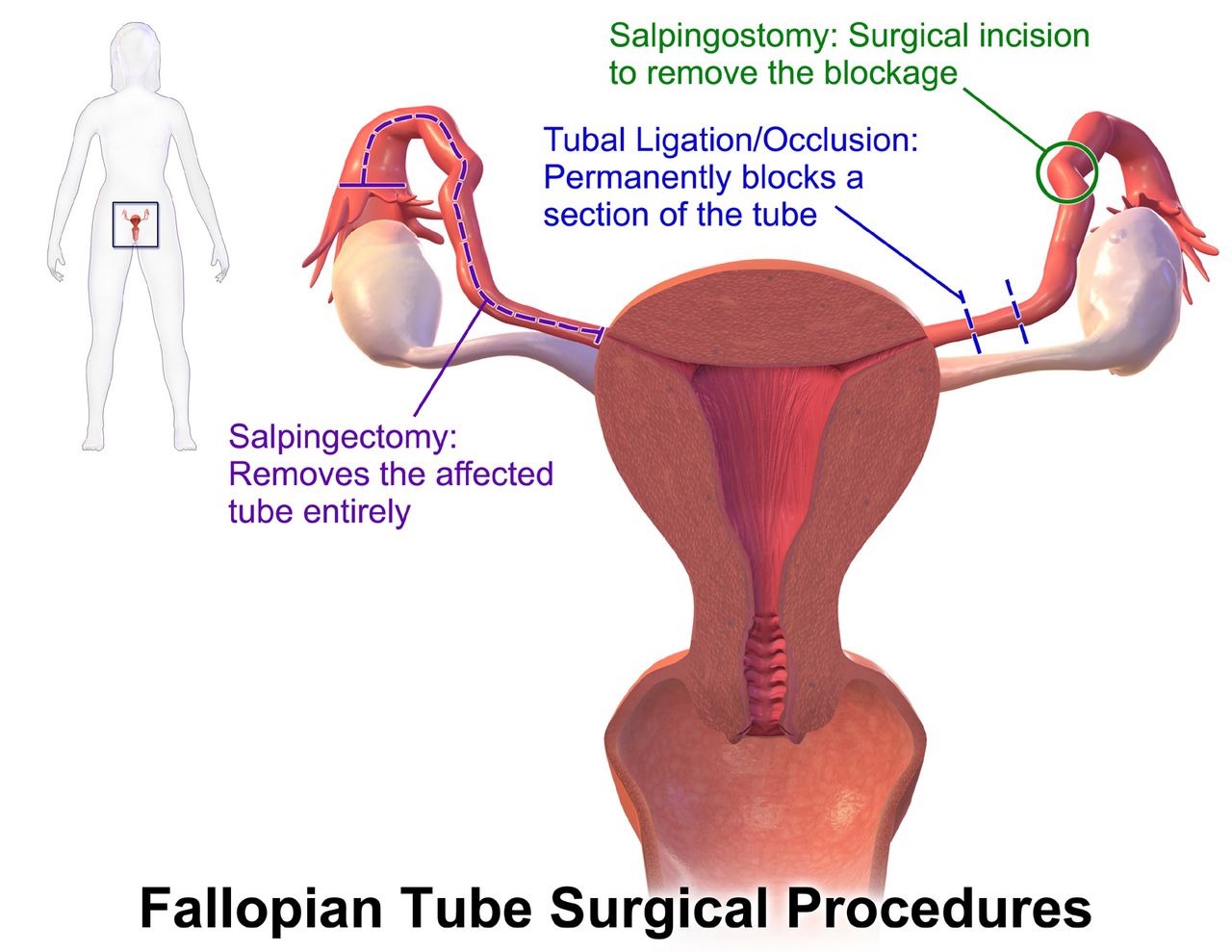
The uterine tube runs from the fimbriae, which sweep the ovulated ovum from the peritoneal surface, through the wide ampulla where fertilisation normally occurs, then the narrow isthmus and the interstitial segment through the uterine wall. Ciliary motion and peristalsis move the zygote to the uterus over roughly three to four days. Anything that scars the tube — pelvic inflammatory disease, endometriosis, previous tubal surgery — slows that transit and raises the risk of an ectopic implantation.
The cervix is a fibrous canal with an internal and external os, lined by mucus that is thick and hostile for most of the cycle and thin, alkaline and stretchy around ovulation. Pelvic architecture and clinical pelvimetry matter at birth rather than here, and are taken up in the labour and birth chapter.
On the male side, spermatogenesis takes roughly 64 to 74 days from spermatogonium to mature sperm, and the testes sit outside the body because that process needs a temperature about 2 to 3 °C below core. LH stimulates Leydig cells to make testosterone; FSH acts on Sertoli cells to support sperm maturation. That 64-to-74-day production cycle is why an abnormal semen analysis — the first test in an infertility work-up, later in this chapter — is repeated after about three months rather than immediately: a sample taken sooner still reflects the same batch of sperm. Sperm remain viable in the female tract for up to five days; the ovum is fertilisable for only 12 to 24 hours. That asymmetry is why the “fertile window” is the five days before ovulation plus the day of ovulation, and why fertility-awareness methods must predict ovulation in advance rather than detect it afterwards.
The ovarian cycle: FSH, LH and the surge
Pulsatile GnRH from the hypothalamus drives anterior pituitary release of FSH and LH. In the follicular phase, FSH recruits a cohort of follicles; one becomes dominant and its granulosa cells secrete rising oestradiol. Early on, oestradiol feeds back negatively and holds LH down. Once oestradiol is high and sustained for roughly two days, the feedback flips to positive and triggers the LH surge. Ovulation follows about 24 to 36 hours after the surge begins, roughly 10 to 12 hours after the LH peak — which is exactly what a urinary LH predictor kit detects, and why the kit tells a couple to have intercourse now rather than tomorrow.
The endometrial cycle runs underneath in lockstep: menstrual shedding, then the oestradiol-driven proliferative phase that regrows the functional layer during the follicular phase, then the progesterone-driven secretory phase after ovulation. The ruptured follicle becomes the corpus luteum and secretes progesterone, which raises basal body temperature by about 0.3 to 0.6 °C and converts the endometrium to that secretory, implantation-ready surface. If fertilisation does not occur, the corpus luteum involutes after a remarkably fixed 14 ± 2 days, progesterone and oestrogen fall, the spiral arterioles constrict, the functional layer becomes ischaemic and sheds. If implantation does occur, trophoblastic hCG rescues the corpus luteum, which sustains the pregnancy until the placenta takes over hormone production at around 8 to 10 weeks.
Because the luteal phase is fixed and the follicular phase is variable, cycle-length differences come from the follicular phase. A person with a 35-day cycle ovulates around day 21, not day 14 as the 28-day assumption would predict. Teach it this way — ovulation is 14 days before the next period, not 14 days after the last one — and both contraception counselling and infertility timing become intuitive.
| Parameter | Typical range | Why the nurse cares |
|---|---|---|
| Age at menarche | 12–13 years (normal 9–15) | No menses by 15 with breast development, or by 13 with none, warrants evaluation |
| Cycle length | 24–38 days (average 28) | Outside this range is abnormal uterine bleeding, not “just irregular” |
| Duration of flow | 8 days or less | More than 8 days suggests anovulation, fibroids or a bleeding disorder |
| Blood loss per cycle | 5–80 mL | Soaking a pad or tampon hourly for 2 or more hours is heavy bleeding — check haemoglobin and ferritin |
| Luteal phase length | 14 ± 2 days (fixed) | Anchors ovulation timing and pregnancy dating |
| Age at menopause | Average 51 (normal 45–55) | Diagnosed retrospectively after 12 consecutive months without menses |
Reading the cycle at the bedside
Three findings let a patient track their own cycle. Cervical mucus shifts from scant and tacky to abundant, clear and stretchy at ovulation — spinnbarkeit, stretching 6 cm or more between the fingers, and ferning in a dried smear under the microscope, both driven by oestrogen. Basal body temperature taken before rising each morning steps up after ovulation under progesterone and stays up; it confirms ovulation has happened but cannot predict it, which is why BBT alone is a poor contraceptive. Mittelschmerz, a brief unilateral mid-cycle pelvic pain, marks follicular rupture in some people.
Anything that disrupts pulsatile GnRH stops the cycle: significant weight loss, low energy availability in athletes and high stress produce functional hypothalamic amenorrhoea. That label is a diagnosis of exclusion — thyroid disease and hyperprolactinaemia are separate endocrine causes of amenorrhoea and must be ruled out first with a TSH and a prolactin level, with pituitary imaging if the prolactin is raised. The nursing point is that amenorrhoea in an adolescent athlete is not a training success — it belongs to the female athlete triad with low energy availability and low bone density, and it needs nutrition and endocrine evaluation.
Ovulation occurs 14 days before the next menses, driven by an oestrogen-triggered LH surge; the fixed 14-day luteal phase is what makes cycle tracking, pregnancy dating and contraceptive counselling calculable.
Section 3Menstrual Disorders, Contraception and Fertility
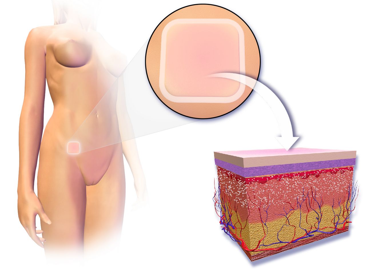
Menstrual disorders and common gynaecological conditions
Primary dysmenorrhoea is prostaglandin-mediated cramping that begins with ovulatory cycles in adolescence, peaks in the first 24 to 48 hours of flow and has no underlying pathology. NSAIDs are first line and work best when started at the first sign of bleeding or 24 to 48 hours before it, because they inhibit prostaglandin synthesis rather than block prostaglandin already made. Heat, exercise and combined hormonal contraception all help. Secondary dysmenorrhoea starts later, worsens over time and points to endometriosis, adenomyosis, fibroids or pelvic inflammatory disease.
Endometriosis is endometrial-like tissue outside the uterus that bleeds cyclically into a closed space, producing the classic triad of dysmenorrhoea, dyspareunia and infertility, often with dyschezia. Diagnosis has historically required laparoscopy, and the average delay from symptom onset to diagnosis is measured in years — largely because adolescents reporting severe period pain are told it is normal. Believe them and refer. Polycystic ovary syndrome is diagnosed by the Rotterdam criteria when two of three are present: oligo- or anovulation, clinical or biochemical hyperandrogenism, and polycystic ovarian morphology on ultrasound. In adolescents that third limb does not apply: multifollicular ovaries are physiological after menarche, so neither ovarian morphology nor anti-Müllerian hormone may be used to diagnose PCOS within 8 years of menarche — in that window both irregular cycles and hyperandrogenism are required, and everyone else is designated “at risk” and re-assessed later. It carries insulin resistance, so screening for impaired glucose tolerance and counselling on weight-neutral metabolic health matter as much as cycle regulation.
Abnormal uterine bleeding is classified by PALM-COEIN: structural causes are Polyp, Adenomyosis, Leiomyoma and Malignancy; non-structural are Coagulopathy, Ovulatory dysfunction, Endometrial, Iatrogenic and Not otherwise classified. In an adolescent with very heavy bleeding from menarche, think coagulopathy — von Willebrand disease is present in a meaningful minority and is routinely missed.
Contraception: counsel by effectiveness tier
| Tier | Method | Typical-use failure per year | Key teaching or caution |
|---|---|---|---|
| Tier 1 — most effective | Etonogestrel implant | Under 1 in 100 (about 0.1%) | Subdermal, now approved for up to 5 years of use (originally 3) — check the current label for the product in use; irregular spotting is the top reason for removal, so pre-counsel it |
| Levonorgestrel IUD | Under 1 in 100 | 3–8 years by product; lightens or stops bleeding, useful for heavy menses | |
| Copper IUD | Under 1 in 100 | 10 years; hormone-free but heavier, crampier periods; also the emergency contraceptive with the longest evidence base | |
| Tubal ligation / vasectomy | Under 1 in 100 | Considered permanent; vasectomy needs a confirmatory semen analysis before relying on it | |
| Tier 2 — effective if used consistently | Combined pill, patch, ring | About 7 in 100 | Oestrogen-containing; screen contraindications; patch changed weekly, ring monthly |
| Progestin-only pill | About 7 in 100 | No oestrogen, so usable where combined methods are contraindicated; timing-sensitive — the traditional norethindrone pill must be taken within the same 3-hour window daily | |
| Depot medroxyprogesterone acetate | About 4–6 in 100 | 150 mg IM or 104 mg SC every 13 weeks; reversible bone density loss — ensure calcium and vitamin D | |
| Tier 3 — least effective | External condom | About 13 in 100 | Only method that also reduces STI transmission; no oil-based lubricants with latex |
| Withdrawal | About 20 in 100 | Requires perfect control every single time | |
| Fertility-awareness methods | About 11–23 in 100 by method | Needs regular cycles, daily tracking and abstinence or backup during the fertile window |
Combined hormonal contraception is contraindicated whenever the oestrogen’s cardiovascular risk outweighs the benefit: age 35 or older and smoking at all — generally avoid at any daily amount, and an absolute contraindication at 15 or more cigarettes daily; migraine with aura at any age; personal history of venous thromboembolism or known thrombophilia; major surgery with prolonged immobilisation; a history of ischaemic heart disease or of stroke; complicated valvular heart disease (pulmonary hypertension, atrial fibrillation, a history of subacute bacterial endocarditis); peripartum cardiomyopathy; systemic lupus erythematosus with positive or unknown antiphospholipid antibodies; diabetes with nephropathy, retinopathy, neuropathy or other vascular disease, or of more than 20 years’ duration; uncontrolled hypertension — systolic 160 mmHg or above or diastolic 100 mmHg or above, either value on its own being enough; active liver disease; current breast cancer; and the first 21 days postpartum, with the method also generally avoided from 21 to 42 days postpartum when any additional venous thromboembolism risk factor is present (obesity, caesarean birth, haemorrhage or transfusion, pre-eclampsia, immobility, smoking). The mechanisms behind that list are not all the same, and separating them is what makes it memorable rather than arbitrary. Four entries are thrombotic: previous venous thromboembolism or thrombophilia, major surgery with immobilisation, the postpartum window, and systemic lupus erythematosus with positive or unknown antiphospholipid antibodies, which carry a thrombotic risk in the venous and the arterial bed alike. Six are arterial — migraine with aura, smoking at 35 or older, uncontrolled hypertension, established ischaemic heart disease or stroke, complicated valvular disease and long-standing or vascular diabetes all raise the risk of ischaemic stroke and myocardial infarction, not of clot in a vein. The remaining three turn on neither: current breast cancer is a hormone-responsive tumour, active liver disease is a question of hepatic metabolism and decompensation, and peripartum cardiomyopathy is a question of cardiac function — a ventricle that is already failing does not tolerate the added thrombotic and volume load. Teach the ACHES warning signs to every patient who starts one — Abdominal pain, Chest pain or shortness of breath, Headache that is severe or new, Eye changes or visual loss, Severe leg pain or swelling — and tell them to stop the method and seek care rather than wait for the next appointment.
Fertility, infection and preventive screening
Infertility is 12 months of regular unprotected intercourse without conception, or 6 months if the person with ovaries is 35 or older, or immediately when there is known pathology such as amenorrhoea or bilateral tubal disease. Causes distribute roughly evenly — about a third female factor, about a third male factor, and the remainder combined or unexplained — so evaluate both partners together. The first test is a semen analysis, because it is cheap, fast and non-invasive, and it is unreasonable to put one partner through a hysterosalpingogram before the other has produced a sample. Ovarian reserve is assessed with day-3 FSH and anti-Müllerian hormone; tubal patency with hysterosalpingography. Infertility care is grief work as much as procedure work: name the losses, ask about the treatment burden, and screen for depression.
Sexually transmitted infection screening is a routine nursing responsibility. Screen for chlamydia and gonorrhoea annually in all sexually active women under 25 and in older women with risk factors; screen for HIV at least once for everyone aged 15 to 65 on an opt-out basis; screen for HIV, syphilis, hepatitis B and hepatitis C at the first prenatal visit in every pregnancy, repeat syphilis at about 28 weeks and again at delivery for every pregnant patient, and repeat HIV in the third trimester where risk or local prevalence indicates. Chlamydia in a non-pregnant adult is treated with doxycycline 100 mg orally twice daily for 7 days; azithromycin 1 g as a single dose is used in pregnancy, with a test of cure 4 weeks after treatment in pregnancy and retesting of everyone at about 3 months. Gonorrhoea is treated with ceftriaxone 500 mg IM as a single dose (1 g IM at 150 kg or more), plus doxycycline 100 mg orally twice daily for 7 days if chlamydial infection has not been excluded — azithromycin 1 g in pregnancy. Syphilis is treated with benzathine penicillin G (Bicillin L-A, never the combination Bicillin C-R, a dispensing error that undertreats): 2.4 million units IM as a single dose for primary, secondary or early latent disease, and 2.4 million units IM weekly for 3 doses for late latent syphilis or latent syphilis of unknown duration; in pregnancy with early syphilis a second dose one week later is recommended. And a pregnant patient with a penicillin allergy must be desensitised and given penicillin, because no alternative reliably treats the fetus.
Cervical cancer screening begins at 21 regardless of sexual debut. From 21 to 29, cytology every 3 years; from 30 to 65, cytology every 3 years, primary HPV testing every 5 years, or co-testing every 5 years. HPV vaccination is routine at ages 11 to 12, may start at 9, and is a two-dose series if begun before the 15th birthday and a three-dose series if begun at 15 or later or if immunocompromised. Preconception care is the highest-yield visit in obstetrics: start folic acid 400 micrograms daily at least one month before conception and continue through the first trimester, or 4 mg daily for someone with a previous neural-tube-defect-affected pregnancy; confirm rubella and varicella immunity and vaccinate before conception, since these are live vaccines — give them at least 4 weeks before conception and advise reliable contraception for those 28 days, and if non-immunity is found during pregnancy, defer and vaccinate immediately postpartum, which is compatible with breastfeeding; optimise glycaemic control toward an HbA1c below 6.5%; and review medications for teratogens including isotretinoin, ACE inhibitors and ARBs, warfarin and valproate.
Counsel contraception by typical-use effectiveness tier and screen every oestrogen-containing method against the absolute contraindications, and start folic acid before conception rather than after the pregnancy test — the neural tube closes by 28 days after conception, about 6 weeks from the LMP — roughly two weeks after the first missed period, and usually before the pregnancy has been confirmed or a first antenatal visit has happened.
Section 4Cultural Humility, Equity and the Social Determinants of Maternal-Child Health

Humility is the practice; competence is the illusion
Cultural competence treats culture as a syllabus: learn what “this group” believes about birth, and you are finished. It reliably produces the nurse who has memorised a list and applies it to a patient who does not recognise herself in it — stereotyping with a certificate attached. Cultural humility begins from the opposite premise: you cannot finish learning a person, so you ask, and keep asking. Its three parts are lifelong examination of your own assumptions, deliberate correction of the power imbalance between the person in the uniform and the person in the gown, and accountability to the communities you serve.
At the bedside that converts statements into questions. Ask who this family wants in the room and who kept out; what they eat, avoid or need warm after birth; whether the placenta is kept; when a child is named; and who actually makes decisions, which is not always the person doing the talking. Record the answers so the family is not re-interviewed every shift, and honour what you were told unless it is unsafe — where it is unsafe, negotiate the smallest possible change rather than issuing a prohibition.
Working across language is the same discipline with a legal floor beneath it: in facilities receiving federal funding, meaningful language access is a civil rights obligation, and clinically it is the difference between a consent and a signature. Use a trained interpreter whenever the patient’s preferred language for health care is not yours, and for the whole encounter — consent, medication teaching and discharge instructions, not merely the history. The interpreter may be in person, by video or by telephone. Never a family member, and never a child. Relatives edit: they soften a prognosis, omit the questions about sexual history and intimate partner violence, and answer on the patient’s behalf. A child asked to interpret her mother’s miscarriage is harmed by the task itself.
Many patients here carry a history of trauma, and this specialty is full of what reawakens it: intimate examination, loss of control, pain, exposure, being spoken about rather than to. Trauma-informed care does not require you to know the history; it asks you to assume it may be present and to deliver care that does not add to the injury. Its principles are safety, trustworthiness and transparency, peer support, collaboration and mutuality, empowerment through voice and choice, and attention to cultural, historical and gender issues. In practice: explain before you touch and ask permission every time rather than once, offer real choices about position and pace, stop when asked and mean it, and read the frightened or “difficult” patient as a nervous system doing what it learned to do.
The social determinants arrive with the patient
The social determinants of health — economic stability, education, health care access, neighbourhood and built environment, and social and community context — are the conditions in which people are born, grow, work, live and age. In this specialty they are not background colour. They help set birth weight; they decide whether the antibiotic is collected, whether the baby has a safe surface to sleep on, whether the immunisation is given on time, and whether the patient comes back at all. They explain most of what gets recorded as non-adherence.
So the language discipline from Section 1 earns its keep: a parent who has missed two appointments has a barrier, not a character flaw, and barriers are solvable by nurses in a way that character flaws are not. Ask about the determinants as you would anything else clinical — but observe the rule that separates screening from voyeurism: never ask about a need you have no route to address. Two specifics are worth carrying. A family stretching a tin of formula with extra water is a cause of hyponatraemic seizures, so the answer to a family who cannot afford formula is exact mixing instructions and a same-day referral for supply, never a warning. And Medicaid pays for roughly four in ten United States births, with postpartum coverage that once ended at 60 days now extendable to twelve months in most states — confirm coverage and book the postpartum contact before discharge. The table below sets out those same five domains in the forms a nurse actually meets, which is why it runs to more rows than there are domains: income and employment and food security sit inside economic stability, and housing and transport inside neighbourhood and built environment.
| Determinant | How it shows up in maternity or paediatrics | What the nurse does about it |
|---|---|---|
| Income and employment | No paid leave, early return to work, milk supply falls, medicine competes with rent | Ask about leave at the first postpartum contact; refer to social work; teach expression and storage |
| Housing | No safe sleep surface, overcrowding, damp and pests driving asthma, lead in older housing | Ask where the baby sleeps; arrange a cot; screen for lead on schedule; refer to a housing navigator |
| Food security | Anaemia in pregnancy, formula stretched with water, the parent skipping meals first | Screen with the two-question Hunger Vital Sign — worried the food would run out, and the food bought did not last; refer to WIC and SNAP; teach exact mixing, never dilution |
| Transport | Missed antenatal, postnatal and well-child visits, recorded as non-adherence | Chart the barrier as the reason; arrange the transport benefit; consolidate visits; offer telehealth |
| Education and health literacy | Instructions given at a reading level nobody checked | Plain language, teach-back, written material in her language, pictures for dosing |
| Health care access and insurance | Coverage gaps after birth, no usual source of care, long waits for the postpartum visit | Confirm coverage; book the postpartum contact before discharge; teach the warning signs to everyone |
| Neighbourhood and built environment | Pharmacy and grocery deserts, unsafe streets, poor air quality | Check the prescription can be filled nearby; prefer once-daily options; ask before advising exercise |
| Social and community context | Isolation, intimate partner violence, discrimination, fear related to immigration status | Screen privately with the patient alone; have the advocate’s number before you ask |
Disparities are produced, and bias is how they reach the bedside
In the United States, Black birthing people die of pregnancy-related causes at roughly three times the rate of white birthing people, and American Indian and Alaska Native people are also substantially over-represented; preterm birth is about half again as common among Black birthing people, and Black infants die in the first year at more than twice the rate of white infants. Severe maternal morbidity — surviving an event that could have killed you, such as massive transfusion, eclampsia or intensive care admission — follows the same pattern and affects many times more patients than death does.
The tempting explanation is poverty, and it is insufficient: the gap does not close at the top of the income or education distribution, where a Black patient with a degree, a job and insurance still fares worse than a white patient with none of those. Race is a social category, not a biological one, and no gene produces a difference of that size. What produces it is structural racism and unequal access — delivery concentrated in lower-resourced hospitals, different treatment inside the same hospital by the same staff, the physiological cost of living with discrimination, and generations of policy deciding which neighbourhoods received clinics, groceries, clean air and mortgages. Teach it, and chart it, as an exposure rather than an attribute. Two findings make it actionable: state review committees judge more than four in five pregnancy-related deaths preventable, and most occur after the birth, roughly a third between six weeks and one year — so the postpartum period is not one visit at six weeks.
Implicit bias carries all of this into a single room: automatic associations operating outside awareness that can contradict what you sincerely believe. It shows itself most reliably in pain — patients of colour are less likely to be offered analgesia for the same presentation, and in one widely cited study half of a sample of white medical students and residents endorsed at least one false belief about biological differences in how Black and white patients feel pain. The failure mode is repeatable: a patient says something is wrong, the observations are near enough to normal, the report is written down as anxiety, and the next contact is the emergency department.
So take her report as a clinical finding and document it verbatim in quotation marks. Perform the assessment the symptom demands rather than the one the chart’s labels suggest. Escalate on the concern and not only the number, using SBAR — Situation, Background, Assessment, Recommendation — to name the red flag and make a specific, time-bound request. In one sentence: “Twelve days postpartum, calling about a headache (situation); normal pressures at discharge and a note describing her as anxious (background); three days of headache that paracetamol is not touching, and she says she knows something is wrong (assessment); I want her seen today with a blood pressure, a urine protein and bloods (recommendation).” Ask whether you would arrange this same assessment for a different patient. Teach the urgent maternal warning signs before discharge — a headache that will not go away, visual change, chest pain or breathlessness, seizure, fever, heavy bleeding, swelling or pain in one leg, and thoughts of harming herself or the baby — and know the referral routes, because advocacy that stops at sympathy is not advocacy.
Cultural humility asks rather than assumes and a trained interpreter is a clinical requirement rather than a courtesy; the racial gaps in maternal mortality, preterm birth and infant mortality are produced by structural racism and unequal access rather than by race — which is why a patient’s own report of pain or of something being wrong is data you document verbatim and act on, not anxiety you reassure.
Section 5Consent, Confidentiality and the Nurse’s Role
What makes consent informed, and what your signature says
Consent is a conversation; the form is only its receipt. It is informed when the patient has been told, in language she genuinely understands, the diagnosis or nature of the problem; what is proposed and why; the material risks and expected benefits; the reasonable alternatives with their own risks; and what happens if nothing is done. She must also know that the decision is voluntary and revocable at any point — including after the form is signed. A signature without those elements is paper, not consent, and it protects nobody.
Two conditions sit underneath. She must have capacity, and the information must actually reach her — a trained interpreter if her preferred language is not yours, the form read aloud if she cannot read. Timing matters as much as content: consent taken after an opioid or a sedative, or from a patient in severe pain, is fragile at best, so planned procedures are discussed while she is comfortable and unhurried. In a genuine emergency, where delay would threaten life or limb and no decision-maker is reachable, consent is implied; document the emergency and the attempts you made.
Now the distinction students most often get wrong. The duty to obtain consent belongs to the clinician performing the procedure and cannot be handed to nursing. When the nurse signs she is witnessing three narrow things: that the signer is who she says she is, that the signature is voluntary, and that she appeared to have capacity. She is not attesting that the patient understood the medicine. The real work happens before the pen moves — ask her to say in her own words what will be done and why, and whether she knows any alternative including doing nothing. If the answer is wrong, vague or absent, stop, do not have her sign, and call the clinician back. That is not obstruction; it is the only mechanism the system has for catching a consent that never took place.
Autonomy, refusal and the adolescent patient
The right to refuse follows from the right to consent, and it is the harder half. A patient with capacity may decline any recommended treatment, and the pregnant patient is no exception: she remains the decision-maker for her own body through pregnancy, labour and birth. Professional guidance in obstetrics is explicit that coercion — threats, court orders, child-protection referral used as leverage — is ethically unjustified in all but the rarest circumstances, and it drives patients out of antenatal care, falling hardest on those already described in Section 4. Keep the relationship: explore what the refusal is really about, offer alternatives, and make clear she may change her mind. Then document the recommendation, the risks explained, her decision in her own words, that the refusal was informed, and that care is still offered. Escalate to an ethics consultation, not a judge.
Adolescent care adds a second axis: Section 1 covered who may consent, and the question here is who may be told. Every United States jurisdiction allows a minor to consent to diagnosis and treatment of sexually transmitted infections, though ages and conditions differ; consent for contraception, prenatal care and mental health or substance use care varies far more widely, and abortion law differs by state and keeps changing — so know your own jurisdiction rather than recite a national answer. Where she lawfully consents she generally controls that part of the record, and a parent is not automatically entitled to it.
Two habits follow. Interview every adolescent alone for part of every visit, so that it reads as routine rather than as suspicion of the family. And state the limits of confidentiality before she tells you anything: what she says stays between you except where someone is being hurt or she is at risk of serious harm. Promise only what you can keep, and warn her that an explanation-of-benefits statement mailed to the policyholder can disclose the very visit confidentiality was meant to protect. Tell her in advance and offer what workarounds exist: a confidential-communication request to the insurer, care through a Title X or school-based clinic, or self-payment.
Privacy, documentation and the duties that stay with you
Privacy here is unusually hard because the room is unusually full. The patient decides who receives information about her — her partner, her mother, the baby’s other parent, the friend at the door. Ask on admission who may be told what and who may visit, record it, and follow it without editorialising; in maternity, one reason a patient gives no reason is that the person asking is the source of the harm. Offer exclusion from the facility directory, and watch the ordinary leaks: the whiteboard, the corridor handover, the open screen. In paediatrics the record belongs to the parent or guardian, except for care an adolescent consented to independently and where a court has restricted a parent’s rights — read the custody paperwork rather than assuming.
Documentation is a clinical act before it is a legal one, and it is both. Chart contemporaneously, objectively and specifically: what you assessed, what you found in measurable terms, what you did, whom you notified, at what time, and what followed. Use her own words in quotation marks for symptoms, concerns and refusals — that is the entry that survives a later dispute about whether anybody listened. Never chart in advance or for another nurse, and correct an error with a single line through it, the correction, your initials, time and date. A concern you raised and did not document is one you cannot prove you raised.
Your scope of practice is set by your jurisdiction’s nurse practice act, not by the habits of the unit: policy may narrow it and can never widen it, and “the provider told me to” is no defence for working outside it. The five rights of delegation from Section 1 apply every time you hand a task on, and accountability stays with the RN who delegated.
Some duties cannot be delegated, referred or shared. Nurses are mandated reporters of suspected child abuse and neglect in every United States jurisdiction; the threshold is reasonable suspicion rather than proof, and the duty belongs personally to the nurse who formed the suspicion — telling the charge nurse does not discharge it unless a report is actually made. Good-faith reports carry statutory immunity. Federal child-welfare law separately requires that an infant identified as affected by substance exposure or withdrawal be notified to child protective services and given a plan of safe care, a support mechanism rather than a finding of abuse. Rules for intimate partner violence against a competent adult differ by jurisdiction — in most places the adult decides, and reporting without her consent can increase the danger she is in — so learn your own rule, and tell the patient before you make a report about her.
Finally, the conflict that most often leaves nurses stuck: the patient wants one thing and the family another. If the adult patient has capacity she decides, and your task is to build the conditions in which she can — usually by emptying the room and asking her alone, never with the person she may fear. In paediatrics the parents decide, but within limits: the choice must be one a reasonable parent could make for that child, assent should be sought from around seven, and a sustained refusal by an older child is a finding rather than a nuisance. When a parental decision risks serious harm, escalate rather than argue — charge nurse, treating clinician, chain of command, ethics — and in a genuine emergency treat under implied consent.
| Situation | Who holds the decision | What the nurse does |
|---|---|---|
| Adult with capacity, any recommended treatment | The patient | Make sure the information and an interpreter actually reached her; support the decision; document |
| Pregnant patient refusing a recommended caesarean or transfusion | The pregnant patient | Document informed refusal in her own words; keep offering care; request ethics support, never coercion |
| Adult who has lost capacity, no advance directive | Surrogate under the jurisdiction’s hierarchy | Confirm who the legal surrogate is; do not default to whoever speaks loudest |
| Child below the age of consent | Parent or legal guardian, with the child’s assent from about age 7 | Seek assent; escalate a parental refusal that risks serious harm |
| Adolescent consenting independently under a minor-consent statute | The adolescent | Protect that part of the record; warn her that billing statements can disclose the visit |
| Emancipated minor | The minor | Verify and document the basis once, then treat the consent as an adult’s |
| Life-threatening emergency, no decision-maker reachable | Consent is implied | Treat; document the emergency and the attempts made to reach a decision-maker |
| Child in foster or kinship placement | Varies — agency, court, or birth parent by order | Read the placement paperwork before any non-emergency consent |
Consent is the conversation and the form is only its receipt: the clinician performing the procedure must obtain it, the nurse witnesses identity, voluntariness and apparent capacity, and a patient with capacity — pregnant or not — may refuse. Confidentiality is defined by the patient, documentation is contemporaneous and in her own words, and scope of practice and mandatory reporting are duties owed by you personally that cannot be handed to anyone else.
The chapter in one breath
Maternal-newborn and paediatric nursing treats the family as the unit of care, built on dignity and respect, information sharing, participation and collaboration, and delivered through atraumatic care, cultural humility and language that describes rather than blames. The RN retains assessment, teaching, evaluation and the unstable patient no matter how busy the shift. Reproductive physiology is the grammar of the specialty: pulsatile GnRH drives FSH and LH, rising oestradiol triggers the LH surge, ovulation follows 24 to 36 hours later and about 14 days (14 ± 2) before the next menses, and progesterone from the corpus luteum prepares the endometrium until hCG rescues it or the lining sheds; on the male side a 64-to-74-day spermatogenic cycle sets how soon an abnormal semen analysis can usefully be repeated. That grammar underwrites pregnancy dating by Nägele’s rule, contraceptive counselling by effectiveness tier, and preconception folic acid started before conception rather than after. The same grammar organises the rest of Section 3: dysmenorrhoea, endometriosis and polycystic ovary syndrome, abnormal uterine bleeding classified by PALM-COEIN, an infertility work-up that begins with a semen analysis and evaluates both partners together, and the routine screening that surrounds all of it — sexually transmitted infection screening and treatment, cervical cytology and HPV testing, and HPV vaccination. Two foundations then decide whether any of that knowledge reaches the patient. The first is humility: culture is asked about rather than memorised, language access runs through a trained interpreter and never a family member or a child, the social determinants are screened for only where a referral route already exists, and the racial gaps in maternal mortality, preterm birth and infant mortality are taught as products of structural racism and unequal access rather than of race — which is why a patient’s own report that something is wrong is documented verbatim and acted on rather than filed as anxiety. The second is accountability: consent is the conversation and the form only its receipt, the provider obtains it while the nurse witnesses identity, voluntariness and apparent capacity, a patient with capacity may refuse and a pregnant patient remains the decision-maker for her own body, confidentiality is defined by the patient and complicated by adolescence, and documentation, scope of practice and mandatory reporting are duties the individual nurse cannot hand to anyone else.
Words worth knowing
| Term | What it means |
|---|---|
| Family-centred care | Practice model built on dignity and respect, information sharing, participation and collaboration with the family as partner |
| Assent | A child’s developmentally appropriate agreement to treatment, expected from about age 7, alongside parental permission |
| Emancipated minor | A minor who may consent independently by virtue of marriage, military service, independent living or court declaration |
| Mature minor | A minor judged by a clinician or a court to have sufficient maturity to consent to a particular treatment |
| LH surge | Oestradiol-triggered pituitary release that causes ovulation 24 to 36 hours later |
| Corpus luteum | Post-ovulatory follicular remnant secreting progesterone for a fixed 14 ± 2 days unless rescued by hCG |
| Spinnbarkeit | The stretchy, clear, oestrogen-driven quality of cervical mucus at ovulation |
| Nägele’s rule | Estimated date of birth = first day of LMP − 3 months + 7 days + 1 year |
| PALM-COEIN | Classification of abnormal uterine bleeding into structural and non-structural causes |
| Rotterdam criteria | Two of three findings — anovulation, hyperandrogenism, polycystic morphology — diagnose PCOS in adults; within 8 years of menarche the morphology limb does not apply and both of the other two are required |
| ACHES | Warning signs on combined hormonal contraception: abdominal pain, chest pain, headache, eye changes, severe leg pain |
| Trained interpreter | A qualified interpreter used for the whole encounter whenever the patient’s preferred language for health care is not the nurse’s — never a family member and never a child |
| SBAR | Escalation format — situation, background, assessment, recommendation — used to name a red flag and make a specific, time-bound request |
| Cultural humility | Lifelong self-examination, correction of the clinician–patient power imbalance and accountability to communities — asking rather than assuming, in contrast to cultural competence |
| Social determinants of health | Economic stability, education, health care access, neighbourhood and social context — the conditions that decide whether a plan of care is achievable |
| Structural racism | Policies and systems that distribute resources and treatment unequally by race, producing the disparities that race itself does not explain |
| Implicit bias | Automatic associations operating outside awareness that can contradict stated values; classically expressed as under-treated pain |
| Trauma-informed care | Assuming trauma may be present and building safety, transparency, collaboration and choice into every interaction so care does not re-injure |
| Capacity | Ability to understand, appreciate, reason and communicate a consistent choice; decision-specific, and not lost by disagreeing with the clinician |
| Mandated reporter | Nurse’s personal, non-delegable duty to report suspected child abuse or neglect on reasonable suspicion rather than proof |
Check yourself
CONCEPTION, FETAL DEVELOPMENT AND GENETICS
Every patient you will ever meet began as a single cell that had to divide correctly, travel correctly, implant correctly and then build an entire human body in thirty-eight weeks. Most of the time this happens without anyone watching. When it does not, the consequences arrive in your care — as a newborn who cannot be roused, a toddler with an unexplained developmental delay, a family holding a genetic report they cannot read. This chapter follows the biology in the order the body performs it: gametes and fertilisation, implantation, the embryonic window when organs form and teratogens do their worst, the placenta and the fetal circulation that sustain the pregnancy, and the inheritance patterns that explain why a condition appears in one sibling and not another. Then it turns practical — what each prenatal test can and cannot say, what you do first in a cord prolapse, and how to speak with a family about genetics without ever implying that anyone is at fault.
By the end of this chapter you can
- Trace the sequence from gametogenesis through fertilisation, cleavage, blastocyst formation and implantation, and state the day range for each step.
- Distinguish gestational age from fertilisation age and calculate an estimated date of birth using Naegele rule.
- Identify the embryonic period as the window of maximum teratogenic vulnerability and match named teratogens to the structures they damage.
- Explain placental transfer, the placental hormones, amniotic fluid function and the three fetal shunts, including how each closes after birth.
- Sequence the immediate nursing actions for umbilical cord prolapse, magnesium sulfate toxicity and early postpartum haemorrhage, naming the first action in each.
- Predict recurrence risk for autosomal dominant, autosomal recessive and X-linked recessive conditions, and describe nondisjunction, translocation, imprinting and trinucleotide repeat expansion.
- Compare prenatal screening with diagnostic testing, including timing, accuracy and the meaning of a positive cell-free DNA result.
- Demonstrate non-directive, family-centred communication about genetic risk, and state which nursing actions may not be delegated.
Section 1From Gametes to Implantation
Gametogenesis and the chromosomal arithmetic of a new person
Human somatic cells carry forty-six chromosomes arranged in twenty-three pairs: twenty-two pairs of autosomes and one pair of sex chromosomes. Gametes cannot carry forty-six, or every generation would double the count, so germ cells undergo meiosis — two divisions that halve the chromosome number to twenty-three and, along the way, shuffle the parental chromosomes through crossing over. Fertilisation restores the diploid number. That single sentence contains most of what goes wrong in clinical genetics: if meiosis distributes the chromosomes unevenly, the resulting gamete carries twenty-four or twenty-two, and the conceptus is trisomic or monosomic from the first cell onward.
Oogenesis and spermatogenesis are not symmetrical, and the difference matters at the bedside. Oogenesis begins before birth: a female fetus contains six to seven million primary oocytes at around twenty weeks of gestation, roughly one to two million at birth, and about three hundred thousand to four hundred thousand at puberty, of which only four hundred or so will ever be ovulated. Those primary oocytes sit arrested in prophase of the first meiotic division for decades, and the spindle apparatus that must eventually separate the chromosomes ages with the person. This is the accepted explanation for the steep rise in nondisjunction with maternal age — the risk of a liveborn infant with trisomy 21 is approximately 1 in 1250 at age twenty-five, 1 in 350 at thirty-five, 1 in 100 at forty and 1 in 30 at forty-five. Note how the language should be framed for families: the ovum did not fail, and the person carrying it did nothing wrong.
Spermatogenesis is continuous from puberty, with each cycle taking roughly sixty-four to seventy-four days — which is why a change in health, temperature, illness or medication shows up in semen parameters about three months later, and why preconception counselling for the sperm-producing partner needs at least that much lead time. Continuous division carries its own cost: advanced paternal age is associated with new (de novo) autosomal dominant mutations such as achondroplasia, Marfan syndrome and some craniosynostosis syndromes. The sperm also determines chromosomal sex, since the ovum always contributes an X, and the SRY gene on the short arm of the Y chromosome initiates testicular differentiation from about week seven.
Fertilisation, cleavage and implantation: the first ten days
Ovulation releases a secondary oocyte that remains capable of fertilisation for roughly twelve to twenty-four hours. Sperm, once in the female reproductive tract, survive up to five days, so the fertile window in a cycle is about six days — the five days before ovulation plus the day of ovulation itself. Fertilisation almost always occurs in the ampulla, the outer third of the fallopian tube. Sperm must first undergo capacitation, several hours of enzymatic change in the female tract that makes the acrosome reactive; the acrosomal enzymes then digest a path through the corona radiata and zona pellucida. The instant one sperm nucleus enters, the zona reaction hardens the outer coat and blocks polyspermy. The fused nuclei form the zygote, a complete forty-six chromosome cell with a unique genome.
Cleavage begins within about thirty hours: two cells, then four, then a solid sixteen-cell ball called the morula that enters the uterine cavity around day three to four. By day five, fluid has collected inside and the structure is a blastocyst with two distinct populations — the inner cell mass (embryoblast), which becomes the embryo and amnion, and the outer trophoblast, which becomes the chorion and placenta. The blastocyst hatches from the zona pellucida on day five or six and burrows into the secretory endometrium, usually in the upper posterior uterine wall, between days six and ten. Roughly a quarter of patients notice light implantation bleeding around this time and may mistake it for a scant period, which is one reason self-reported dating is unreliable.
The trophoblast differentiates into cytotrophoblast and syncytiotrophoblast, and the syncytiotrophoblast begins secreting human chorionic gonadotropin (hCG) almost immediately. hCG rescues the corpus luteum so that it keeps producing progesterone and the endometrium is not shed; it is detectable in maternal serum about eight to eleven days after conception and in urine a few days later. Levels roughly double every forty-eight to seventy-two hours in early normal pregnancy and peak at eight to ten weeks. By weeks eight to ten the placenta assumes progesterone production and the corpus luteum regresses — the so-called luteal-placental shift. When implantation occurs anywhere other than the uterine cavity, most often in a fallopian tube, the pregnancy is ectopic; this is covered in the high-risk chapter, but recognise here that the biology of implantation is what makes it possible.
Twinning: the timing of division decides the risk
Dizygotic (fraternal) twins arise from two ova fertilised by two sperm. They are genetically ordinary siblings who happen to share a uterus, they may be of different sexes, and they always have two chorions and two amnions. Incidence rises with maternal age, with a family history on the maternal side, and markedly with ovulation induction and assisted reproduction. Monozygotic (identical) twins arise from a single zygote that splits, occur at a fairly constant rate worldwide of about 1 in 250 births, and are genetically identical apart from later somatic mutations and differences in gene expression.
For monozygotic twins the clinically decisive variable is when the split occurred, because that determines how many membranes and how many placentas the twins share. Shared circulation is the danger. Monochorionic twins are at risk of twin-to-twin transfusion syndrome, in which vascular anastomoses in the shared placenta shunt blood from one fetus to the other, leaving a plethoric, polyhydramniotic recipient and an anaemic, oligohydramniotic donor. Monoamniotic twins add the risk of cord entanglement, and are typically monitored intensively and delivered by caesarean before term.
| Day of division (post-fertilisation) | Result | Placentas / sacs | Clinical significance |
|---|---|---|---|
| Days 1–3 (before morula) | Dichorionic diamniotic | 2 placentas, 2 sacs | Lowest risk of the monozygotic group |
| Days 4–8 (blastocyst) | Monochorionic diamniotic | 1 placenta, 2 sacs | Most common; risk of twin-to-twin transfusion |
| Days 8–13 (after amnion forms) | Monochorionic monoamniotic | 1 placenta, 1 sac | Cord entanglement; high fetal loss; planned caesarean |
| After day 13 | Incomplete division | 1 placenta, 1 sac | Conjoined twins |
Fertilisation occurs in the ampulla within twenty-four hours of ovulation, the blastocyst implants on days six to ten while secreting hCG to sustain the corpus luteum, and for monozygotic twins the day of division determines chorionicity and therefore risk.
Section 2The Embryo, the Fetus and the Windows of Vulnerability
The embryonic period: weeks three to eight, when everything is built
Development divides into three stages with very different vulnerabilities. The pre-embryonic period covers the first two weeks after fertilisation, when the conceptus is a dividing ball of undifferentiated cells. Injury here tends to follow an all-or-none pattern: either enough cells are destroyed that the pregnancy is lost, often before it is recognised, or the remaining totipotent cells replace the losses and development proceeds normally. Structural malformation from a first-fortnight exposure is uncommon, which is a genuinely reassuring thing to be able to tell a patient who drank alcohol before she knew she was pregnant — paired, always, with counselling to stop now.
The embryonic period runs from week three through week eight post-conception, and it is the period of organogenesis. In these six weeks the flat embryonic disc folds into a body, the neural tube closes, the heart begins to beat, limbs bud and rotate, the face and palate fuse, and every major organ system is at least sketched. This is why the embryonic period is the window of maximum teratogenic sensitivity: a structure can only be malformed while it is being formed. The neural tube closes by about day twenty-six to twenty-eight post-conception, that is, roughly six weeks gestational age — frequently before a person has missed a second period or booked a first prenatal visit. The heart begins contracting around day twenty-one to twenty-two and cardiac septation is complete by about day forty. The palate fuses between roughly days forty-two and fifty-five, which is why cleft palate can result from an exposure that occurs weeks after the heart and neural tube are already safe.
Each structure therefore has its own critical period, and the same drug taken at two different gestational ages produces two different outcomes. Angiotensin-converting enzyme inhibitors, for example, are relatively less damaging in the first trimester but cause fetal renal failure, oligohydramnios and skull hypoplasia when taken in the second and third trimesters, because the injury is to a functioning kidney rather than to a forming one. When a patient asks whether an exposure has harmed the pregnancy, the two questions that determine the answer are what and exactly when — and a teratogen information service, not an internet search, should supply the risk estimate.
The fetal period: growth, maturation and the edge of viability
From week nine post-conception until birth, the fetus grows and its organs mature functionally. Nothing new is built, but a great deal becomes capable of working. Kidneys produce urine from about week nine to twelve, and fetal urine becomes the dominant source of amniotic fluid after roughly sixteen weeks — which is why renal agenesis or urinary obstruction produces severe oligohydramnios. External genitalia are distinguishable on ultrasound by around twelve to fourteen weeks. Fetal movement is usually first felt (quickening) at sixteen to eighteen weeks by someone who has been pregnant before, and eighteen to twenty weeks in a first pregnancy, because experience is what makes the sensation recognisable.
Lung maturation dominates the third trimester and dominates neonatal outcome. Type II pneumocytes begin producing surfactant at around twenty-four weeks, but production is scanty and unstable until roughly thirty-two to thirty-four weeks and functionally mature at about thirty-five weeks, when the lecithin-to-sphingomyelin ratio reaches 2:1. Periviability begins around twenty-two to twenty-five weeks and survival improves week by week within that band; antenatal corticosteroids (betamethasone 12 mg intramuscularly, two doses twenty-four hours apart, or dexamethasone 6 mg every twelve hours for four doses) accelerate surfactant production and reduce respiratory distress syndrome, intraventricular haemorrhage and necrotising enterocolitis. Brown fat, the tissue the newborn will later draw on for non-shivering thermogenesis, is deposited mostly in these last weeks, which is another reason the preterm infant cannot hold a temperature.
| Gestational age | Approximate size | Landmark developments |
|---|---|---|
| 6 weeks (4 post-conception) | 4–5 mm | Neural tube closing; heart tube begins to beat; limb buds appear |
| 10 weeks (8 post-conception) | 3 cm, 2 g | All organ systems present; recognisably human; embryonic period ends |
| 12 weeks | 8 cm, 45 g | Fetal heart audible by Doppler; kidneys produce urine; sex distinguishable |
| 16 weeks | 14 cm, 200 g | Lanugo appears; meconium forms; quickening may be felt (multipara) |
| 20 weeks | 25 cm, 300–450 g | Vernix caseosa; fetal heart audible by fetoscope; anatomy survey performed |
| 24 weeks | 30 cm, 600–800 g | Surfactant production begins; alveolar ducts form; edge of viability |
| 28 weeks | 35–38 cm, 1100–1300 g | Eyes open; strong respiratory effort possible with support; testes descending |
| 32 weeks | 40 cm, 1800–2000 g | Bone fully developed; more subcutaneous fat; better temperature control |
| 36 weeks | 45 cm, 2500–2750 g | Lanugo disappearing; sole creases forming; lecithin/sphingomyelin approaching 2:1 |
| 40 weeks | 48–53 cm, 3000–3600 g | Full term at 39 0/7 to 40 6/7; vernix in creases only; ready for extrauterine life |
Teratogens, TORCH infections and the conversation that prevents harm
A teratogen is any agent — drug, infection, chemical, radiation, maternal metabolic state — that disturbs development and produces a permanent structural or functional abnormality. Teratogenic effect depends on the agent, the dose, the timing relative to organogenesis and the genetic susceptibility of both fetus and pregnant person. The old letter categories (A, B, C, D, X) were retired in favour of the Pregnancy and Lactation Labeling Rule in 2015, which replaced a falsely precise letter with a narrative risk summary; the letters still circulate in older references and in student memory, so recognise them but do not rely on them.
Alcohol deserves separate emphasis because it is the most common preventable cause of intellectual disability in the developed world, and because fetal alcohol spectrum disorder includes central nervous system effects that occur across all three trimesters, not only during organogenesis. No safe amount, no safe trimester and no safe type of alcohol has ever been established. The counselling stance is non-judgemental and forward-facing: what is drunk today is what matters now, screening with a validated tool is routine for everyone rather than targeted at people who look like they might drink, and referral is offered rather than imposed.
The TORCH infections — toxoplasmosis, other (syphilis, varicella, parvovirus B19, Zika, hepatitis, HIV), rubella, cytomegalovirus and herpes simplex — cross the placenta and cause a shared pattern of microcephaly, intracranial calcification, hearing loss, chorioretinitis, hepatosplenomegaly and growth restriction. Cytomegalovirus is the most common congenital infection and the leading infectious cause of congenital sensorineural hearing loss; it is spread mainly by the saliva and urine of young children, so hand hygiene teaching for pregnant people who work in childcare or already have toddlers is genuinely preventive nursing. Rubella vaccine is a live vaccine and cannot be given during pregnancy; a non-immune patient is vaccinated postpartum, before discharge, and advised to avoid pregnancy for four weeks.
| Agent | Characteristic effect | Highest-risk window | Nursing action |
|---|---|---|---|
| Alcohol | Fetal alcohol spectrum disorder: growth restriction, smooth philtrum, thin upper lip, short palpebral fissures, lifelong cognitive and behavioural effects | All trimesters | Universal screening; non-blaming counselling; stopping at any point helps |
| Isotretinoin | Craniofacial, cardiac, thymic and CNS malformations; high loss rate | Weeks 3–8 | Pregnancy prevention programme; two contraceptive methods; negative tests before, during and after |
| Warfarin | Nasal hypoplasia, stippled epiphyses, CNS anomalies, fetal bleeding | Weeks 6–12 and third trimester | Transition to low-molecular-weight heparin, which does not cross the placenta |
| ACE inhibitors and ARBs | Renal failure, oligohydramnios, skull hypoplasia, limb contracture | Second and third trimesters | Discontinue and substitute a pregnancy-compatible antihypertensive |
| Valproate / carbamazepine | Neural tube defects (1–2 percent with valproate), craniofacial anomalies, reduced IQ | Weeks 3–4 for the neural tube | Preconception neurology review; high-dose folic acid; never stop an antiepileptic abruptly |
| Lithium | Ebstein anomaly of the tricuspid valve | First trimester | Risk-benefit review with psychiatry; fetal echocardiography |
| Tetracyclines | Permanent tooth discoloration, enamel hypoplasia | After 18 weeks | Substitute a safe antibiotic class |
| Methotrexate | Craniofacial and limb anomalies; pregnancy loss | Weeks 6–8 | Contraception counselling before prescribing |
| Uncontrolled maternal diabetes | Caudal regression, cardiac and neural tube defects; macrosomia later | Weeks 3–8 | Preconception glycaemic control; target HbA1c below 6.5 percent where achievable |
| Cytomegalovirus | Microcephaly, sensorineural hearing loss, chorioretinitis | Any, worst in first half | Hand hygiene after nappy changes and toddler saliva contact |
| Toxoplasma gondii | Hydrocephalus, intracranial calcification, chorioretinitis | Any; severity worst early | Avoid cat litter, undercooked meat, unwashed produce |
| Parvovirus B19 | Severe fetal anaemia and hydrops fetalis | First half of pregnancy | Serology after exposure; serial Doppler for anaemia |
Organs are built in weeks three to eight post-conception, so that is when structural teratogens do their damage; the fetal period grows and matures what already exists, and the timing of an exposure predicts the injury more reliably than the agent alone.
Section 3The Placenta, Membranes, Cord and Fetal Circulation
The placenta as endocrine organ and imperfect filter
The placenta forms where the chorion frondosum meets the decidua basalis; it is structurally established by the end of week twelve and continues to enlarge until term, when it weighs roughly 500 grams — about one-sixth of the fetal weight — and contains fifteen to twenty cotyledons on the dull, red maternal surface. The fetal surface is smooth and glistening with radiating vessels under the amnion. At term the placenta receives roughly 500 to 750 mL of maternal blood per minute. Maternal and fetal blood do not mix; they are separated by the placental membrane, across which exchange occurs by four mechanisms. Simple diffusion moves oxygen, carbon dioxide, water, electrolytes and most drugs; facilitated diffusion moves glucose; active transport moves amino acids, calcium, iron and water-soluble vitamins against a gradient; and pinocytosis moves large molecules including immunoglobulin G.
That last mechanism has a clinical corollary worth memorising. Maternal IgG crosses the placenta, mainly in the third trimester, and gives the newborn passive immunity for the first months of life — which is why the preterm infant, who missed that transfer, is more susceptible to infection, and why maternal pertussis and influenza vaccination protects the newborn. Maternal IgM does not cross. Therefore elevated IgM in a newborn was made by the newborn and indicates intrauterine infection. The placenta is often described to patients as a barrier; it is more honestly described as a selective filter that alcohol, nicotine, most medications and many viruses cross freely.
The placenta is also the largest endocrine organ the body ever produces. hCG maintains the corpus luteum and is the basis of pregnancy testing. Progesterone maintains the endometrium and keeps the myometrium quiescent. Oestrogen drives uterine growth, breast ductal development and increased uteroplacental blood flow. Human placental lactogen (hPL) is an insulin antagonist that deliberately raises maternal blood glucose so that more glucose is available to the fetus — the physiological basis of gestational diabetes, and the reason screening is timed at twenty-four to twenty-eight weeks when hPL is rising steeply. Relaxin softens the cervix and pelvic ligaments, and also explains the altered balance and joint discomfort patients report in later pregnancy.
Amniotic fluid, membranes and the cord
Amniotic fluid is derived early from maternal plasma and later, after about sixteen weeks, predominantly from fetal urine; it is cleared chiefly by fetal swallowing and by absorption across the membranes. Volume rises from about 30 mL at ten weeks to 800 to 1000 mL at thirty-six to thirty-eight weeks, then declines. It cushions the fetus from trauma, maintains a stable temperature, prevents adhesion and permits symmetrical growth, allows the free movement that develops the musculoskeletal system, prevents cord compression and — critically — is required for lung development, because the fetus breathes fluid in and out to expand the developing airways.
Volume is measured either as the amniotic fluid index (the sum of the deepest vertical pocket in four quadrants, normal 5 to 24 cm) or as the single deepest pocket (normal 2 to 8 cm). Oligohydramnios suggests reduced fetal urine output or fluid loss — renal agenesis, posterior urethral valves, ruptured membranes, placental insufficiency with growth restriction, post-term pregnancy, or exposure to an ACE inhibitor — and predisposes to cord compression, pulmonary hypoplasia and limb deformity. Polyhydramnios suggests the fetus cannot swallow or is producing excess urine: oesophageal atresia, tracheo-oesophageal fistula, anencephaly, poorly controlled maternal diabetes, multiple gestation or fetal anaemia. Green or greenish-brown fluid is meconium-stained and must be reported, since it signals a fetus that has passed stool in utero and raises the possibility of meconium aspiration.
The umbilical cord contains two arteries and one vein wrapped in Wharton jelly, which resists compression and kinking. Remember the direction, because it is counterintuitive: the single vein carries oxygenated blood to the fetus and the two arteries carry deoxygenated blood away to the placenta. Average cord length is about 55 cm, with a normal range of roughly 30 to 90 cm. A two-vessel cord (single umbilical artery) occurs in about one percent of births and is associated with renal and cardiac anomalies, so its identification obliges a careful newborn assessment and often a renal ultrasound. Nuchal cord — the cord looped around the neck — is found in about a quarter of births and is usually managed simply by slipping it over the head at delivery.
Fetal circulation and the switch that happens at the first breath
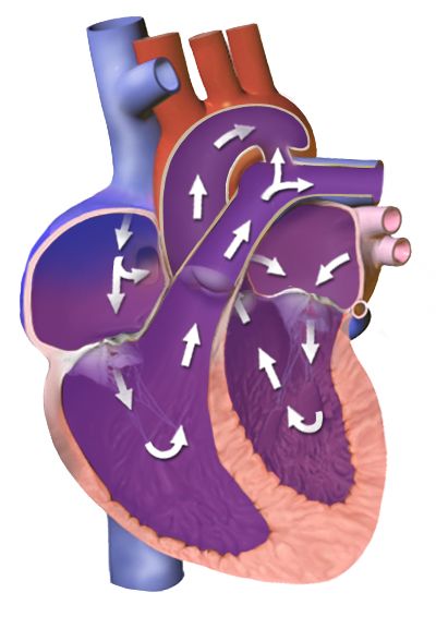
In utero the lungs are fluid-filled and pulmonary vascular resistance is high, while the placenta provides a low-resistance circuit; three shunts exploit this arrangement. The ductus venosus carries the well-oxygenated umbilical vein blood past the liver directly into the inferior vena cava. The foramen ovale lets that blood cross from right atrium to left atrium, so the best-oxygenated blood goes preferentially to the coronary arteries and brain. The ductus arteriosus shunts blood from the pulmonary artery into the descending aorta, bypassing the lungs. Fetal oxygen saturation is highest in the umbilical vein at roughly eighty percent, and the fetus compensates for the modest saturation with fetal haemoglobin, which has a higher affinity for oxygen, and with a haemoglobin concentration around 15 to 20 g/dL.
At birth the sequence reverses in seconds. The first breaths expand the alveoli and drop pulmonary vascular resistance; clamping the cord removes the low-resistance placenta and raises systemic vascular resistance. Left atrial pressure now exceeds right atrial pressure and the foramen ovale closes functionally, becoming the fossa ovalis over the following months. Rising arterial oxygen tension and falling prostaglandin levels constrict the ductus arteriosus, usually within twelve to twenty-four hours and almost always by seventy-two hours, leaving the ligamentum arteriosum. The ductus venosus closes within days to become the ligamentum venosum. This physiology is directly actionable: an infant with a ductal-dependent cardiac lesion deteriorates as the duct closes, and prostaglandin E1 is infused at 0.05 to 0.1 micrograms per kilogram per minute to keep it open — for a 3 kg newborn that is 3 multiplied by 0.05, or 0.15 micrograms per minute at the starting rate. Apnoea is a common and expected adverse effect, so airway equipment stays at the bedside.
| Structure | Function before birth | Closure | Becomes |
|---|---|---|---|
| Ductus venosus | Umbilical vein blood bypasses the liver | Days after cord clamping | Ligamentum venosum |
| Foramen ovale | Right atrium to left atrium, bypassing the lungs | Functional at first breaths; anatomic over months | Fossa ovalis |
| Ductus arteriosus | Pulmonary artery to aorta, bypassing the lungs | Functional 12–24 hours, usually complete by 72 hours | Ligamentum arteriosum |
| Umbilical vein | Carries oxygenated blood to the fetus | Obliterates after birth | Ligamentum teres (round ligament of the liver) |
| Umbilical arteries | Carry deoxygenated blood to the placenta | Obliterate after birth | Medial umbilical ligaments |
When the cord or placenta fails: sequences that save the life
Umbilical cord prolapse occurs when the cord slips below the presenting part, most often after spontaneous or artificial rupture of membranes with an unengaged presenting part, in malpresentation, prematurity, polyhydramnios or multiple gestation. The cord is then compressed between the presenting part and the pelvis, and fetal oxygenation stops. The presentation is a sudden prolonged deceleration or bradycardia immediately after membranes rupture, sometimes with a palpable or visible cord. The instinctive action — to push the cord back in — is exactly wrong, because handling the cord causes vasospasm and worsens the very hypoxia you are trying to relieve.
Vasa previa is the related catastrophe: unprotected fetal vessels crossing the internal os rupture when membranes rupture, producing painless vaginal bleeding with immediate fetal bradycardia or a sinusoidal fetal heart rate pattern. The blood lost is fetal blood, and a fetus has only about 250 mL in total, so exsanguination is measured in minutes. This is an immediate caesarean birth. Uterine rupture presents with a sudden tearing abdominal pain, loss of the fetal station on examination, an abnormal fetal heart rate pattern, cessation of contractions and signs of maternal hypovolaemia; it demands immediate surgical delivery and volume resuscitation.
When the placenta separates and the uterus does not contract down onto the open spiral arteries, the result is early postpartum haemorrhage — defined as cumulative blood loss of 1000 mL or more, or any loss accompanied by signs of hypovolaemia, within twenty-four hours of birth. Uterine atony causes seventy to eighty percent of cases, so the first action is to massage the fundus, not to start a drug and not to call the provider. Massage while a second nurse establishes access and empties the bladder, since a full bladder displaces the uterus and prevents contraction. Only then do the uterotonics follow: oxytocin 10 to 40 units in 500 to 1000 mL of crystalloid intravenously or 10 units intramuscularly; methylergonovine 0.2 mg intramuscularly, contraindicated in hypertension; carboprost 250 micrograms intramuscularly, avoided in asthma; misoprostol 600 to 1000 micrograms rectally or sublingually; and tranexamic acid 1 g intravenously, which works best given within three hours of birth. Quantify the loss by weighing, do not estimate it by eye.
The placenta is a selective filter and the largest endocrine organ of pregnancy, three shunts close within hours to days of the first breath, and in cord prolapse, magnesium toxicity and postpartum haemorrhage the first action — lift the presenting part, stop the infusion, massage the fundus — is what changes the outcome.
Section 4Patterns of Inheritance and Chromosomal Variation

Mendelian patterns: dominant, recessive and X-linked
In autosomal dominant inheritance a single altered copy of a gene is enough to produce the condition. An affected person has a fifty percent chance of passing the altered allele to each child, independent of sex, and the condition typically appears in every generation of a pedigree. Two features complicate the picture. Penetrance describes what proportion of people carrying the allele show any signs at all, and expressivity describes how severely they show them — which is why one family member with neurofibromatosis type 1 has a few café-au-lait macules and a relative has plexiform neurofibromas. Familiar autosomal dominant conditions include Huntington disease, Marfan syndrome, achondroplasia, neurofibromatosis type 1, familial hypercholesterolaemia, autosomal dominant polycystic kidney disease and hereditary breast and ovarian cancer syndrome caused by pathogenic BRCA1 or BRCA2 variants.
In autosomal recessive inheritance two altered copies are required, one from each parent. Carriers have one copy and are clinically unaffected, so the condition appears to skip generations and often surprises a family with no known history. When both parents are carriers, each pregnancy carries a twenty-five percent chance of an affected child, a fifty percent chance of an unaffected carrier and a twenty-five percent chance of a child with two typical copies — so two-thirds of the unaffected children are carriers. Consanguinity raises the risk because relatives share alleles. Carrier frequencies differ by ancestry: cystic fibrosis carriers are roughly 1 in 25 among people of Northern European descent, sickle cell trait affects roughly 1 in 12 Black Americans, and Tay-Sachs carriers are about 1 in 30 in Ashkenazi Jewish and certain French-Canadian and Cajun populations. Ancestry-based figures guide screening offers but must never be used to assume a person’s ancestry or to decline screening a family requests.
X-linked recessive inheritance is the pattern students most often get backwards, so work it from the chromosomes. A carrier mother has one altered X and one typical X. Each son receives one of her X chromosomes, so fifty percent of sons are affected; each daughter receives one of her X chromosomes plus a typical X from her father, so fifty percent of daughters are carriers and essentially none are affected. An affected father passes his X to every daughter, so all his daughters are carriers, and passes his Y to every son, so no sons are affected — there is no father-to-son transmission of an X-linked condition, and finding it in a pedigree rules the pattern out. Haemophilia A and B, Duchenne muscular dystrophy, glucose-6-phosphate dehydrogenase deficiency and red-green colour vision deficiency follow this pattern.
| Pattern | Risk per pregnancy | Pedigree clue | Examples |
|---|---|---|---|
| Autosomal dominant | 50 percent of children affected if one parent affected | Present in every generation; male and female equally | Huntington disease, Marfan syndrome, achondroplasia, BRCA1/2 |
| Autosomal recessive | 25 percent affected, 50 percent carrier when both parents carry | Skips generations; consanguinity increases risk | Cystic fibrosis, sickle cell disease, phenylketonuria, Tay-Sachs |
| X-linked recessive | Carrier mother: 50 percent of sons affected, 50 percent of daughters carriers | Males affected; no father-to-son transmission | Haemophilia A, Duchenne muscular dystrophy, G6PD deficiency |
| X-linked dominant | Affected father: all daughters affected, no sons | Rare; may be lethal in males | Rett syndrome, X-linked hypophosphataemic rickets |
| Multifactorial | 2–5 percent recurrence after one affected child | Clusters in families without a clean pattern | Neural tube defects, cleft lip and palate, pyloric stenosis, clubfoot |
| Mitochondrial | Affected mother: all children at risk; affected father: none | Transmitted only through the maternal line | MELAS, Leber hereditary optic neuropathy |
Numerical and structural chromosome changes
Nondisjunction — failure of a chromosome pair to separate during meiosis — produces a gamete with an extra or a missing chromosome, and therefore a trisomic or monosomic conceptus. Most autosomal trisomies are lethal early; three are compatible with live birth. Trisomy 21 (Down syndrome) occurs in roughly 1 in 700 live births and results from nondisjunction in about ninety-five percent of cases, from an unbalanced Robertsonian translocation in three to four percent and from mosaicism in one to two percent. The clinical picture includes generalised hypotonia, upslanting palpebral fissures with epicanthal folds, a flat nasal bridge and small low-set ears, a single transverse palmar crease, brachycephaly, and a wide sandal gap between the first and second toes. Nursing priorities follow the associated conditions rather than the appearance: forty to fifty percent have a congenital heart defect, most often an atrioventricular septal defect, so every infant needs an echocardiogram; duodenal atresia presents with bilious vomiting in the first day; congenital hypothyroidism and later thyroid disease are common; there is an increased leukaemia risk; and atlantoaxial instability requires evaluation before contact sports and careful neck positioning during intubation or transfer.
Trisomy 18 (Edwards syndrome), roughly 1 in 6000 to 8000 births, presents with severe growth restriction, a prominent occiput, micrognathia, clenched hands with overlapping index and fifth fingers, rocker-bottom feet and major cardiac defects. Trisomy 13 (Patau syndrome), roughly 1 in 10,000 to 16,000, produces midline defects — holoprosencephaly, cleft lip and palate, microphthalmia — along with polydactyly and cardiac anomalies. Both carry high early mortality, and care is a shared decision with the family that may reasonably range from full intervention to comfort-focused care; the nurse’s role is to make sure the family understands the options, that the infant is treated as a person with a name, and that memory-making and palliative support are offered without being imposed.
Sex chromosome variations behave differently because a missing or extra sex chromosome is better tolerated. Turner syndrome (45,X), about 1 in 2500 female births, presents with short stature, a webbed neck, a low posterior hairline, lymphoedema of the hands and feet in the newborn, coarctation of the aorta or a bicuspid aortic valve, streak ovaries with primary amenorrhoea and infertility, and typically normal intelligence with a specific weakness in spatial and mathematical reasoning; growth hormone and later oestrogen replacement are standard. Klinefelter syndrome (47,XXY), about 1 in 500 to 1000 male births, is often not recognised until adolescence or an infertility evaluation: tall stature with long limbs, small firm testes, gynaecomastia, sparse body hair and language-based learning difficulties, treated with testosterone from puberty. Structural rearrangements complete the picture — deletions such as cri-du-chat (5p minus) and the 22q11.2 deletion (conotruncal cardiac defect, abnormal facies, thymic hypoplasia with immunodeficiency, cleft palate and hypocalcaemia), duplications, inversions, and balanced translocations in which the carrier is clinically well but has recurrent pregnancy loss.
Beyond Mendel: multifactorial, mitochondrial, imprinting and repeat expansion
Multifactorial conditions arise from several genes interacting with the environment and account for most common congenital anomalies: neural tube defects, cleft lip and palate, congenital heart disease, pyloric stenosis, clubfoot and developmental dysplasia of the hip. They cluster in families without following a clean pattern, and empiric recurrence risk after one affected child is generally two to five percent, rising with each additional affected relative and with the severity of the defect. This is exactly the situation in which folic acid supplementation, glycaemic control and avoidance of specific teratogens genuinely lower risk.
Mitochondrial inheritance follows the ovum. Mitochondria carry their own small genome and the sperm contributes essentially none, so an affected mother may pass the condition to all of her children while an affected father passes it to none — a pattern that instantly identifies mitochondrial disease in a pedigree. Because cells carry many mitochondria with a variable proportion of altered copies (heteroplasmy), severity varies widely even within one family, and tissues with the highest energy demand — brain, heart, skeletal muscle — are affected first.
Genomic imprinting means some genes are expressed only from the maternally inherited copy and others only from the paternal copy, so the parent of origin changes the phenotype. The classic pair involves the same region of chromosome 15: loss of the paternal contribution produces Prader-Willi syndrome, which presents in the newborn as profound hypotonia and poor feeding requiring gavage, then converts in early childhood to insatiable hyperphagia with obesity; loss of the maternal contribution produces Angelman syndrome, with severe intellectual disability, minimal speech, ataxic movements and a happy, excitable demeanour. Finally, trinucleotide repeat expansion explains conditions that worsen down the generations (anticipation): fragile X syndrome from CGG expansion in FMR1 is the most common inherited cause of intellectual disability, with normal alleles below 45 repeats, premutation at 55 to 200 and full mutation above 200; Huntington disease from CAG expansion; and myotonic dystrophy from CTG expansion. Premutation carriers are clinically important in their own right, being at risk of primary ovarian insufficiency and of a late-onset tremor-ataxia syndrome.
Dominant conditions carry a fifty percent risk per pregnancy, recessive conditions twenty-five percent when both parents carry, X-linked recessive conditions affect half of a carrier’s sons with no father-to-son transmission, and trisomies arise from nondisjunction — except when a parental translocation raises recurrence risk enough to change counselling.
Section 5Screening, Diagnosis and Family-Centred Genetic Nursing
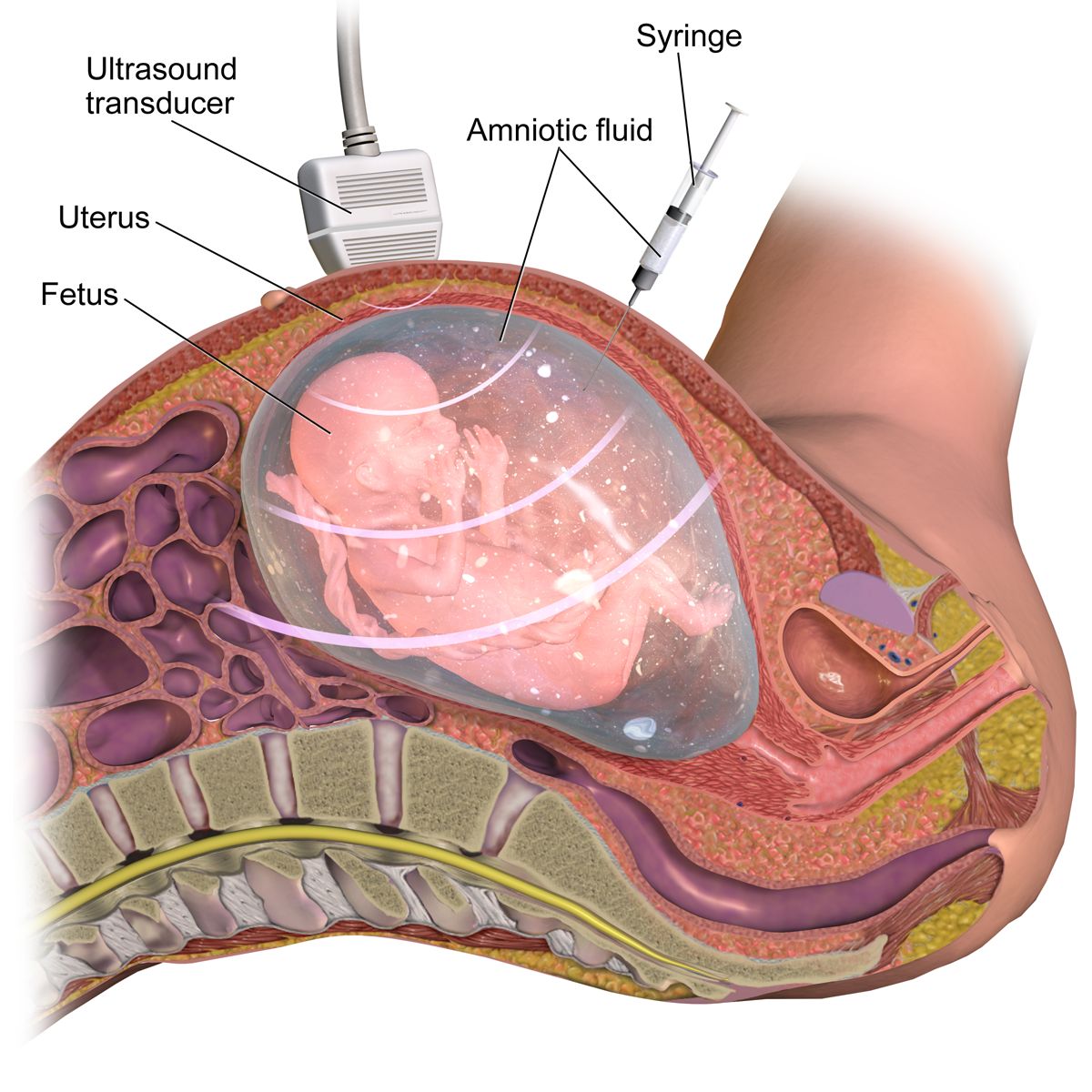
Screening tests: what they can and cannot say
The single most consequential teaching point in prenatal genetics is the difference between a screen and a diagnosis. A screening test estimates probability in a healthy population and can never be positive or negative for a condition — it returns a risk. A diagnostic test examines fetal cells and answers the question. Patients routinely hear a high-risk screen as a diagnosis, make irreversible decisions on it, and are then devastated or falsely reassured. Every conversation about a screening result should include the sentence: this is a screen, and it must be confirmed by a diagnostic test before anything is decided.
The first-trimester combined screen is performed at 11 0/7 to 13 6/7 weeks and pairs an ultrasound measurement of nuchal translucency with maternal serum PAPP-A and free beta-hCG, detecting roughly eighty-five percent of trisomy 21. Cell-free DNA screening (non-invasive prenatal testing) analyses placental DNA fragments circulating in maternal blood from ten weeks onward; for trisomy 21 it detects over ninety-nine percent with a false-positive rate under one in a thousand, and it also reports fetal sex and fetal RhD status. Even so, its positive predictive value depends on how common the condition is in that population, so a positive cell-free DNA result in a young patient screening for a rare trisomy may still be more likely to be a false positive than a true one. Discordant results also arise from confined placental mosaicism, a vanished twin or a maternal chromosomal finding. Cell-free DNA does not screen for neural tube defects.
Second-trimester serum screening is performed at 15 to 20 weeks. Maternal serum alpha-fetoprotein is elevated in open neural tube defects, abdominal wall defects, multiple gestation and simple underestimation of gestational age — and the first step after a high result is always to confirm dates and fetal number by ultrasound, because wrong dating is the commonest explanation. The quadruple screen adds hCG, unconjugated oestriol and inhibin A; the trisomy 21 pattern is low AFP, low oestriol, high hCG and high inhibin A. The detailed anatomy ultrasound at 18 to 22 weeks then evaluates structure directly. Preconceptionally and in early pregnancy, expanded carrier screening panels test the pregnant person first and the partner if a variant is found; a three-generation family history remains the cheapest and most informative genetic tool available and takes about ten minutes to take.
Diagnostic testing and the nursing care around it
Chorionic villus sampling is performed at 10 0/7 to 13 6/7 weeks, transcervically or transabdominally under ultrasound guidance, and samples placental tissue for karyotype, microarray or targeted gene testing. Its advantage is early results; its limitations are that it cannot detect neural tube defects because there is no amniotic fluid to assay, that it carries a procedure-related loss risk of roughly one in five hundred in experienced hands, and that it must not be performed before ten weeks because of an association with limb reduction defects.
Amniocentesis for genetic indications is performed from 15 to 20 weeks, when there is enough fluid to sample safely, with a procedure-related loss risk of about one in five hundred to one in a thousand. Later in pregnancy the same procedure assesses fetal lung maturity or bilirubin in alloimmunisation, or decompresses polyhydramnios. Nursing care: confirm informed consent has been obtained by the provider and that the patient can explain in her own words what is being tested and what she will do with the result; obtain baseline vital signs and fetal heart rate; ask the patient to empty her bladder before a second- or third-trimester procedure so the bladder is not punctured; support positioning and, more importantly, support the patient, because this is frightening. Afterwards, monitor fetal heart rate and contractions for thirty to sixty minutes, and teach the patient to report leaking of fluid, vaginal bleeding, contractions, fever or decreased fetal movement. For any Rh-negative unsensitised patient, Rh immune globulin is indicated after an invasive procedure and after any potential sensitising event: 300 micrograms intramuscularly covers 30 mL of fetal whole blood and is given within seventy-two hours; a 50 microgram microdose is used for events before thirteen weeks.
| Test | Timing | Screen or diagnostic | What it detects / limits |
|---|---|---|---|
| Cell-free DNA (NIPT) | From 10 weeks | Screen | Trisomies 21, 18, 13, sex chromosomes, RhD; does not detect neural tube defects; must be confirmed |
| Nuchal translucency plus PAPP-A and free beta-hCG | 11 0/7 – 13 6/7 weeks | Screen | Approximately 85 percent detection for trisomy 21 |
| Chorionic villus sampling | 10 0/7 – 13 6/7 weeks | Diagnostic | Karyotype and microarray; no neural tube information; loss risk about 1 in 500 |
| Maternal serum AFP / quad screen | 15 – 20 weeks | Screen | High AFP: neural tube or abdominal wall defect, twins, wrong dates. Low AFP with high hCG and inhibin A: trisomy 21 |
| Amniocentesis | 15 – 20 weeks (genetic) | Diagnostic | Karyotype, microarray, AFP, infection, later lung maturity; loss risk about 1 in 500 to 1 in 1000 |
| Anatomy ultrasound | 18 – 22 weeks | Screen (structural) | Direct visualisation of structure, placenta, fluid volume and growth |
| Newborn blood spot screen | 24 – 48 hours of age | Screen | Metabolic, endocrine, haemoglobin and CF disorders; repeat if drawn before 24 hours |
The first hours after birth: newborn screening
Newborn screening is the largest and most successful public-health genetics programme in existence, and the nurse performs it. The heel-stick blood spot is collected after at least twenty-four hours of feeding, ideally at twenty-four to forty-eight hours of age, because several analytes require protein intake to become abnormal; a specimen drawn before twenty-four hours — increasingly common with early discharge — must be repeated. Technique matters: warm the heel, puncture the lateral or medial aspect of the plantar surface to avoid the calcaneus, allow a large drop to form, fill each printed circle completely from one side of the card only, and never layer successive drops or touch the paper, because layering invalidates the assay and a rejected card delays a diagnosis by days. Panels vary by jurisdiction but generally include phenylketonuria (treated with a phenylalanine-restricted formula, which prevents intellectual disability entirely if started in the first weeks), congenital hypothyroidism (levothyroxine begun within the first two weeks preserves cognition), cystic fibrosis, sickle cell disease, galactosaemia, medium-chain acyl-CoA dehydrogenase deficiency, congenital adrenal hyperplasia and biotinidase deficiency.
Two more screens are completed before discharge. Hearing is screened by otoacoustic emissions or automated auditory brainstem response, because early identification and amplification preserve language development. Critical congenital heart disease is screened by pulse oximetry at twenty-four hours of age or later, measured in the right hand (pre-ductal) and either foot (post-ductal): the infant passes if the saturation is ninety-five percent or higher in the hand or the foot and the difference between the two is three percent or less. A saturation below ninety percent in either limb is an immediate fail requiring urgent evaluation with no repeat; a reading of ninety to ninety-four percent in both limbs, or a difference greater than three percent, is repeated in one hour and becomes a fail if it persists on the third measurement. A fail leads to echocardiography, not to discharge.
Non-directive counselling, privacy, delegation and the family
Genetic counselling in nursing is non-directive. Your task is to make sure the family understands the information, not to steer them toward a decision, and this is harder than it sounds because families frequently and directly ask what you would do. A workable reply acknowledges the question rather than deflecting it: “I can tell you what the test can and cannot show, and what families in your situation usually think about. What matters is what fits your family.” Language carries judgement whether or not it is intended — prefer “the chance is one in four” to “the risk is twenty-five percent”, “a condition” or “a difference” to “an abnormality”, and person-first phrasing such as “an infant with Down syndrome” rather than “a Down baby”. Never describe a family as non-compliant; describe what was recommended, what happened and what barrier the family identified.
Genetic information is family information, which creates the privacy problem unique to this field: a result belongs to the patient, yet it may be medically important to relatives who have not consented to know. The patient decides what is shared. Patients also have a right not to know — a person at risk for Huntington disease may reasonably decline predictive testing, and that decision is respected without repeated offers. In the United States the Genetic Information Nondiscrimination Act of 2008 prohibits the use of genetic information in health insurance and employment decisions, but it does not extend to life, disability or long-term care insurance, and patients considering predictive testing deserve to be told that gap plainly before the blood is drawn.
Finally, delegation. The registered nurse retains assessment, teaching, evaluation and the care of any unstable patient, and in this chapter that means the RN takes the family history, explains what a screening result means, obtains and evaluates post-procedure fetal monitoring, and performs and interprets the newborn assessment. Assistive personnel may weigh and measure the infant, position and comfort the patient, gather supplies, transport specimens and take vital signs on a stable patient and report them — but interpreting a pulse oximetry screening result, teaching a family about recurrence risk, or evaluating a patient who is leaking fluid after an amniocentesis are never delegated. Licensed practical or vocational nurse scope varies by jurisdiction; know yours before you assign.
A screen returns a probability and a diagnostic test returns an answer, newborn screening only works if the blood spot is collected correctly after twenty-four hours of feeding, and genetic counselling stays non-directive with assessment, teaching and evaluation firmly held by the registered nurse.
The chapter in one breath
Meiosis halves forty-six chromosomes to twenty-three so that fertilisation in the ampulla can restore the diploid number, and from that moment the timetable governs everything. The blastocyst implants on days six to ten while secreting hCG to sustain the corpus luteum; weeks three to eight post-conception build every organ, which is why teratogens do structural damage then and why folic acid at 400 micrograms — or 4 mg after a previously affected pregnancy — must already be on board before the neural tube closes at day twenty-eight. The placenta then feeds, filters imperfectly and manufactures hCG, progesterone, oestrogen and human placental lactogen, while the ductus venosus, foramen ovale and ductus arteriosus divert blood past the liver and lungs until the first breath reverses the pressures and closes them. Inheritance explains recurrence: fifty percent for dominant, twenty-five percent for recessive with two carriers, half of a carrier’s sons for X-linked recessive, and a translocation karyotype that changes the counselling entirely. Screens estimate; only chorionic villus sampling and amniocentesis diagnose. And in the emergencies, sequence is survival — lift the presenting part off a prolapsed cord, stop the magnesium before reaching for calcium gluconate, and massage the fundus before any uterotonic.
Words worth knowing
| Term | What it means |
|---|---|
| Blastocyst | Fluid-filled stage reached about day five, with an inner cell mass that becomes the embryo and a trophoblast that becomes the placenta; the stage that implants. |
| Capacitation | Enzymatic conditioning of sperm within the female reproductive tract that must occur before the acrosome can release its enzymes and fertilisation can happen. |
| Cell-free DNA screening | Analysis of placental DNA fragments in maternal blood from ten weeks; highly sensitive for common trisomies but a screen, never a diagnosis. |
| Chorionicity | The number of chorions (and therefore placentas) in a multiple pregnancy; determined by when a monozygotic zygote divides and the strongest predictor of twin risk. |
| Ductus arteriosus | Fetal shunt from pulmonary artery to aorta that bypasses the lungs; closes functionally within 12 to 24 hours of birth to become the ligamentum arteriosum. |
| Foramen ovale | Fetal opening between the right and left atria; closes functionally at the first breaths when left atrial pressure exceeds right. |
| Gestational age | Age counted from the first day of the last menstrual period, approximately two weeks more than fertilisation age; the basis of all obstetric documentation. |
| Human placental lactogen | Placental hormone that antagonises insulin to divert glucose to the fetus; the physiological driver of gestational diabetes. |
| Imprinting | Parent-of-origin-specific gene expression; loss of the paternal chromosome 15 region causes Prader-Willi syndrome and loss of the maternal causes Angelman syndrome. |
| Nondisjunction | Failure of chromosomes to separate during meiosis, producing a gamete with an extra or missing chromosome; the mechanism of most trisomies. |
| Oligohydramnios | Amniotic fluid index below 5 cm or deepest pocket below 2 cm; suggests reduced fetal urine output or ruptured membranes and risks cord compression and pulmonary hypoplasia. |
| Organogenesis | Formation of organ systems during weeks three to eight post-conception; the window of maximum teratogenic vulnerability. |
| Penetrance | The proportion of people carrying a genetic variant who show any clinical sign of it; distinct from expressivity, which describes severity. |
| Polyhydramnios | Amniotic fluid index above 24 cm or deepest pocket above 8 cm; associated with impaired fetal swallowing, anencephaly, maternal diabetes and fetal anaemia. |
| Robertsonian translocation | Fusion of two acrocentric chromosomes; a balanced carrier parent is healthy but recurrence risk for translocation Down syndrome is far higher than the age-related risk. |
| Surfactant | Phospholipid produced by type II pneumocytes from about 24 weeks that lowers alveolar surface tension; functionally mature near 35 weeks at a lecithin/sphingomyelin ratio of 2:1. |
| Teratogen | Any agent that disturbs development and causes permanent structural or functional abnormality; effect depends on agent, dose, timing and genetic susceptibility. |
| Trinucleotide repeat expansion | Growth of a repeated three-base sequence across generations, causing anticipation; the mechanism of fragile X syndrome, Huntington disease and myotonic dystrophy. |
| Wharton jelly | Gelatinous connective tissue surrounding the umbilical vessels that resists compression and kinking of the cord. |
Check yourself
The Antepartum Period: Adaptation and Prenatal Care
Pregnancy is not a disease, and the antepartum period is not a waiting room. Across roughly forty weeks a healthy body remodels nearly every organ system: blood volume climbs by almost half, the kidneys filter faster than they ever will again, the diaphragm rises about four centimetres, and insulin resistance is deliberately manufactured so that glucose crosses the placenta to the fetus. Prenatal care exists because those adaptations look, on paper, exactly like pathology — the physiologic anaemia, the glycosuria, the dependent oedema, the soft systolic murmur. The nurse’s work is to know which changes are expected, which are early warnings, and which demand escalation within minutes. This chapter builds that discrimination: dating and history first, then adaptation system by system, then the schedule of visits and screens that turns adaptation into surveillance, then the teaching that keeps a family safe between visits, and finally the danger signs whose sequence of response saves a life.
By the end of this chapter you can
- Classify the presumptive, probable and positive signs of pregnancy and explain why only the positive signs confirm a fetus.
- Calculate an estimated date of birth using Naegele’s rule and record an obstetric history accurately in GTPAL format.
- Explain the mechanism behind at least eight maternal physiological adaptations and distinguish each expected finding from its pathological look-alike.
- Describe the standard prenatal visit schedule and state what is measured at every visit, including fundal height and fetal heart rate.
- Match each routine antepartum screen — cell-free DNA, the anatomy ultrasound, the glucose challenge, the group B streptococcus culture, Rh immune globulin — to its correct gestational window.
- Apply weight-gain, energy and micronutrient recommendations by pre-pregnancy body mass index, including the ten-fold difference between routine and high-risk folic acid dosing.
- Teach the antepartum danger signs and escalate an abnormal finding using SBAR, naming the correct first action for cord prolapse, magnesium toxicity, postpartum haemorrhage, shoulder dystocia and uterine rupture.
- Apply delegation limits, keeping assessment, teaching, evaluation and the unstable patient with the registered nurse.
Section 1Confirming, Dating and Beginning: The First Prenatal Visit
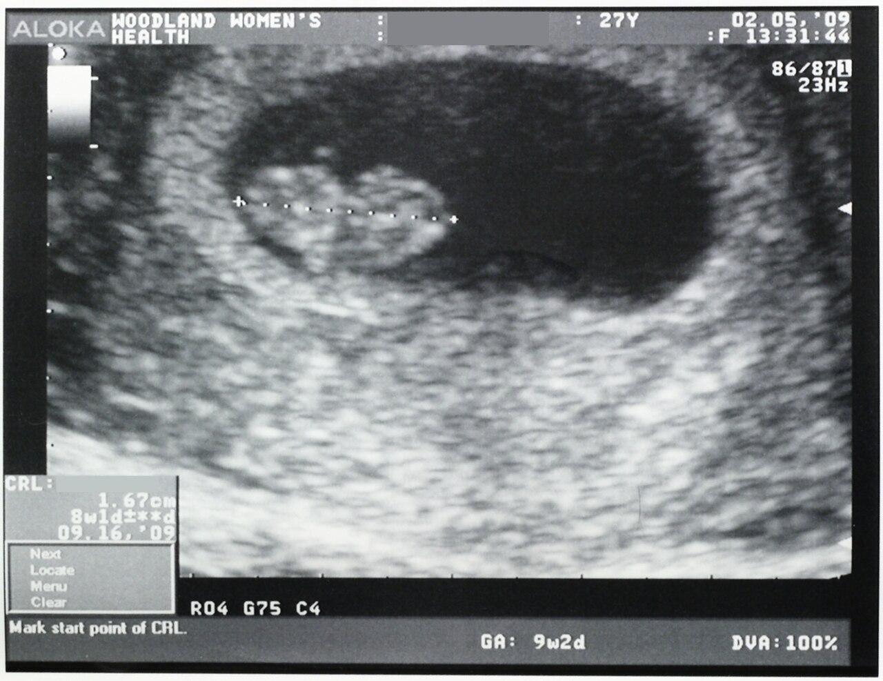
Presumptive, probable and positive signs
Pregnancy is confirmed in tiers, and the tiers exist because each lower tier has impostors. Presumptive signs are what the pregnant person notices: amenorrhoea, nausea and vomiting, breast tenderness and darkening of the areolae, urinary frequency, fatigue, and quickening — the first perceived fetal movement, typically at sixteen to twenty weeks in a first pregnancy and as early as fourteen to sixteen weeks in a person who has been pregnant before. Every one of these has another explanation. Amenorrhoea follows stress, thyroid disease, extreme exercise and perimenopause; nausea follows gastroenteritis; quickening can be intestinal gas. Presumptive signs are subjective, and they belong in the history, not in the diagnosis.
Probable signs are what the examiner detects. Goodell sign is softening of the cervix at about six weeks. Chadwick sign is the blue-violet discolouration of the cervix and vaginal wall from venous congestion, at six to eight weeks. Hegar sign is softening of the lower uterine segment, palpable at six to twelve weeks. Ballottement, a positive pregnancy test on urine or serum, uterine enlargement and Braxton Hicks contractions also sit here. A serum human chorionic gonadotrophin level is still only probable: hydatidiform mole and some germ-cell tumours secrete the same hormone. Early in a normal intrauterine pregnancy, human chorionic gonadotrophin roughly doubles every forty-eight to seventy-two hours, peaks around eight to ten weeks, then falls and plateaus — a doubling pattern that flattens is a reason to look for ectopic implantation or early pregnancy loss.
Only three findings are positive signs, and all three come directly from the fetus: visualisation of the fetus by ultrasound (a gestational sac from about five weeks by transvaginal scan, cardiac activity from about six weeks), auscultation of the fetal heart (Doppler at ten to twelve weeks, fetoscope at eighteen to twenty weeks), and fetal movement palpated by a trained examiner. Fetal heart rate baseline is 110 to 160 beats per minute. If what you hear matches the pregnant person’s radial pulse, you are listening to the uterine souffle, not the fetus — palpate the radial pulse while you auscultate, every time.
Dating, gravidity and parity
Dating drives everything downstream: when to screen, when to worry about growth, when a birth is preterm, when an induction is safe. Naegele’s rule estimates the date of birth from the first day of the last menstrual period: subtract three months, add seven days, add one year. A last menstrual period beginning 10 September 2025 gives 10 June 2025, then 17 June 2025, then an estimated date of birth of 17 June 2026 — the seven days are the step most often dropped. The rule assumes a regular twenty-eight-day cycle with ovulation on day fourteen, so it drifts in anyone with irregular or long cycles. A first-trimester crown-rump length by ultrasound is accurate to within five to seven days and supersedes menstrual dating when the two disagree by more than that margin; accuracy degrades to one to two weeks in the second trimester and two to three weeks in the third, which is why an early scan is worth more than a late one.
Obstetric history is recorded as GTPAL. Gravida is the total number of pregnancies including the current one, counting a multiple gestation once. Term births are those at thirty-seven weeks zero days and beyond; Preterm births are from twenty weeks zero days through thirty-six weeks six days; Abortions covers all losses, spontaneous or induced, before twenty weeks; Living is the number of children currently alive. A person pregnant now, with one birth at thirty-nine weeks, one set of twins born at thirty-four weeks, and one miscarriage at nine weeks, with all three children alive, is G4 T1 P1 A1 L3. Note that twins counted once in gravidity and once in parity still contribute two living children — parity counts births, not babies.
What the first visit actually contains
The first prenatal visit is the longest of the pregnancy and is ideally completed before ten weeks. It gathers a menstrual, obstetric, medical, surgical, medication, substance, genetic and psychosocial history; establishes dating; performs a full physical including a pelvic examination; and draws a baseline laboratory panel. That panel routinely includes blood type and Rh with antibody screen, complete blood count, rubella immunity, varicella immunity, hepatitis B surface antigen, syphilis serology, human immunodeficiency virus (opt-out testing), urinalysis with culture, and chlamydia and gonorrhoea screening. Hepatitis C screening is now recommended in every pregnancy. Tuberculosis, haemoglobinopathy and thyroid testing are added by risk.
Two of those results carry immediate action. Asymptomatic bacteriuria — a positive urine culture with no symptoms — is treated in pregnancy even though it would be left alone outside pregnancy, because the dilated, progesterone-relaxed ureters let bacteria ascend, and untreated bacteriuria progresses to pyelonephritis in a substantial minority and is associated with preterm birth and low birth weight. A negative rubella titre means the person is susceptible; the vaccine is a live virus and is contraindicated during pregnancy, so the intervention is to document susceptibility, counsel on avoiding exposure, and give the vaccine in the immediate postpartum period, ideally before hospital discharge.
Only ultrasound visualisation, auscultated fetal heart tones and examiner-palpated fetal movement confirm a pregnancy; everything else — including a positive hormone test — is presumptive or probable and must be dated by first-trimester ultrasound to be clinically useful.
Section 2Maternal Physiological Adaptation, System by System
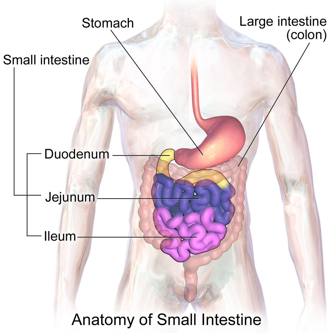
Cardiovascular and haematological adaptation
Total blood volume rises forty to fifty per cent by about thirty-two weeks. The rise is disproportionate: plasma volume increases roughly fifty per cent while red cell mass increases only twenty to thirty per cent. Dilution follows, and the haemoglobin and haematocrit fall — the physiologic anaemia of pregnancy. This is a laboratory appearance, not a disease; oxygen-carrying capacity has actually increased in absolute terms. True anaemia is diagnosed at a haemoglobin below 11 g/dL in the first and third trimesters and below 10.5 g/dL in the second. The extra volume is not waste. It perfuses the placental bed, it dissipates the heat of fetal metabolism, and it is the reserve that lets a person lose 500 mL at a vaginal birth without becoming unstable.
Cardiac output rises thirty to fifty per cent, peaking around twenty-five to thirty weeks, driven early by stroke volume and later by heart rate, which climbs ten to twenty beats per minute above baseline. Systemic vascular resistance falls under the influence of progesterone, relaxin, nitric oxide and prostacyclin, so blood pressure — particularly diastolic — drops five to fifteen millimetres of mercury, reaching its nadir around twenty-four to twenty-eight weeks before returning toward baseline at term. A blood pressure that rises in the second trimester is therefore moving against the physiology and deserves attention. The heart is displaced upward and to the left, the point of maximal impulse moves laterally, and a soft systolic ejection murmur appears in most pregnancies; a diastolic murmur never becomes normal in pregnancy.
From about twenty weeks, lying supine allows the uterus to compress the inferior vena cava, cutting venous return and cardiac output. Supine hypotensive syndrome presents as dizziness, pallor, clamminess, nausea and hypotension, and it compromises uteroplacental perfusion before the pregnant person feels much at all. The intervention is immediate and mechanical: turn the patient to the left lateral position, or wedge the right hip so the uterus falls off the vessels. Pregnancy is also a hypercoagulable state — fibrinogen rises by about fifty per cent, clotting factors VII, VIII, IX and X rise, and fibrinolysis is suppressed — which protects against haemorrhage at birth and raises venous thromboembolism risk roughly four to five-fold. Unilateral calf pain and swelling in a pregnant patient is a thrombosis until proven otherwise, not a muscle strain.
Respiratory, renal and gastrointestinal adaptation
Progesterone acts directly on the respiratory centre, so tidal volume rises thirty to fifty per cent while respiratory rate barely changes. Minute ventilation rises with it and produces a compensated respiratory alkalosis: arterial carbon dioxide falls to about twenty-seven to thirty-two millimetres of mercury, and the kidney excretes bicarbonate to hold pH near 7.40 to 7.45. That gradient drives carbon dioxide from fetus to parent. Meanwhile the growing uterus elevates the diaphragm about four centimetres, the subcostal angle widens, functional residual capacity falls about twenty per cent, and oxygen consumption rises twenty to thirty per cent. The practical consequence is dangerous: a pregnant patient desaturates far faster during apnoea than a non-pregnant one. Physiologic dyspnoea of pregnancy is mild, gradual and does not disturb sleep or accompany chest pain; dyspnoea that is sudden, positional at night, or paired with chest pain, cough or tachycardia is not physiologic.
Renal plasma flow and glomerular filtration rate rise forty to fifty per cent by the end of the first trimester. Serum creatinine and blood urea nitrogen therefore fall: a normal pregnant creatinine is about 0.4 to 0.8 mg/dL, so a creatinine of 1.0 mg/dL — unremarkable in a non-pregnant adult — signals renal impairment in pregnancy. Progesterone-mediated smooth muscle relaxation and mechanical compression by the dextrorotated uterus dilate the collecting system, right side more than left, producing the physiologic hydronephrosis that makes ascending infection easy. Glycosuria is common because filtered glucose exceeds tubular reabsorption; trace glycosuria is expected, but persistent or heavy glycosuria still prompts glucose evaluation. Proteinuria is not in the same category: a dipstick of 1+ or more after twenty weeks must be measured properly and interpreted alongside blood pressure.
In the gastrointestinal tract, progesterone relaxes the lower oesophageal sphincter and slows transit. Reflux, constipation, bloating and haemorrhoids follow, and gallbladder stasis raises the risk of cholelithiasis. Nausea and vomiting affect up to seventy per cent of pregnancies and usually resolve by sixteen to twenty weeks; hyperemesis gravidarum is different in kind, defined by persistent vomiting with weight loss of more than five per cent of pre-pregnancy weight, dehydration, ketonuria and electrolyte disturbance, and it requires intravenous rehydration rather than dietary advice. Gum hypertrophy and bleeding (epulis gravidarum) are oestrogen effects; dental care is safe and should be encouraged, not deferred.
Endocrine, musculoskeletal and integumentary adaptation
The placenta is an endocrine organ. Human placental lactogen, along with placental growth hormone, progesterone and cortisol, is deliberately diabetogenic: it antagonises insulin so that maternal glucose stays available for placental transfer. Insulin requirements consequently fall in early pregnancy — a person with pre-existing type 1 diabetes is at real risk of hypoglycaemia in the first trimester — and then rise steeply in the second half. Thyroid-binding globulin rises under oestrogen so total thyroxine rises, while free thyroxine stays near normal; the thyroid enlarges modestly but a frankly nodular or markedly enlarged gland is not a normal finding. Human chorionic gonadotrophin has weak thyroid-stimulating activity, which is one reason severe early vomiting sometimes travels with transient biochemical hyperthyroidism.
Relaxin and progesterone loosen ligaments, including the sacroiliac joints and pubic symphysis. Combined with the forward shift of the centre of gravity, this produces the lordosis, waddling gait, low back pain and increased fall risk of late pregnancy, and it explains diastasis recti, the midline separation of the rectus abdominis. Fluid retention in confined spaces produces carpal tunnel symptoms and nasal stuffiness with epistaxis. Skin changes are melanocyte-stimulating: linea nigra down the midline, melasma (the “mask of pregnancy”) over the face, darkened areolae, and striae gravidarum. Palmar erythema and spider angiomas reflect the oestrogen-driven rise in cutaneous blood flow. Most of these fade postpartum, though striae persist as silvery lines and melasma improves with sun protection.
| System change | Expected finding | Not normal — investigate |
|---|---|---|
| Plasma volume up ~50 per cent, red cell mass up ~25 per cent | Haemoglobin 11–12 g/dL; mild dilutional fall | Haemoglobin below 11 g/dL (first or third trimester) or below 10.5 g/dL (second) |
| Systemic vascular resistance falls | Diastolic pressure drops 5–15 mm Hg, nadir 24–28 weeks | Blood pressure 140/90 mm Hg or higher on two readings four hours apart after 20 weeks |
| Cardiac output up 30–50 per cent; heart displaced left | Soft systolic ejection murmur; heart rate up 10–20 beats/min | Any diastolic murmur; resting heart rate above 110; chest pain |
| Tidal volume up 30–50 per cent; diaphragm rises 4 cm | Mild gradual dyspnoea; carbon dioxide 27–32 mm Hg | Sudden dyspnoea, orthopnoea, haemoptysis, oxygen saturation below 95 per cent |
| Glomerular filtration rate up 40–50 per cent | Creatinine 0.4–0.8 mg/dL; trace glycosuria | Creatinine 1.0 mg/dL or higher; proteinuria 1+ or more after 20 weeks |
| Ureteral dilation, urinary stasis | Urinary frequency without dysuria | Positive urine culture (treat even without symptoms); flank pain with fever |
| Hypercoagulability; fibrinogen up ~50 per cent | White cell count 5,000–15,000/mm³; dependent oedema of feet and ankles | Unilateral calf pain or swelling; sudden facial or hand oedema |
| Insulin antagonism by human placental lactogen | Rising insulin needs after 20 weeks | Fasting glucose 92 mg/dL or higher; abnormal glucose challenge at 24–28 weeks |
Nearly every antepartum adaptation has a pathological look-alike, so the assessment question is never “is this abnormal?” but “is this the expected magnitude, in the expected direction, at the expected gestational age?”
Section 3The Rhythm of Prenatal Care: Visits, Screening and Fetal Surveillance
The visit schedule and the four measurements taken every time
For an uncomplicated pregnancy the traditional schedule is every four weeks until twenty-eight weeks, every two weeks from twenty-eight to thirty-six weeks, and weekly from thirty-six weeks until birth — about twelve to fourteen visits. The spacing is not arbitrary: it tightens exactly when hypertensive disease, growth restriction and preterm labour become most likely. Reduced-visit and group prenatal care models are safe in low-risk pregnancies and often improve attendance and satisfaction; what matters is not the number of visits but that the surveillance items happen in their windows.
Four things are checked at essentially every return visit: blood pressure, weight, urine dipstick for protein and glucose, and fetal heart rate. From about eighteen or twenty weeks a fifth is added, fundal height: measured in centimetres from the upper border of the symphysis pubis over the curve of the abdomen to the top of the fundus, with the patient supine and the bladder empty. Landmarks worth memorising are the fundus at the symphysis at about twelve weeks and at the umbilicus at about twenty weeks. Between eighteen and thirty-six weeks the measurement in centimetres approximates the gestational age in weeks within about two centimetres. A discrepancy greater than two to three centimetres prompts an ultrasound: too small suggests growth restriction, oligohydramnios or a dating error; too large suggests multiple gestation, polyhydramnios, macrosomia, fibroids or, again, a dating error.
Leopold manoeuvres — four systematic palpations to determine fetal lie, presentation, position and engagement — are performed from about the third trimester, and they tell you where to place the Doppler. Fetal heart tones are best heard through the fetal back: in a cephalic presentation, below the umbilicus on the side where the back lies; in a breech presentation, at or above the umbilicus. Always count the pregnant person’s pulse simultaneously so that a maternal tachycardia is not mistaken for a fetal bradycardia.
Screening, trimester by trimester
Genetic screening is offered, not imposed, and the distinction between screening and diagnosis must be explicit in the teaching. Screening tests estimate risk; diagnostic tests give a karyotype. Cell-free DNA (non-invasive prenatal screening) analyses placental DNA fragments in maternal blood from ten weeks and has very high sensitivity for trisomy 21, but a “positive” result is still a probability and must be confirmed by chorionic villus sampling at ten to thirteen weeks or amniocentesis at fifteen to twenty weeks. A patient who hears “positive” and believes it is a diagnosis has been failed by the counselling, not by the test.
Maternal serum alpha-fetoprotein, drawn as part of the quad screen at fifteen to twenty-two weeks, is a useful mental model for how these markers behave. A high value suggests an open neural tube defect, abdominal wall defect, multiple gestation or, most commonly, inaccurate dating. A low value is associated with trisomy 21 and trisomy 18. The single most frequent cause of an abnormal alpha-fetoprotein is a wrong gestational age, which is one more argument for early dating.
| Window | Screen or intervention | What the nurse teaches or watches |
|---|---|---|
| First visit, ideally before 10 weeks | Blood type, Rh, antibody screen, complete blood count, rubella and varicella immunity, hepatitis B and C, syphilis, human immunodeficiency virus, urine culture, chlamydia and gonorrhoea | Treat asymptomatic bacteriuria; defer live vaccines to postpartum; opt-out human immunodeficiency virus testing |
| From 10 weeks | Cell-free DNA screening (optional) | Screening, not diagnosis; a positive result requires confirmatory testing |
| 10–13 weeks | Chorionic villus sampling (diagnostic, optional) | Rh-negative patients need Rh immune globulin after the procedure |
| 11–13 weeks 6 days | First-trimester combined screen: nuchal translucency plus serum markers | Requires precise dating; results are risk ratios |
| 12–28 weeks (start before 16 if possible) | Low-dose aspirin 81 mg daily for those at elevated pre-eclampsia risk | Continue daily until birth; explain it is prophylaxis, not treatment |
| 15–22 weeks | Quad screen; amniocentesis from 15 weeks if diagnostic testing is chosen | Post-amniocentesis: report leaking fluid, bleeding, fever, contractions |
| 18–22 weeks | Anatomy ultrasound | Full bladder may be requested; fetal sex disclosure is the family’s choice |
| 24–28 weeks | Gestational diabetes screening; repeat complete blood count and antibody screen | See the glucose criteria below |
| 28 weeks | Rh immune globulin 300 micrograms intramuscularly if Rh-negative and antibody-screen negative | Repeat within 72 hours of birth if the newborn is Rh-positive; also give after bleeding, trauma, amniocentesis, ectopic or loss |
| 27–36 weeks | Tdap in every pregnancy; maternal respiratory syncytial virus vaccine at 32–36 weeks in season | Tdap is repeated each pregnancy to transfer pertussis antibody to the newborn |
| 36 weeks 0 days – 37 weeks 6 days | Rectovaginal group B streptococcus culture | A positive culture means intrapartum intravenous antibiotics, not treatment now |
| Any trimester, seasonally | Inactivated influenza vaccine; COVID-19 vaccine per current guidance | Inactivated vaccines are safe in pregnancy; live attenuated influenza vaccine is not |
Gestational diabetes screening at twenty-four to twenty-eight weeks uses one of two approaches. In the two-step method, a non-fasting 50 g glucose challenge is followed by a plasma glucose at one hour; a value at or above 130 to 140 mg/dL (institutional threshold) leads to a fasting 100 g three-hour tolerance test, and gestational diabetes is diagnosed when two or more values are abnormal: fasting 95 mg/dL or higher, one hour 180 or higher, two hours 155 or higher, three hours 140 or higher. In the one-step method, a fasting 75 g two-hour test is diagnostic when any one value is abnormal: fasting 92 or higher, one hour 180 or higher, two hours 153 or higher. Patients with risk factors — prior gestational diabetes, obesity, a previous macrosomic newborn, polycystic ovary syndrome, strong family history — are screened at the first visit as well as at twenty-four to twenty-eight weeks.
Fetal surveillance in the third trimester
The simplest and cheapest surveillance tool belongs to the pregnant person. Fetal movement counting is taught from about twenty-eight weeks: lie on the left side after a meal at roughly the same time each day and count distinct movements; ten movements within two hours is reassuring. Teach the pattern, not just the number — a fetus has sleep cycles of twenty to forty minutes, so a quiet half-hour is not an alarm, but a definite decrease from that fetus’s own established pattern is a reason to be assessed the same day. Never advise a patient to “wait until tomorrow and see”.
The non-stress test records fetal heart rate and movement for twenty minutes. It is reactive at thirty-two weeks or more when there are two accelerations of at least fifteen beats per minute above baseline lasting at least fifteen seconds within twenty minutes; before thirty-two weeks the criterion is ten beats for ten seconds, because an immature autonomic nervous system produces smaller accelerations. A non-reactive test is most often a sleeping fetus, so the test is extended to forty minutes and vibroacoustic stimulation may be used before escalating. The biophysical profile adds ultrasound assessment of fetal breathing movements, gross body movement, tone and amniotic fluid volume to the non-stress test; each of the five components scores 0 or 2. A score of 8 to 10 is reassuring, 6 is equivocal and warrants repeat or delivery depending on gestation, and 4 or less indicates significant risk. Amniotic fluid volume is the component that reflects chronic placental function, because a hypoxic fetus shunts blood from the kidneys and makes less urine.
Every antepartum screen has a gestational window, and the window is the point: cell-free DNA from ten weeks, anatomy scan at eighteen to twenty-two, glucose at twenty-four to twenty-eight, Rh immune globulin at twenty-eight, Tdap at twenty-seven to thirty-six, and group B streptococcus culture at thirty-six weeks zero days to thirty-seven weeks six days.
Section 4Nutrition, Weight, Discomforts and Medication Safety
Weight gain and energy needs
Recommended gestational weight gain is set by pre-pregnancy body mass index, not by a single number, because the risks at either extreme differ. Too little gain is associated with growth restriction, preterm birth and low birth weight; too much is associated with macrosomia, caesarean birth, gestational diabetes, hypertensive disease and postpartum weight retention. Discuss weight in terms of fetal growth and health, never as a moral matter, and be aware that weight conversations are a common reason patients disengage from prenatal care.
| Pre-pregnancy body mass index | Total gain, singleton | Rate in 2nd and 3rd trimesters | Total gain, twins |
|---|---|---|---|
| Underweight, below 18.5 | 12.5–18 kg (28–40 lb) | ~0.5 kg (1 lb) per week | Individualised |
| Normal, 18.5–24.9 | 11.5–16 kg (25–35 lb) | ~0.4–0.5 kg (1 lb) per week | 17–25 kg (37–54 lb) |
| Overweight, 25.0–29.9 | 7–11.5 kg (15–25 lb) | ~0.3 kg (0.6 lb) per week | 14–23 kg (31–50 lb) |
| Obese, 30.0 or higher | 5–9 kg (11–20 lb) | ~0.2 kg (0.5 lb) per week | 11–19 kg (25–42 lb) |
Energy needs do not rise meaningfully in the first trimester. They rise by about 340 kcal per day in the second trimester and about 452 kcal per day in the third, above the pre-pregnancy requirement — roughly a sandwich and a glass of milk, not a second dinner. Protein requirement rises to about 71 g per day. Fluid intake of about 2.3 to 3 litres daily supports the expanded plasma volume and reduces constipation. “Eating for two” is a phrase to retire in teaching; the fetus is small and the surplus required is modest.
The micronutrients that change outcomes
Folic acid is the single clearest example of prevention in obstetrics, and it carries a dosing trap. Routine supplementation is 400 to 800 micrograms daily, started at least one month before conception and continued through the first trimester, because the neural tube closes by day twenty-eight — often before a pregnancy is recognised. A person with a previous pregnancy affected by a neural tube defect is prescribed 4 mg (4000 micrograms) daily, a ten-fold higher dose, beginning one to three months before conception and continuing through the first trimester. Certain anticonvulsants and other folate antagonists also raise the requirement. The trap is the unit: 4 mg is not 400 micrograms, and the high dose is never achieved by taking more prenatal vitamins, because that would deliver a toxic amount of preformed vitamin A.
Iron requirement rises to 27 mg elemental iron daily, and therapeutic dosing for established iron-deficiency anaemia is higher, typically 60 to 200 mg of elemental iron per day in divided doses. Teach absorption: take on an empty stomach or with a vitamin C source such as orange juice; do not take with milk, calcium supplements, antacids, tea or coffee; expect dark stools, and manage constipation with fluid, fibre and a stool softener rather than stopping the iron. Calcium is 1000 mg daily (1300 mg if the pregnant person is eighteen or younger, whose own skeleton is still mineralising). Iodine is 220 micrograms daily and matters for fetal neurodevelopment. Vitamin D deficiency is common and is screened by risk. Preformed vitamin A (retinol) above about 10,000 international units daily is teratogenic; beta-carotene from food is not.
Food safety teaching is concrete. Avoid unpasteurised dairy and juices, soft cheeses made from unpasteurised milk, deli meats and refrigerated pâtés unless heated until steaming, raw sprouts, and undercooked meat, eggs and fish — the target is Listeria monocytogenes, which crosses the placenta and causes loss, preterm birth and neonatal sepsis. Avoid high-mercury fish (shark, swordfish, king mackerel, tilefish, bigeye tuna) but do eat eight to twelve ounces weekly of low-mercury fish, because omega-3 fatty acids support fetal brain and retinal development. Limit caffeine to under 200 mg daily — about one 12-ounce brewed coffee. Avoid changing cat litter and wear gloves for gardening because of Toxoplasma gondii. There is no known safe amount or safe timing of alcohol.
Discomforts and self-care
Most antepartum complaints are managed with positioning, timing and food before they are managed with drugs. Nausea responds to small frequent dry carbohydrate meals, avoiding an empty stomach, ginger, vitamin B6 (pyridoxine) 10 to 25 mg three or four times daily, and doxylamine added if needed. Heartburn responds to small meals, remaining upright for an hour after eating, and avoiding late meals, fatty and spicy food; sodium bicarbonate is avoided because of the sodium load. Constipation responds to fluid, fibre and walking; docusate is preferred to stimulant laxatives, and mineral oil is avoided because it blocks fat-soluble vitamin absorption. Backache responds to pelvic tilt exercises, supportive low-heeled shoes, side-lying with a pillow between the knees and squatting rather than bending. Varicosities and dependent oedema respond to elevation, support hose applied before rising, and avoiding crossed legs and constrictive clothing. Leg cramps respond to dorsiflexing the foot with the knee extended. Supine hypotension responds to the left lateral position, always.
Medications, substances and immunisation
The old letter categories (A, B, C, D, X) have been replaced by narrative Pregnancy and Lactation Labeling Rule summaries describing risk, clinical considerations and data. The teaching point survives the change: no medication, prescription or over-the-counter or herbal, is taken in pregnancy without checking. Known teratogens include isotretinoin, warfarin, angiotensin-converting enzyme inhibitors and angiotensin receptor blockers, methotrexate, valproic acid, thalidomide, lithium (Ebstein anomaly), tetracyclines, live vaccines and high-dose retinol. Non-steroidal anti-inflammatory drugs are avoided from twenty weeks because of oligohydramnios and premature ductus arteriosus constriction; paracetamol/acetaminophen is the usual analgesic of choice at the lowest effective dose. Antidepressants are frequently continued because untreated perinatal depression carries its own substantial risk — that decision belongs to a shared conversation, not to a blanket rule.
Substance use is screened with a validated tool at the first visit and at least once each trimester, using the same non-judgemental, universal script for every patient. Opioid use disorder in pregnancy is treated with buprenorphine or methadone; medically supervised withdrawal is not recommended because relapse risk and fetal risk both rise. Smoking cessation support at any gestation improves birth weight. Tobacco, alcohol and stimulant use are health conditions, and the language used about them determines whether a patient tells you the truth at the next visit.
Routine folic acid is 400 to 800 micrograms daily, but a prior neural-tube-defect-affected pregnancy requires 4 mg daily — a ten-fold difference that is never met by taking extra prenatal vitamins, because that would deliver a teratogenic amount of preformed vitamin A.
Section 5Danger Signs, Escalation and the Sequences That Save Lives

Teaching the danger signs and escalating what you find
Between visits, the family is the monitoring system, so danger-sign teaching is a clinical intervention, not a courtesy. Teach these as “call us today, do not wait for your appointment”: any vaginal bleeding; a gush or steady trickle of fluid; a severe or persistent headache that does not respond to paracetamol; visual changes such as blurring, spots or flashes; epigastric or right upper quadrant pain; sudden swelling of the face or hands; persistent vomiting with inability to keep fluids down; fever of 38 °C (100.4 °F) or higher; painful urination; regular contractions before thirty-seven weeks; a definite decrease in fetal movement; and any abdominal trauma, including a fall or a motor vehicle crash even at low speed. Give the teaching in writing and in the family’s language, and confirm understanding by asking what they would do rather than asking whether they understood.
Three of these carry particular weight. Headache, visual change, epigastric pain and sudden facial oedema cluster as the warning signature of pre-eclampsia with severe features, which is diagnosed by blood pressure at or above 140/90 mm Hg on two occasions four hours apart after twenty weeks with proteinuria (300 mg per 24 hours, a protein-to-creatinine ratio of 0.3 or more, or dipstick 2+), and which becomes severe at 160/110 mm Hg or with platelets below 100,000/mm³, creatinine above 1.1 mg/dL, transaminases at twice normal, pulmonary oedema, or new neurological symptoms. Any abdominal trauma in an Rh-negative patient requires Rh immune globulin and a minimum of four hours of electronic fetal monitoring, because placental abruption may declare itself hours after the impact. And a decrease in fetal movement is assessed today, not tomorrow.
Escalate with SBAR, and put the ask in the recommendation. Situation: “Ms Oyelaran, 34 weeks, in triage, blood pressure 168 over 112 on two readings fifteen minutes apart.” Background: “Gravida 2 para 1, no chronic hypertension, on aspirin 81 mg since 13 weeks; she has a frontal headache and sees spots.” Assessment: “This looks like pre-eclampsia with severe features; fetal heart rate is 145 with moderate variability, reflexes 3+, urine dipstick 3+ protein.” Recommendation: “I have her in left lateral with a large-bore line and I need an order now for magnesium sulfate seizure prophylaxis and an antihypertensive, plus severe-range preeclampsia labs. Can you come to the bedside?” Say the number, name the concern, request the specific order, and state a timeframe.
The emergencies where the sequence itself is the treatment
In obstetrics the correct first action is frequently not the intuitive one, and doing the intuitive thing first costs time that a fetus does not have. These sequences are introduced here and developed in the intrapartum and postpartum chapters; learn the first action now.
| Emergency | Recognition | What you do FIRST | Then, and never |
|---|---|---|---|
| Postpartum haemorrhage | Blood loss 1000 mL or more, or bleeding with signs of hypovolaemia; soft “boggy” fundus, often displaced upward and to the right by a full bladder | Massage the fundus — one hand supporting the lower uterine segment, the other massaging the fundus. Uterine atony causes the great majority of cases and massage treats it in seconds | Then empty the bladder, call for help, start oxytocin (10–40 units in 500–1000 mL crystalloid), add second-line uterotonics and tranexamic acid within 3 hours. Never give methylergonovine in hypertension, never carboprost in asthma, never start the intravenous line before you have massaged |
| Magnesium sulfate toxicity | The therapeutic serum magnesium range for seizure prophylaxis is 4–7 mEq/L (about 5–8 mg/dL). Above it the signs appear in order: flushing, warmth, lethargy and slurred speech; then loss of deep tendon reflexes (about 9–12 mg/dL); then respiratory rate below 12 (about 12–18 mg/dL); then cardiac arrest (about 24–30 mg/dL). Reflexes disappear before breathing slows, which is why the patellar reflex is the earliest bedside warning. Urine output below 30 mL/hour is not a stage of toxicity but a cause of it — magnesium is cleared renally, so oliguria lets the level climb | Stop the infusion immediately, then assess airway and support ventilation | Then give the antidote, calcium gluconate 1 g (10 mL of a 10 per cent solution) intravenously over at least 3 minutes, draw a magnesium level and notify the provider. Never continue the infusion “until the level comes back”. Hold the infusion if reflexes are absent, respirations are under 12, or urine output is under 30 mL/hour |
| Umbilical cord prolapse | Sudden prolonged fetal deceleration or bradycardia, usually right after membranes rupture; a pulsating cord felt or seen at the introitus | Relieve the pressure of the presenting part on the cord: insert a gloved hand and lift the presenting part off the cord, and keep it there until birth | Then place the patient in knee-chest or steep Trendelenburg or modified Sims, give oxygen at 10 L/min by non-rebreather, call for immediate caesarean, and cover an exposed cord with sterile saline-moistened gauze. Never push the cord back in and never leave the bedside to call — use the call light or send someone |
| Shoulder dystocia | The head delivers and retracts against the perineum (“turtle sign”); the shoulders do not follow with the next contraction | McRoberts manoeuvre — two people hyperflex the thighs sharply onto the abdomen, which rotates the symphysis and flattens the sacral promontory | Then add suprapubic pressure, then internal rotational manoeuvres and delivery of the posterior arm; call for help and assign someone to announce elapsed time. Never apply fundal pressure — it drives the shoulder harder into the symphysis and risks uterine rupture |
| Uterine rupture | Abnormal fetal heart rate — usually a prolonged deceleration or bradycardia — is often the earliest sign; then sudden sharp “tearing” pain, loss of fetal station, palpable fetal parts through the abdominal wall, cessation of contractions, and signs of hypovolaemic shock | Prepare for immediate surgical birth: call for help, stop oxytocin, give oxygen, position for uteroplacental perfusion, and start volume resuscitation through large-bore access | Then notify the surgical and neonatal teams and initiate the massive transfusion protocol. Never continue oxytocin, and never treat a prolonged deceleration after a previous caesarean as merely positional |
Psychosocial adaptation, safety and delegation
Psychological work runs alongside the physiological. Classically the first trimester is ambivalence — even a deeply wanted pregnancy provokes mixed feelings, and normalising that is genuinely therapeutic; the second trimester brings acceptance and the fantasy of the baby as quickening makes the fetus real; the third brings preparation, nesting, and the anticipatory fear of labour. The developmental tasks are to seek safe passage for self and child, to secure acceptance of the child by significant others, to bind in to the unknown child, and to learn to give of oneself. Partners adapt too, and some experience couvade syndrome — nausea, weight gain and backache mirroring the pregnancy. Family-centred care means the pregnant person defines who the family is, and that definition is honoured on the chart and at the bedside.
Screen for intimate partner violence at the first visit and at least once per trimester, always with the partner out of the room and never using a family member as interpreter. Pregnancy is a period of elevated risk, and abuse in pregnancy is associated with abruption, low birth weight, preterm birth and homicide. Screen for perinatal depression and anxiety with a validated instrument such as the Edinburgh Postnatal Depression Scale at least once antenatally and again postpartum, and act on any positive response to the self-harm item immediately. Also assess social determinants directly: food security, housing, transport to appointments, interpersonal racism in care, and immigration-related fear of accessing services all shape outcomes as powerfully as any laboratory result.
Delegation in the antepartum setting follows the rule that runs through this whole book. Assessment, teaching, evaluation, clinical judgement and the unstable patient stay with the registered nurse. Unlicensed assistive personnel may take vital signs on stable patients, obtain weights and clean-catch urine specimens, assist with positioning and ambulation, record intake and output, and provide comfort measures — and they must be told exactly what to report and by when (“tell me immediately if the systolic is 140 or higher”). A licensed practical or vocational nurse may reinforce teaching the registered nurse has already given and administer many medications within scope. Titrating a magnesium sulfate infusion, performing the admission assessment, interpreting a fetal heart tracing, teaching danger signs for the first time and caring for the patient with severe-range blood pressures are not delegated.
In every obstetric emergency the first action is the mechanical one at the bedside — massage the fundus, lift the presenting part off the cord, hyperflex the thighs, stop the magnesium — and the registered nurse never delegates assessment, first teaching, evaluation or the unstable patient.
The chapter in one breath
The antepartum period is a physiological renovation that the nurse must be able to read. Pregnancy is confirmed only by ultrasound visualisation, auscultated fetal heart tones or examiner-palpated movement, dated most accurately by first-trimester crown-rump length, and recorded in GTPAL. Then almost every system changes in a way that mimics disease: plasma volume outruns red cell mass to produce physiologic anaemia; systemic vascular resistance falls so diastolic pressure dips to a nadir at twenty-four to twenty-eight weeks; tidal volume and glomerular filtration rate rise while carbon dioxide and creatinine fall; progesterone slows the gut and dilates the ureters; the placenta manufactures insulin resistance. Prenatal care converts those adaptations into surveillance on a schedule — blood pressure, weight, urine, fetal heart rate and fundal height at every visit, with cell-free DNA from ten weeks, the anatomy scan at eighteen to twenty-two, glucose at twenty-four to twenty-eight, Rh immune globulin at twenty-eight, Tdap at twenty-seven to thirty-six and group B streptococcus culture at thirty-six to thirty-seven weeks. Teaching closes the gap between visits: gain by body mass index, 340 then 452 extra kilocalories, folic acid 400 to 800 micrograms — or 4 mg after a prior neural tube defect — and a written list of danger signs. And when the emergency comes, the hands move first: massage, lift, hyperflex, stop the pump.
Words worth knowing
| Term | What it means |
|---|---|
| Antepartum | The period from conception to the onset of labour; also called the prenatal period. |
| Naegele’s rule | Estimated date of birth from the first day of the last menstrual period: minus three months, plus seven days, plus one year. |
| GTPAL | Gravida, Term, Preterm, Abortions, Living — the standard shorthand for obstetric history; parity counts births, not babies. |
| Quickening | The pregnant person’s first perception of fetal movement, usually 16–20 weeks in a first pregnancy, 14–16 weeks thereafter; a presumptive sign. |
| Goodell, Chadwick and Hegar signs | Probable signs: cervical softening, blue-violet vaginal and cervical discolouration, and softening of the lower uterine segment. |
| Physiologic anaemia of pregnancy | Apparent fall in haemoglobin because plasma volume rises ~50 per cent while red cell mass rises only ~25 per cent; true anaemia is below 11 g/dL (below 10.5 in the second trimester). |
| Supine hypotensive syndrome | Vena caval compression by the uterus when supine after ~20 weeks, reducing cardiac output and uteroplacental perfusion; treated by left lateral position or right hip wedge. |
| Human placental lactogen | Placental hormone that antagonises insulin so glucose crosses to the fetus; the basis of gestational insulin resistance. |
| Fundal height | Symphysis-to-fundus measurement in centimetres, approximating gestational age in weeks from about 18–36 weeks within 2 cm. |
| Leopold manoeuvres | Four systematic abdominal palpations determining fetal lie, presentation, position and engagement, and guiding Doppler placement. |
| Cell-free DNA screening | Analysis of placental DNA fragments in maternal blood from 10 weeks; a screening test that estimates risk and always requires diagnostic confirmation. |
| Non-stress test | Twenty-minute fetal heart tracing; reactive at 32 weeks or more with two accelerations of 15 beats per minute for 15 seconds (10 by 10 before 32 weeks). |
| Biophysical profile | Non-stress test plus ultrasound assessment of fetal breathing, movement, tone and amniotic fluid; each scores 0 or 2, with 8–10 reassuring and 4 or less concerning. |
| Rh immune globulin | 300 micrograms intramuscularly at 28 weeks and within 72 hours of birth of an Rh-positive newborn, plus after bleeding, trauma or invasive procedures, to prevent alloimmunisation. |
| Asymptomatic bacteriuria | A positive urine culture without symptoms; always treated in pregnancy because of the risk of pyelonephritis and preterm birth. |
| Hyperemesis gravidarum | Persistent pregnancy vomiting with more than five per cent weight loss, dehydration, ketonuria and electrolyte disturbance; treated with intravenous rehydration rather than dietary advice. |
| McRoberts manoeuvre | Sharp hyperflexion of the thighs onto the abdomen, the first action in shoulder dystocia; suprapubic — never fundal — pressure is added. |
| Couvade syndrome | Physical symptoms experienced by a partner that mirror the pregnancy, such as nausea, weight gain and backache. |
Check yourself
High-Risk Pregnancy and Antepartum Complications
Most pregnancies are ordinary until, quite suddenly, one is not. A high-risk pregnancy is not a different kind of pregnancy so much as an ordinary one carrying a named threat — a placenta in the wrong place, an endothelium that has stopped behaving, a pancreas that cannot keep pace, a cervix that opens without pain. The nursing work is to know which threats announce themselves loudly and which arrive silently, to recognise the pattern before the numbers collapse, and to act in the correct order. Sequence matters enormously here. In obstetrics the intuitive first action is frequently the wrong one: you do not examine the bleeding cervix, you do not push a prolapsed cord back, you do not reach for the antidote before you stop the infusion. This chapter builds the mechanism first, then the assessment, then the management, then the complication that follows if the management fails — and it insists throughout on family-centred, non-blaming language, because a frightened person hears every word you choose.
By the end of this chapter you can
- Classify a pregnancy by risk using history, current findings and gestational age, and explain why risk is dynamic rather than assigned once at booking.
- Interpret the results of a non-stress test, biophysical profile, contraction stress test, amniotic fluid assessment and umbilical artery Doppler, and state the nursing action each result demands.
- Map early, variable and late fetal heart rate decelerations to head compression, cord compression and uteroplacental insufficiency, and carry out the intrauterine resuscitation sequence in order.
- Differentiate placenta praevia, abruptio placentae, vasa praevia and uterine rupture by pain, bleeding character, uterine tone and fetal heart rate, and name the first action for each.
- Apply the diagnostic criteria for gestational hypertension, pre-eclampsia with severe features, eclampsia and HELLP syndrome, including the severe-range blood-pressure treatment window.
- Monitor a person receiving magnesium sulfate, recognise the ordered sequence of toxicity, and carry out the antidote protocol with calcium gluconate.
- Manage gestational diabetes, pre-existing diabetes and hyperemesis gravidarum using target glucose values, insulin as first-line pharmacotherapy and thiamine-before-dextrose safety.
- Explain Rh alloimmunisation prophylaxis, PPROM latency care, group B Streptococcus prophylaxis and antenatal corticosteroid timing.
- Calculate weight-based doses in pregnancy, showing the arithmetic and checking every calculated dose against the maximum adult dose.
- Escalate a deteriorating antepartum patient using SBAR and delegate appropriately, retaining assessment, teaching, evaluation and the unstable patient for the registered nurse.
Section 1Naming the Risk and Watching the Fetus We Cannot See
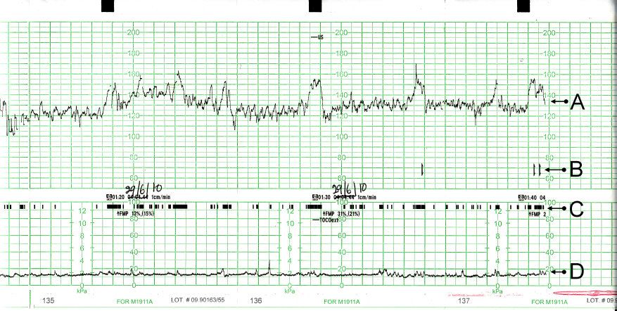
Risk is a moving target, not a label
A high-risk pregnancy is one in which the pregnant person, the fetus or the newborn carries a greater-than-background probability of illness or death. That definition is deliberately broad because risk arrives from four directions: pre-existing medical conditions carried into the pregnancy (chronic hypertension, type 1 or type 2 diabetes, epilepsy, lupus, cardiac disease, thrombophilia, chronic kidney disease), obstetric history (prior preterm birth, prior stillbirth, prior pre-eclampsia, recurrent pregnancy loss, prior caesarean birth, cervical insufficiency), conditions arising during this pregnancy (pre-eclampsia, gestational diabetes, placenta praevia, multiple gestation, fetal growth restriction, infection), and the social and structural determinants that shape whether a person can reach care at all — distance, cost, insurance status, immigration status, intimate partner violence, food insecurity, racism inside the health system.
That last category is not a footnote. Outcome disparities in maternity care are large, persistent, and are not explained by individual behaviour. Black birthing people in the United States die of pregnancy-related causes at roughly three times the rate of white birthing people, and a substantial share of those deaths are judged preventable, most often because a symptom was reported and not believed. The nursing implication is concrete and repeatable: when a pregnant person tells you something is wrong, that report is data. Document it in their words, escalate it, and do not let it be resolved by reassurance alone.
Risk is also dynamic. A pregnancy classed as low risk at booking becomes high risk the afternoon the blood pressure reads 158/104, and a pregnancy flagged high risk for chronic hypertension may sail through with no superimposed disease at all. Reassess risk at every contact rather than treating the booking classification as permanent. Practically, that means every antenatal encounter includes blood pressure, weight, urine testing when indicated, fundal height after 24 weeks, fetal heart tones, and an open question about fetal movement, headache, visual change, epigastric pain, bleeding, fluid leakage and contractions.
Antepartum fetal surveillance and what each test actually measures
We cannot see the fetus, so we infer its oxygenation from its behaviour. A fetus with adequate oxygen and an intact autonomic nervous system moves, breathes, holds flexor tone, accelerates its heart rate and produces urine (and therefore amniotic fluid). Hypoxia switches these off in roughly the reverse order of their development: heart rate reactivity and fetal breathing are lost early, gross movement next, tone last, and reduced amniotic fluid signals chronic rather than acute compromise because it takes days of shunted renal blood flow to drop the volume. Knowing that hierarchy tells you what a given abnormal test means.
The non-stress test (NST) asks only whether the fetal heart rate accelerates with movement. At 32 weeks or more, reactive means at least two accelerations of at least 15 beats per minute above baseline lasting at least 15 seconds within a 20-minute window. Before 32 weeks the criterion drops to 10 beats per minute for 10 seconds because the autonomic nervous system is immature. A non-reactive tracing is far more often a sleeping fetus than a dying one — fetal sleep cycles run 20 to 40 minutes — so extend the test to 40 minutes and consider vibroacoustic stimulation before escalating. The NST has a high negative predictive value and a poor positive predictive value: a reactive test is genuinely reassuring, a non-reactive test mostly means do another test.
The biophysical profile (BPP) adds ultrasound to the NST and scores five parameters two points each. The contraction stress test (CST) asks a different question — whether the placenta has enough reserve to survive the transient perfusion drop of a contraction — and its scoring is counter-intuitive: negative (no late decelerations) is the good result. It is contraindicated when labour is dangerous, so avoid it in placenta praevia, PPROM, prior classical uterine incision or myomectomy entering the cavity, and cervical insufficiency. Umbilical artery Doppler is used in fetal growth restriction; absent or reversed end-diastolic flow signals critically raised placental resistance and prompts urgent obstetric review.
Three deceleration patterns carry almost the whole of intrapartum decision-making, and the mapping has to be automatic because the wrong label produces the wrong action. An early deceleration is gradual, mirrors the contraction with its nadir at the contraction peak, and reflects head compression — it is benign and requires no intervention. A variable deceleration is abrupt and V-shaped with no fixed relationship to the contraction, and reflects cord compression — reposition the patient and consider amnioinfusion; a sudden deep variable or a prolonged deceleration immediately after membranes rupture is umbilical cord prolapse until proven otherwise. A late deceleration is gradual, begins after the contraction has started and returns to baseline after it has finished, and reflects uteroplacental insufficiency — the placenta cannot meet oxygen demand, and this is the most ominous of the three. Recurrent late decelerations, recurrent variables, or a prolonged deceleration all trigger the same intrauterine resuscitation sequence, in order: reposition to the left or right lateral side, stop the oxytocin, give an intravenous bolus of isotonic crystalloid, apply oxygen at 10 litres per minute by non-rebreather mask, and notify the provider — then reassess, and prepare for expedited birth if the pattern does not recover.
| Test | What is measured | Normal / reassuring | Abnormal result and nursing action |
|---|---|---|---|
| Non-stress test | FHR accelerations with movement | ≥32 wk: 2 accels ≥15 bpm × ≥15 s in 20 min. <32 wk: ≥10 bpm × ≥10 s | Non-reactive → extend to 40 min, reposition left lateral, vibroacoustic stimulation, then BPP or CST |
| Biophysical profile | NST + breathing + movement + tone + fluid (2 points each) | 8–10 with normal fluid | 6 = equivocal, repeat in 24 h or deliver if term; ≤4 = deliver |
| Modified BPP | NST + amniotic fluid volume | Reactive NST + AFI >5 cm | Either abnormal → full BPP |
| Contraction stress test | Placental reserve under contractions (3 in 10 min, ≥40 s) | Negative = no late decelerations (good) | Positive = late decels with ≥50% of contractions → obstetric review, likely delivery |
| Amniotic fluid index | Sum of 4 quadrant pockets | 5–24 cm; single deepest pocket 2–8 cm | ≤5 cm oligohydramnios (renal, PPROM, placental insufficiency); ≥24 cm polyhydramnios (diabetes, anomaly) |
| Umbilical artery Doppler | Placental vascular resistance in FGR | Forward flow throughout diastole | Absent or reversed end-diastolic flow → urgent review, admission, steroids, timed delivery |
| Fetal movement counting | Maternal perception of movement | 10 distinct movements within 2 h, same time daily | Fewer than 10 in 2 h → come in the same day for NST; never advise waiting until morning |
Escalation, documentation and what you may not hand off
High-risk antepartum care generates a stream of borderline findings, and the failure mode is not usually ignorance — it is delay. Structured escalation shortens the delay. SBAR gives the receiving clinician the picture in the order they need it. A worked example for a patient at 33 weeks with new severe-range blood pressure sounds like this. Situation: “This is the RN on antepartum. Room 4, Ms Okoye, 33 weeks and 2 days, gravida 2 para 1, her blood pressure is 168 over 112 repeated at four minutes and she now has a headache that did not respond to paracetamol.” Background: “Admitted yesterday for gestational hypertension, no proteinuria on admission, platelets were 148.” Assessment: “I think this is pre-eclampsia with severe features. Reflexes are 3+, no clonus, urine output 25 millilitres over the last hour, fetal heart rate baseline 150 with moderate variability.” Recommendation: “I need you at the bedside now, I would like orders for IV labetalol and magnesium sulfate, and I have started repeat pre-eclampsia labs.” Notice that the recommendation is specific and time-bound. “Can you come see her when you get a chance” is not escalation.
Delegation on a high-risk antepartum unit follows one rule with no exceptions: the registered nurse retains assessment, teaching, evaluation and the unstable patient. Assistive personnel may take vital signs on stable patients, record intake and output, obtain daily weights, assist with hygiene and ambulation, and in many facilities perform capillary glucose checks — but the RN interprets every one of those numbers. A licensed practical or vocational nurse may reinforce teaching the RN has already delivered and give many routine medications, but titrated high-alert infusions such as magnesium sulfate and insulin, blood product administration, and the initial assessment of a patient who is bleeding, seizing or hypotensive remain with the RN. Delegating a task never delegates accountability for the outcome.
Documentation carries the same weight. Record the actual numbers, the time, who was notified, what was requested, what response was received, and what you did next. “Provider aware” is not a plan. If the response you get does not match the severity of the finding, use the chain of command — charge nurse, then the next clinician up — and document that you did. Every maternal mortality review that reads “the nurse recognised the deterioration” and still ends badly turns on the gap between recognition and effective escalation.
Antepartum surveillance tests have strong negative predictive value and weak positive predictive value — a reassuring result is trustworthy, an abnormal one is a prompt to escalate and test further, never a reason to wait and watch.
Section 2Bleeding in Pregnancy: Early Loss and Late Haemorrhage
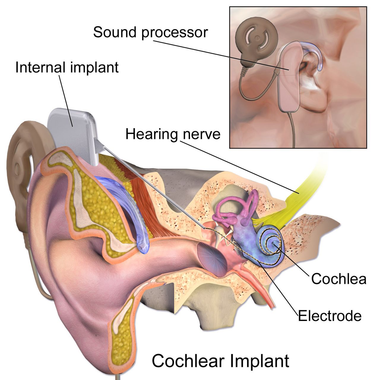
Early pregnancy loss and ectopic pregnancy
Bleeding before 20 weeks occurs in roughly one in four pregnancies, and about half of those pregnancies continue normally. The clinical task is to sort threatened loss from established loss from ectopic implantation, because one of these three can kill within hours. Classification depends on the cervical os and on what has passed: threatened (bleeding, cramping, closed os, viable intrauterine pregnancy), inevitable (open os, bleeding, cramping), incomplete (some products passed, os open, bleeding often heavy), complete (all products passed, os closed, bleeding and cramping settling), missed (fetal demise retained, os closed, symptoms may have quietly disappeared), and septic (any of the above plus fever, foul discharge, uterine tenderness — a surgical and antibiotic emergency).
Ectopic pregnancy is implantation outside the uterine cavity, most often in the ampulla of the fallopian tube. Classic presentation is amenorrhoea of six to eight weeks, unilateral lower abdominal pain and light vaginal spotting; the spotting is decidual, so it is typically far less than the internal bleeding. Serum human chorionic gonadotropin rises more slowly than in a normal intrauterine pregnancy (a normal early rise is at least 35 to 50 percent over 48 hours), and transvaginal ultrasound should show an intrauterine gestational sac once hCG is around 3,500 milli-international units per millilitre. Rupture announces itself with sudden severe pain, signs of hypovolaemic shock, and referred shoulder pain from diaphragmatic irritation by blood in the peritoneum — the Kehr sign. Cervical motion tenderness is common.
Management depends entirely on stability. A stable, unruptured ectopic with a mass under about 3.5 to 4 centimetres, no fetal cardiac activity and hCG below roughly 5,000 may be treated medically with methotrexate 50 milligrams per square metre of body surface area intramuscularly, with hCG followed on days 4 and 7 expecting a fall of at least 15 percent. Teach the patient to avoid folic acid supplements (they antagonise the drug), avoid NSAIDs and alcohol, use sun protection, avoid gas-forming foods that confuse abdominal assessment, and to report severe abdominal pain immediately — some transient pain is expected, escalating pain with dizziness is rupture. A ruptured ectopic or an unstable patient goes to surgery; nursing priority is two large-bore intravenous lines, type and crossmatch, oxygen, and continuous monitoring while the theatre is readied.
Gestational trophoblastic disease — the hydatidiform mole — presents with bleeding that may contain grape-like vesicles, a uterus large for dates, markedly elevated hCG often above 100,000, severe nausea and vomiting, and pre-eclampsia appearing before 20 weeks, which is otherwise almost unheard of. Ultrasound shows the classic snowstorm pattern with no fetus in a complete mole. Treatment is suction curettage, and then the part that nursing follow-up must protect: serial hCG levels weekly until three consecutive negatives, then monthly for six months, with reliable contraception throughout, because a rising hCG signals choriocarcinoma and a new pregnancy would make the marker uninterpretable.
Late-pregnancy bleeding: praevia, abruption and vasa praevia
Bleeding after 20 weeks is treated as haemorrhage until proven otherwise. The three named causes separate cleanly on four features: pain, blood colour, uterine tone and fetal status. Placenta praevia is implantation of the placenta over the internal cervical os; a placental edge that lies within two centimetres of the os without covering it is termed low-lying, and both carry the same nursing prohibition. The classic presentation is painless, bright red bleeding, often in the late second or third trimester, with a soft, non-tender, relaxed uterus and a normal fetal heart rate, because the blood being lost is maternal and the placenta is still functioning. Malpresentation is common because the placenta blocks engagement. The single most important nursing rule is absolute: no digital vaginal examination and nothing in the vagina — no speculum from nursing, no cervical check, no intercourse, no tampons. A digital exam can perforate the placenta and cause exsanguinating haemorrhage in minutes. Transabdominal and transvaginal ultrasound are diagnostic and transvaginal scanning is safe because the probe is angled away from the os.
Abruptio placentae is premature separation of a normally implanted placenta. It presents with painful bleeding that is dark red, a uterus that is tender, rigid or board-like with elevated resting tone and frequent low-amplitude contractions, and a fetal heart rate that frequently deteriorates because the exchange surface has been lost. Roughly one in five abruptions is concealed: the blood collects behind the placenta and the external loss badly understates the true loss, so a patient can be in shock with very little visible bleeding. Any mismatch between visible blood and haemodynamic status should make you think concealed haemorrhage. Risk factors include hypertension in any form, blunt abdominal trauma including intimate partner violence, cocaine and other stimulant use, smoking, prior abruption, PPROM and sudden uterine decompression. Disseminated intravascular coagulation is the classic complication because thromboplastin from the decidua enters the circulation.
Vasa praevia is rarer and faster than either. Unprotected fetal vessels cross the internal os, and when the membranes rupture the vessels tear. The presentation is a small volume of painless bleeding coinciding with rupture of membranes, immediately followed by fetal bradycardia or a sinusoidal fetal heart rate pattern. The blood is fetal, and total fetal blood volume is only about 80 to 100 millilitres per kilogram — a term fetus has roughly 250 to 350 millilitres in total, so a loss that would barely register in an adult is exsanguinating. Vasa praevia is an immediate caesarean birth. Placenta accreta spectrum, in which the placenta invades the myometrium, is increasingly common because its dominant risk factor is the combination of a prior caesarean birth with a current praevia; it is planned for with delivery between 34 and 36 weeks in a centre with massive transfusion capability, and caesarean hysterectomy is often expected.
| Feature | Placenta praevia | Abruptio placentae | Vasa praevia | Uterine rupture |
|---|---|---|---|---|
| Pain | Painless | Painful, constant | Painless | Sudden sharp tearing pain, may then ease |
| Bleeding | Bright red, external | Dark red; concealed in ~20% | Small volume, fetal blood, at ROM | Variable, often concealed |
| Uterine tone | Soft, relaxed, non-tender | Rigid, board-like, tender, high resting tone | Normal | Contractions cease; loss of tone |
| Fetal heart rate | Usually normal initially | Late decelerations, bradycardia | Abrupt bradycardia or sinusoidal | Prolonged deceleration; presenting part recedes (station rises) |
| First nursing action | NOTHING in the vagina; left lateral, IV access, type and cross, fetal monitoring | Large-bore IV × 2, oxygen, left lateral, continuous EFM, coagulation studies, prepare for caesarean | Call for immediate caesarean; do not delay for confirmation | Prepare for immediate laparotomy; fluid and blood resuscitation |
Volume, coagulation and the haemorrhage response
Pregnancy hides blood loss. Plasma volume rises by 40 to 50 percent by the third trimester, so a healthy pregnant person can lose 1,000 to 1,500 millilitres — up to 25 percent of blood volume — before the blood pressure falls. Tachycardia, narrowing pulse pressure, rising respiratory rate, restlessness and cool clammy skin appear long before hypotension, and a falling urine output below 30 millilitres per hour is one of the earliest reliable signs of inadequate perfusion. Do not wait for a low blood pressure to declare haemorrhage; by then the compensation has already failed.
The standing antepartum haemorrhage response is: call for help and activate the obstetric haemorrhage protocol, place two large-bore intravenous catheters (16 or 18 gauge), give warmed isotonic crystalloid, send type and crossmatch with a full coagulation panel and fibrinogen, apply oxygen at 10 litres per minute by non-rebreather mask if there is evidence of compromise, position left lateral or with manual displacement of the uterus off the vena cava, insert an indwelling catheter to measure output hourly, monitor the fetus continuously, weigh pads and drapes for quantitative blood loss rather than estimating visually, and keep the patient and family informed in plain sentences. Fibrinogen deserves special attention: it rises physiologically in pregnancy to 400 to 600 milligrams per decilitre, so a “normal” adult value near 200 in a bleeding obstetric patient is a red flag for consumptive coagulopathy.
Although postpartum haemorrhage belongs to a later chapter, the sequence is worth fixing here because it is the single most tested and most misordered response in maternity nursing. When the uterus is boggy and bleeding after birth, the first action is to massage the fundus — not to start the oxytocin, not to call the provider, not to take a blood pressure. Massage stimulates the myometrium to contract and clamp the spiral arteries mechanically, which is faster than any drug can act. Massage first, then empty the bladder (a distended bladder displaces the uterus and prevents contraction), then give uterotonics as ordered while help is on the way.
Painless bright-red bleeding with a soft uterus is praevia and forbids any vaginal examination; painful dark bleeding with a rigid tender uterus is abruption and may conceal most of its blood loss behind the placenta.
Section 3Hypertensive Disorders and Magnesium Sulfate Safety
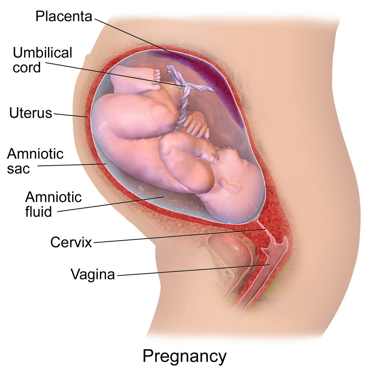
Mechanism and the four diagnostic categories
Pre-eclampsia begins in the placenta and ends in the maternal endothelium. In normal implantation, trophoblast remodels the spiral arteries into wide, low-resistance channels. In pre-eclampsia that remodelling is incomplete, the placenta is under-perfused, and it releases anti-angiogenic factors into the maternal circulation. Those factors injure endothelium everywhere at once, which explains why the disease is multi-system rather than merely hypertensive: vasospasm raises blood pressure, glomerular endothelial injury leaks protein, hepatic sinusoidal injury distends the liver capsule and produces right upper quadrant pain, cerebral vasospasm and oedema produce headache, visual scotomata and ultimately seizure, and platelet consumption drops the count. Fluid leaves the intravascular space, so these patients are simultaneously oedematous and intravascularly depleted — which is why aggressive fluid loading causes pulmonary oedema.
The four categories are distinguished by timing and by findings. Chronic hypertension is blood pressure at or above 140/90 before 20 weeks or persisting beyond 12 weeks postpartum. Gestational hypertension is new hypertension at or after 20 weeks without proteinuria or severe features, resolving postpartum. Pre-eclampsia is new hypertension at or after 20 weeks plus either proteinuria or, in the absence of proteinuria, any severe feature. Chronic hypertension with superimposed pre-eclampsia is a worsening picture on a pre-existing baseline and carries the highest risk. Proteinuria is defined as 300 milligrams or more in 24 hours, a protein-to-creatinine ratio of 0.3 or above, or a dipstick of 2+ when quantitative testing is unavailable. Critically, proteinuria is no longer required for the diagnosis — waiting for it delays treatment and has killed patients.
Prevention is real and under-used. Low-dose aspirin 81 milligrams daily, started between 12 and 28 weeks (ideally before 16 weeks) and continued until birth, meaningfully reduces pre-eclampsia in people with one high-risk factor (prior pre-eclampsia, chronic hypertension, pre-gestational diabetes, chronic kidney disease, autoimmune disease, multifetal gestation) or two moderate-risk factors (first pregnancy, age 35 or older, body mass index above 30, family history, sociodemographic risk, interpregnancy interval over 10 years). Antihypertensives compatible with pregnancy are labetalol, nifedipine, methyldopa and hydralazine; ACE inhibitors, angiotensin receptor blockers and direct renin inhibitors are contraindicated because of fetal renal injury and oligohydramnios.
| Severe feature | Threshold | What it reflects |
|---|---|---|
| Severe-range blood pressure | Systolic ≥160 or diastolic ≥110 on two occasions 4 h apart (or confirmed within minutes to permit treatment) | Risk of maternal stroke — the leading cause of death in pre-eclampsia |
| Thrombocytopenia | Platelets <100,000 per microlitre | Platelet consumption at injured endothelium |
| Impaired liver function | AST or ALT at least twice the upper limit of normal, or severe persistent right upper quadrant or epigastric pain | Hepatic ischaemia, capsular distension, risk of subcapsular haematoma |
| Renal insufficiency | Serum creatinine >1.1 mg/dL or doubling of baseline | Glomerular endothelial injury; normal pregnancy creatinine is LOW (0.4–0.8) |
| Pulmonary oedema | Clinical or radiographic | Capillary leak plus low oncotic pressure |
| New cerebral or visual symptoms | Headache unresponsive to analgesia, scotomata, blurred or double vision, altered mental state | Cerebral vasospasm and oedema — the immediate seizure warning |
Severe-range pressure, eclampsia, and what you do first
A confirmed severe-range blood pressure is a hypertensive emergency with a clock attached. Treatment should be given within 30 to 60 minutes of confirmation, and sooner is better. First-line agents are intravenous labetalol 20 milligrams over 2 minutes, repeated at escalating doses of 40 then 80 milligrams every 10 minutes to a cumulative maximum of 300 milligrams; intravenous hydralazine 5 to 10 milligrams over 2 minutes repeated every 20 minutes; or immediate-release oral nifedipine 10 milligrams repeated at 20 milligrams if intravenous access is not yet established. The goal is not normotension — it is 140 to 150 systolic over 90 to 100 diastolic, because dropping the pressure too far collapses placental perfusion. Avoid labetalol in asthma, decompensated heart failure and bradycardia.
Eclampsia is a generalised tonic-clonic seizure in a person with pre-eclampsia and no other cause. Nursing first actions, in order: stay with the patient and call for help, turn them onto their side, protect the head and clear the immediate environment, and do not restrain the limbs or place anything in the mouth. Do not attempt to assess the fetus during the seizure — the fetal heart rate will be abnormal, and the only useful intervention for the fetus is resuscitating the person carrying it. When the seizure ends, suction the airway, give oxygen at 10 litres per minute by non-rebreather, establish or confirm intravenous access, give the magnesium sulfate bolus as ordered, then assess the fetus and prepare for birth. Delivery is the only definitive treatment for pre-eclampsia, but the patient must be stabilised first; a caesarean during an active seizure endangers both patients.
Two timing facts change discharge teaching. First, roughly a third to a half of eclamptic seizures occur after birth, some days later. Second, postpartum pre-eclampsia can present up to six weeks after birth in someone whose pregnancy was entirely uncomplicated. Every postpartum patient therefore leaves with explicit warning signs: severe headache that does not respond to paracetamol, visual changes, right upper quadrant or epigastric pain, shortness of breath, sudden swelling of face or hands, or a seizure — and an instruction to present to an emergency department and say the words “I gave birth recently”.
Magnesium sulfate: therapeutic window, toxicity order and antidote
Magnesium sulfate is given to prevent and treat eclamptic seizures, not to lower blood pressure. It competes with calcium at the neuromuscular junction and stabilises the cerebral membrane. A typical regimen is a loading dose of 4 to 6 grams intravenously over 15 to 30 minutes followed by a maintenance infusion of 1 to 2 grams per hour, continued for 24 hours after birth. Given before 32 weeks it also confers fetal neuroprotection against cerebral palsy, usually as a 4-gram load followed by 1 gram per hour. It is a high-alert medication: it must run on a dedicated pump with a second-nurse independent double check of the concentration, rate and pump settings, and it must never be given by rapid intravenous push.
Toxicity is entirely predictable because it follows serum level in a fixed order, and that order is the assessment schedule. The therapeutic range is 4 to 7 milliequivalents per litre (about 4.8 to 8.4 milligrams per decilitre). Deep tendon reflexes disappear at roughly 9 to 12 milligrams per decilitre — before respiratory depression, which begins around 12 to 18, and long before cardiac arrest above 25 to 30. This is why hourly patellar reflex checks are not a ritual: the absent reflex is the early warning that buys you the time to act. Magnesium is cleared entirely by the kidney, so urine output is the other essential parameter; oliguria means the drug is accumulating even at an unchanged infusion rate.
Hold parameters before each hourly assessment are: respiratory rate at least 12 per minute, deep tendon reflexes present (at least 1+), urine output at least 30 millilitres per hour (or 100 millilitres over 4 hours), and level of consciousness intact. If any parameter fails, the sequence is fixed: stop the infusion first, then give the antidote — calcium gluconate 1 gram, which is 10 millilitres of a 10 percent solution, intravenously over approximately 3 minutes — then support ventilation with a bag-valve mask and oxygen, call for rapid response and the obstetric team, and send a stat magnesium level. Stopping first matters because giving calcium while magnesium continues to infuse is treating against a running tap. Keep calcium gluconate and a bag-valve mask physically at the bedside of every patient on magnesium.
HELLP syndrome
HELLP is haemolysis, elevated liver enzymes and low platelets — a severe variant of pre-eclampsia that can occur with only modest hypertension and, in a substantial minority, without proteinuria, which is exactly why it gets missed. The laboratory definition is haemolysis (lactate dehydrogenase at or above 600 international units per litre, schistocytes on smear, or bilirubin at or above 1.2 milligrams per decilitre), AST or ALT at least twice the upper limit of normal, and platelets below 100,000 per microlitre. The presenting complaint is often deceptively vague: malaise, nausea and vomiting that feels like influenza, and right upper quadrant or epigastric pain from hepatic capsular distension.
Any pregnant or recently pregnant person describing epigastric pain, especially with nausea, should have a blood pressure, platelet count and liver enzymes checked before the symptom is attributed to reflux or gastritis. Nursing care adds bleeding precautions to standard pre-eclampsia management: avoid intramuscular injections and non-essential venipuncture, apply prolonged pressure to puncture sites, use a soft toothbrush, avoid NSAIDs, and palpate the right upper quadrant gently or not at all because a subcapsular hepatic haematoma can rupture. Sudden shoulder pain, severe abdominal pain and shock in a HELLP patient suggests exactly that rupture and is a surgical emergency. Definitive treatment is birth, with magnesium sulfate for seizure prophylaxis, antihypertensives for severe-range pressures, corticosteroids for fetal lung maturity if gestational age allows, and platelet transfusion generally reserved for counts below 20,000 or for surgery.
On magnesium sulfate, deep tendon reflexes vanish before the respiratory rate falls — so if reflexes are absent, stop the infusion first, then give calcium gluconate 1 gram (10 mL of 10 percent) intravenously over about 3 minutes.
Section 4Metabolic, Endocrine and Nutritional Complications
Diabetes in pregnancy: the mechanism sets the risks
Human placental lactogen, progesterone, cortisol and growth hormone all antagonise insulin, and their concentrations climb through the second and third trimesters. This is a design feature: maternal insulin resistance keeps glucose in the circulation for the fetus. A pancreas with reserve simply secretes more insulin. A pancreas without reserve cannot, and maternal hyperglycaemia results. Glucose crosses the placenta freely; insulin does not. The fetus therefore receives the glucose load and responds by producing its own insulin, which is a powerful growth factor. That single sentence explains nearly every complication: macrosomia and shoulder dystocia, neonatal hypoglycaemia in the minutes after the cord is cut and the glucose supply stops while fetal hyperinsulinaemia persists, delayed surfactant production and respiratory distress, polycythaemia, hypocalcaemia and hyperbilirubinaemia.
Timing separates the two diseases. Pre-gestational diabetes exposes the embryo to hyperglycaemia during organogenesis, so it raises the risk of congenital anomalies — cardiac defects, neural tube defects and the rare but characteristic caudal regression syndrome — and of spontaneous loss. Preconception care aiming for a haemoglobin A1c below 6.5 percent is the single most effective intervention available, along with folic acid supplementation and switching off ACE inhibitors, ARBs and statins. Gestational diabetes appears in the second half of pregnancy, after organogenesis, so it does not raise anomaly risk but does drive macrosomia, hypertensive disorders, polyhydramnios, and a long-term maternal risk of type 2 diabetes of roughly 50 percent within 10 to 20 years — which is why the 4-to-12-week postpartum 75-gram oral glucose tolerance test is not optional follow-up.
Screening happens at 24 to 28 weeks, earlier if risk factors are present. The two-step approach gives a non-fasting 50-gram glucose challenge with a one-hour threshold of 130 to 140 milligrams per decilitre, and those who screen positive proceed to a fasting 100-gram three-hour test where two or more abnormal values diagnose gestational diabetes (fasting 95, one hour 180, two hours 155, three hours 140). The one-step approach uses a fasting 75-gram two-hour test where a single abnormal value diagnoses it (fasting 92, one hour 180, two hours 153). Glycaemic targets in pregnancy are tighter than outside it: fasting and pre-meal below 95, one hour after the start of a meal below 140, two hours after below 120. Nutrition therapy and physical activity are first-line; when pharmacotherapy is needed, insulin is the first-line agent because it does not cross the placenta, whereas metformin and glyburide both do.
The arithmetic habit: weight-based dosing and dose ceilings
Every dose that appears in this book from here forward is expressed per kilogram, because the patients on the other side of the birth are newborns and children, and children are not small adults. The habit starts in the antepartum unit. Enoxaparin for venous thromboembolism in pregnancy is prophylactic at 40 milligrams subcutaneously daily but therapeutic at 1 milligram per kilogram every 12 hours: for a 78-kilogram patient that is 78 milligrams every 12 hours, and a nurse who accepts a pre-printed “80 mg” without checking the documented weight has skipped the calculation. Vancomycin for group B streptococcal prophylaxis in severe penicillin allergy is 20 milligrams per kilogram every 8 hours with a maximum of 2 grams per dose: for a 90-kilogram patient, 20 × 90 = 1,800 milligrams, which is under the cap and therefore given; for a 120-kilogram patient, 20 × 120 = 2,400 milligrams, which exceeds the cap and is given as 2 grams.
That last step is the rule that prevents the most dangerous paediatric error: a weight-based dose must always be checked against the maximum adult dose, and the smaller number wins. Paracetamol for a child is 15 milligrams per kilogram per dose every 4 to 6 hours. For a 30-kilogram child: 15 × 30 = 450 milligrams, well within limits. For a 70-kilogram adolescent: 15 × 70 = 1,050 milligrams — which exceeds the 1,000-milligram adult single dose, so the dose given is 1,000 milligrams. A large adolescent is not entitled to a larger-than-adult dose simply because the multiplication says so.
| Calculation | Rule | Worked example | Safety check |
|---|---|---|---|
| Per-dose medication | mg/kg/dose × weight, then compare with maximum adult dose | Paracetamol 15 mg/kg for 70 kg = 1,050 mg → give 1,000 mg | The smaller of (calculated dose, adult maximum) is the dose |
| Capped antibiotic dose | mg/kg/dose with a stated ceiling per dose | Vancomycin 20 mg/kg for 120 kg = 2,400 mg → give 2 g | Confirm a current, documented weight in kilograms — never a reported weight in pounds |
| Therapeutic enoxaparin | 1 mg/kg subcutaneously every 12 h | 78 kg → 78 mg q12h | Hold 24 h before neuraxial anaesthesia (12 h for prophylactic dosing) |
Hyperemesis gravidarum, anaemia and cholestasis
Hyperemesis gravidarum is not severe morning sickness; it is intractable vomiting producing weight loss greater than 5 percent of pre-pregnancy weight, dehydration, ketonuria and electrolyte disturbance — classically hypokalaemia and a hypochloraemic metabolic alkalosis from loss of gastric acid. It typically begins before 9 weeks. Management escalates from non-pharmacological measures (small frequent dry carbohydrate snacks, eating before rising, separating fluids from solids, ginger, avoiding triggering smells) to pyridoxine (vitamin B6) 10 to 25 milligrams every 6 to 8 hours with or without doxylamine 12.5 milligrams, then to promethazine, metoclopramide or ondansetron, then to intravenous rehydration and, rarely, enteral or parenteral nutrition.
One safety rule governs the intravenous phase: give thiamine before any dextrose-containing fluid in a person who has been vomiting for more than about three weeks. Dextrose consumes the last of a depleted thiamine store and can precipitate Wernicke encephalopathy — confusion, ataxia and ophthalmoplegia — which is preventable and sometimes irreversible. Thiamine 100 milligrams intravenously is the usual dose. Equally important is the language: this condition is frequently dismissed as psychological or exaggerated, and patients arrive expecting not to be believed. Weigh them, check ketones and electrolytes, and treat the physiology.
Iron-deficiency anaemia is defined in pregnancy as haemoglobin below 11 grams per decilitre in the first and third trimesters and below 10.5 in the second, reflecting the physiological dilution of plasma volume expansion. Daily requirement rises to about 27 milligrams of elemental iron; treatment of established deficiency uses 100 to 200 milligrams of elemental iron daily, taken with a vitamin C source and away from milk, calcium supplements, tea and antacids. Warn about dark stools (expected) and constipation (manage with fluid, fibre and a stool softener). Intrahepatic cholestasis of pregnancy presents with intense itching of the palms and soles, worse at night, with no rash; the diagnostic test is serum total bile acids, abnormal at 10 micromoles per litre or above and severe at 100 or above, where stillbirth risk rises sharply. Treatment is ursodeoxycholic acid 10 to 15 milligrams per kilogram per day with antenatal surveillance and planned birth, usually at 36 to 37 weeks and earlier if bile acids are severe.
Insulin is first-line pharmacotherapy in pregnancy because it does not cross the placenta, and every weight-based dose — maternal or paediatric — is calculated per kilogram and then capped at the maximum adult dose.
Section 5Infection, Immune Incompatibility and Preterm Birth
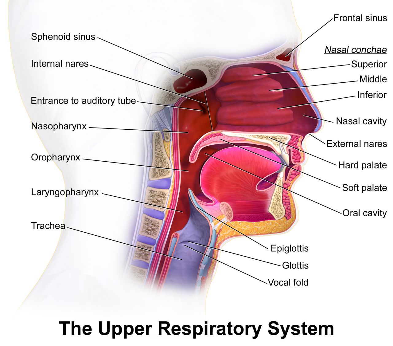
Infection in pregnancy and prophylaxis that works
Pregnancy shifts immune function toward tolerance of the fetus, and that shift makes several infections more severe. Urinary tract infection deserves particular respect: progesterone relaxes ureteral smooth muscle and the growing uterus compresses the ureters, producing physiological hydronephrosis and urinary stasis. Asymptomatic bacteriuria, which would be ignored outside pregnancy, is treated in pregnancy because up to 30 percent of untreated cases ascend to pyelonephritis, and pyelonephritis in pregnancy causes preterm labour, sepsis and acute respiratory distress syndrome. Flank pain, fever and costovertebral angle tenderness in a pregnant patient is an admission, not an outpatient prescription.
Group B Streptococcus colonises the vagina and rectum of about a quarter of pregnant people and is harmless to them, but transmitted during birth it causes early-onset neonatal sepsis, pneumonia and meningitis. Universal screening by vaginal-rectal culture is performed at 36 0/7 to 37 6/7 weeks. Intrapartum prophylaxis — not antenatal treatment, which does not prevent recolonisation — is penicillin G 5 million units intravenously as a load then 2.5 to 3 million units every 4 hours until birth. Prophylaxis is considered adequate once at least 4 hours have elapsed before birth. Indications for prophylaxis without a culture include GBS bacteriuria at any point in this pregnancy, a previous infant with invasive GBS disease, or unknown status with birth before 37 weeks, membranes ruptured 18 hours or more, or intrapartum fever.
The TORCH group — toxoplasmosis, other (syphilis, varicella, parvovirus B19, Zika, hepatitis B), rubella, cytomegalovirus and herpes simplex — matters because these organisms cross the placenta and injure the developing fetus. Teaching is largely preventive: avoid changing cat litter and eating undercooked meat (toxoplasmosis); hand hygiene around toddlers’ saliva and urine, the main source of cytomegalovirus; rubella immunity is checked at booking and the vaccine is given postpartum because it is a live vaccine and contraindicated in pregnancy, with the advice to avoid conception for 28 days. Active genital herpes lesions or prodromal symptoms at the onset of labour indicate caesarean birth. Untreated syphilis causes stillbirth and congenital syphilis and is entirely preventable with penicillin.
Rh alloimmunisation
An Rh-negative pregnant person carrying an Rh-positive fetus can be exposed to fetal red cells at any point where the placental barrier is breached — birth, miscarriage, ectopic pregnancy, chorionic villus sampling, amniocentesis, external cephalic version, abdominal trauma, abruption. The maternal immune system produces anti-D antibodies. The first pregnancy is usually unaffected because sensitisation occurs at delivery, but those IgG antibodies cross the placenta in every subsequent pregnancy and haemolyse fetal red cells, producing fetal anaemia, high-output heart failure, hydrops fetalis and death.
Prevention is one of the great successes of obstetric care. Rh(D) immune globulin 300 micrograms intramuscularly is given at 28 weeks and again within 72 hours of birth if the newborn is Rh-positive, and additionally after any sensitising event; a 50-microgram microdose is used for events before 12 weeks. One 300-microgram dose neutralises approximately 30 millilitres of fetal whole blood (15 millilitres of fetal red cells), so after abruption or trauma a Kleihauer-Betke test quantifies the fetomaternal haemorrhage and determines how many doses are required. Two antibody tests are easily confused: the indirect Coombs (antibody screen) is performed on the pregnant person and detects circulating antibody, while the direct Coombs is performed on the newborn’s cord blood and detects antibody already attached to fetal cells. Rh(D) immune globulin is useless once sensitisation has occurred — a positive indirect Coombs means the window has closed, and management shifts to middle cerebral artery Doppler surveillance for fetal anaemia and, if needed, intrauterine transfusion.
Preterm labour and PPROM
Preterm labour is regular uterine activity with documented cervical change before 37 0/7 weeks. Its symptoms are notoriously vague — low dull backache, pelvic pressure, menstrual-like cramping, increased or changed vaginal discharge, and contractions that may be painless — and patients routinely delay presenting because the sensations do not match the dramatic picture they expect. Teach the specific symptoms, and teach the response: empty the bladder, lie down on the left side, drink two or three glasses of water, and if contractions continue at four or more per hour after an hour, come in. Two tests refine the decision: fetal fibronectin has an excellent negative predictive value, so a negative test makes birth within the next 7 to 14 days very unlikely, and transvaginal cervical length under 25 millimetres before 24 weeks identifies elevated risk. Vaginal progesterone is used for a short cervix; cerclage is used for cervical insufficiency, which classically presents as painless dilation in the second trimester.
Tocolytics are widely misunderstood. They do not meaningfully prolong pregnancy; they buy roughly 48 hours, and that window exists to complete antenatal corticosteroids and to transfer to a facility with an appropriate neonatal unit. Nifedipine and indomethacin are common first choices; indomethacin is restricted to before 32 weeks because it constricts the fetal ductus arteriosus and reduces amniotic fluid. Terbutaline is short-term only and causes maternal tachycardia, tremor and hyperglycaemia — a maternal heart rate above 120 is a hold parameter. Antenatal corticosteroids are the intervention that actually changes newborn survival: betamethasone 12 milligrams intramuscularly every 24 hours for two doses, or dexamethasone 6 milligrams intramuscularly every 12 hours for four doses, given between 24 0/7 and 33 6/7 weeks (and in selected late-preterm situations). Benefit begins about 24 hours after the first dose and is optimal from 48 hours to 7 days. Expect a transient rise in maternal blood glucose — in a patient with diabetes this can be substantial and requires close monitoring. Magnesium sulfate before 32 weeks is added for fetal neuroprotection.
Preterm premature rupture of membranes (PPROM) is rupture before 37 weeks and before labour begins. Confirm with a sterile speculum examination showing pooling, nitrazine paper turning blue (alkaline amniotic fluid) and ferning on a dried slide — and perform no digital examination, because each digital exam shortens latency and introduces organisms. Nursing priorities are temperature every 2 to 4 hours, fetal heart rate for tachycardia above 160 (an early sign of chorioamnionitis), uterine tenderness, the character and odour of fluid, and white cell trend. Latency antibiotics prolong the pregnancy and reduce infection: ampicillin 2 grams intravenously every 6 hours plus erythromycin for 48 hours, followed by 5 days of oral amoxicillin and erythromycin. Amoxicillin-clavulanate is avoided because of an association with neonatal necrotising enterocolitis. Expectant management generally continues to 34 0/7 weeks, and chorioamnionitis, abruption, cord prolapse or non-reassuring fetal status ends it immediately. The greatest immediate hazard after any rupture with an unengaged presenting part is umbilical cord prolapse, so check the fetal heart rate immediately after membranes rupture, every time.
| Emergency | Recognition | FIRST action | Never do this |
|---|---|---|---|
| Umbilical cord prolapse | Sudden variable or prolonged deceleration or bradycardia after ROM; cord palpated or seen | Keep a gloved hand in the vagina and lift the presenting part off the cord; position knee-chest or Trendelenburg; call for immediate caesarean; oxygen 10 L/min | Do not push the cord back in; do not repeatedly handle it; do not remove your hand until the surgeon takes over |
| Magnesium sulfate toxicity | Deep tendon reflexes absent, then RR <12, slurred speech, oliguria | Stop the infusion, then calcium gluconate 1 g (10 mL of 10%) IV over ~3 min; support ventilation | Do not give the antidote while the magnesium continues to run |
| Eclamptic seizure | Generalised tonic-clonic activity in pre-eclampsia | Call for help, turn on the side, protect the head, then oxygen and magnesium bolus | Do not restrain limbs, put anything in the mouth, or leave to fetch equipment |
| Postpartum haemorrhage | Boggy fundus, heavy or continuous bleeding, rising pulse | Massage the fundus, then empty the bladder, then uterotonics as ordered | Do not leave to page the provider before massaging; do not massage a firm fundus |
| Shoulder dystocia | Turtle sign; head delivers and retracts; shoulders will not deliver | Call for help, McRoberts (hyperflex thighs to abdomen) plus suprapubic pressure; note the time | Never apply fundal pressure — it drives the shoulder harder into the symphysis |
| Uterine rupture | Sharp tearing pain, contractions cease, loss of tone, presenting part recedes, prolonged deceleration, shock | Call the obstetric emergency team and prepare for immediate laparotomy; two large-bore IVs, blood, oxygen | Do not give oxytocin; do not delay surgery for imaging |
| Supine hypotension | Dizziness, pallor, nausea, falling BP when lying flat after 20 weeks | Turn to the left side or manually displace the uterus | Do not start with fluids or vasopressors before repositioning |
In every obstetric emergency the life-saving move is the first one — lift the presenting part off a prolapsed cord, stop the magnesium before giving calcium, massage the fundus before anything else — and the intuitive alternative is usually the harmful one.
The chapter in one breath
High-risk pregnancy is ordinary pregnancy plus a named, trackable threat, and nursing owns the surveillance that finds it early. Antepartum testing infers fetal oxygenation from fetal behaviour: the non-stress test, biophysical profile, contraction stress test, amniotic fluid volume and umbilical artery Doppler all reassure powerfully when normal and demand escalation when not. Bleeding sorts by pain and tone — painless bright red with a soft uterus is praevia and forbids vaginal examination, painful dark bleeding with a rigid tender uterus is abruption and may conceal most of its loss, painless bleeding at rupture with sudden fetal bradycardia is vasa praevia and is an immediate caesarean. Hypertensive disease is endothelial injury, diagnosable without proteinuria, treated for severe-range pressures within 30 to 60 minutes, and prevented from seizing with magnesium sulfate — whose toxicity announces itself by absent reflexes before respiratory depression, and whose antidote, calcium gluconate 1 gram, follows stopping the infusion. Diabetes is fetal hyperinsulinaemia driven by maternal glucose, managed with insulin because insulin does not cross the placenta. Every dose is per kilogram, capped at the adult maximum. Every emergency is won or lost on the first action.
Words worth knowing
| Term | What it means |
|---|---|
| Abruptio placentae | Premature separation of a normally implanted placenta; painful dark bleeding with a rigid, tender uterus and up to 20 percent concealed loss. |
| Amniotic fluid index | Sum of the deepest fluid pocket in each of four uterine quadrants; normal 5–24 cm. |
| Biophysical profile | Five-parameter fetal assessment (NST, breathing, movement, tone, fluid) scored 2 points each; 8–10 reassuring, 4 or less means deliver. |
| Calcium gluconate | Antidote to magnesium sulfate toxicity; 1 gram (10 mL of a 10 percent solution) intravenously over about 3 minutes, given after the infusion is stopped. |
| Chorioamnionitis | Intra-amniotic infection; maternal fever, uterine tenderness, foul fluid, maternal and fetal tachycardia — ends expectant management of PPROM. |
| Eclampsia | Generalised tonic-clonic seizure in a person with pre-eclampsia; may occur for the first time postpartum. |
| Ectopic pregnancy | Implantation outside the uterine cavity, usually tubal; unilateral pain, spotting, and shoulder pain (Kehr sign) once ruptured. |
| Fetal fibronectin | Cervicovaginal glycoprotein test whose strength is its negative predictive value for birth within 7 to 14 days. |
| HELLP syndrome | Haemolysis, elevated liver enzymes, low platelets; often presents as epigastric or right upper quadrant pain with only modest hypertension. |
| Hydatidiform mole | Gestational trophoblastic disease with a uterus large for dates, hCG above 100,000, snowstorm ultrasound and pre-eclampsia before 20 weeks. |
| Hyperemesis gravidarum | Intractable vomiting with weight loss over 5 percent, ketonuria and electrolyte disturbance; give thiamine before dextrose-containing fluids. |
| Intrahepatic cholestasis of pregnancy | Nocturnal palmar and plantar itching without rash; bile acids 10 micromoles per litre or more, severe at 100 or more. |
| Kleihauer-Betke test | Quantifies fetal red cells in the maternal circulation to determine how many doses of Rh(D) immune globulin are needed. |
| Macrosomia | Birth weight above 4,000 to 4,500 grams; increases shoulder dystocia, operative birth and neonatal hypoglycaemia risk. |
| Magnesium sulfate | High-alert anticonvulsant for pre-eclampsia and neuroprotectant before 32 weeks; therapeutic 4–7 mEq/L, monitored by reflexes, respiratory rate and urine output. |
| Non-stress test | Fetal heart rate accelerations with movement; reactive at 32 weeks or more is 2 accelerations of 15 bpm for 15 seconds in 20 minutes. |
| Placenta accreta spectrum | Abnormally invasive placenta; strongest risk factor is prior caesarean birth combined with current praevia. |
| Placenta praevia | Placenta covering the internal os (edge within 2 cm but not covering it is low-lying); painless bright red bleeding with a soft uterus and an absolute prohibition on vaginal examination. |
| PPROM | Preterm premature rupture of membranes; confirmed by sterile speculum, pooling, nitrazine and ferning — never by digital examination. |
| Vasa praevia | Unprotected fetal vessels crossing the internal os; painless bleeding at rupture with immediate fetal bradycardia, requiring emergency caesarean. |
Check yourself
Labour and Birth
Labour is the one event in nursing where two patients share a single circulation and a single clock. Everything you assess — the contraction, the cervix, the fetal heart, the colour of the fluid, the tone of the uterus after the placenta delivers — is a window onto whether oxygen is still crossing the placenta and whether the birthing parent is still holding onto their blood volume. This chapter builds labour the way it is practised: first the mechanics that decide whether a fetus can pass through a pelvis, then the stages and what normal progress actually looks like, then fetal surveillance and the interventions that restore oxygen delivery, then comfort and anaesthesia and the complications that follow the needle, and finally the third and fourth stages with the emergency sequences where the order of your actions is the intervention. Nothing here is intuitive by accident. Much of it is counter-intuitive on purpose, and that is exactly why it is memorised as a sequence.
By the end of this chapter you can
- Analyse the five Ps of labour and interpret fetal lie, presentation, attitude, position and station from a documented finding such as LOA at 0 station.
- Differentiate true labour from false labour and describe the first nursing action after spontaneous rupture of membranes.
- Sequence the cardinal movements and describe expected progress and nursing priorities in each of the four stages of labour.
- Interpret an electronic fetal monitoring strip using baseline, variability, accelerations and decelerations, assign a NICHD category, and carry out intrauterine resuscitation in priority order.
- Compare non-pharmacological comfort measures, systemic opioids and neuraxial analgesia, and state the first action for post-epidural hypotension.
- Execute the priority sequence for postpartum haemorrhage, magnesium sulfate toxicity, umbilical cord prolapse, shoulder dystocia and uterine rupture.
- Provide immediate newborn care at birth — warmth, skin-to-skin contact, delayed cord clamping and positive-pressure ventilation — and calculate weight-based delivery-room doses against the maximum recommended dose.
- Escalate an intrapartum red flag using SBAR and apply delegation limits that keep assessment, teaching, evaluation and the unstable patient with the registered nurse.
Section 1The Five Ps: What Decides Whether a Birth Can Happen
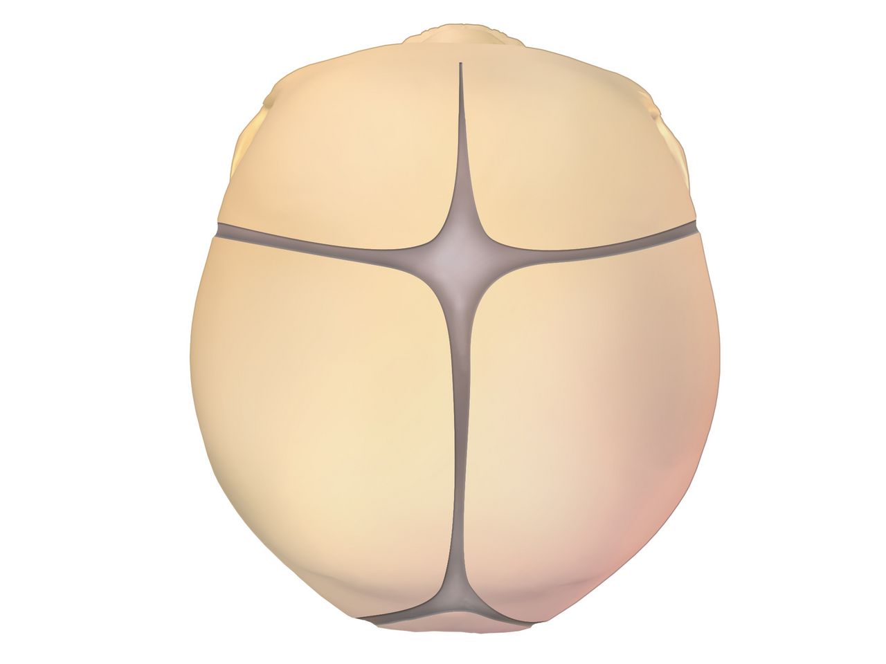
The passenger and the passage
The fetal skull is the widest part of the passenger, and it is deformable. The frontal, sagittal, coronal and lambdoid sutures are membranous seams that allow the parietal bones to overlap under pressure — moulding — which resolves over the first days of life. Where sutures meet, they form fontanelles: the diamond-shaped anterior fontanelle (roughly 2 to 3 cm across, closing at 12 to 18 months) and the small triangular posterior fontanelle (closing by 6 to 8 weeks). On vaginal examination the shape of the fontanelle your fingertip meets tells you the position of the head, which is why the examiner is palpating for a triangle rather than guessing.
Head size depends on attitude, the relationship of fetal parts to one another. With complete flexion — chin to chest — the presenting diameter is the suboccipitobregmatic, about 9.5 cm. Lose that flexion and the presenting diameter grows: a military attitude presents about 12 cm, a brow presentation about 13.5 cm, which is generally too large for the pelvis. Lie is the relationship of the fetal spine to the maternal spine and is longitudinal in about 99 per cent of term labours; transverse or oblique lie cannot be born vaginally. Presentation is the part entering the pelvis first: cephalic in roughly 96 per cent, breech in 3 to 4 per cent, shoulder in the remainder.
Position is recorded as three letters — the side of the maternal pelvis (L or R), the fetal reference point (O for occiput, S for sacrum in breech, M for mentum in face, A for acromion in shoulder), and the part of the pelvis it points toward (A anterior, P posterior, T transverse). LOA, left occiput anterior, is the most common and the most efficient. Occiput posterior positions produce intense back pain, a longer second stage and a greater chance of operative birth; hands-and-knees positioning, counterpressure and lunging help the fetus rotate. Station measures descent in centimetres above or below the ischial spines, from −5 to +5, with 0 station meaning the widest diameter of the head has passed the pelvic inlet — engagement.
The passage is the bony pelvis plus the soft tissues. The gynaecoid pelvis, present in roughly half of people, has a rounded inlet and wide outlet and is the most favourable. The android (heart-shaped inlet, narrow outlet), anthropoid (oval, favours occiput posterior) and platypelloid (flattened, the least favourable) types describe the rest. A diagonal conjugate of 11.5 cm or more on clinical pelvimetry suggests an adequate inlet. Pelvic capacity is not fixed: squatting and McRoberts positioning can increase the outlet diameter measurably, which is the physiological basis for the shoulder-dystocia manoeuvre you will meet in Section 5.
The powers, the position and the psyche
The primary power is the involuntary uterine contraction. Describe it in four terms. Frequency is measured from the beginning of one contraction to the beginning of the next, in minutes. Duration is the beginning to the end of the same contraction, in seconds. Intensity is the peak strength — by palpation graded mild (fundus indents like the tip of the nose), moderate (chin) or strong (forehead), or by intrauterine pressure catheter in mmHg. Resting tone is the pressure between contractions and should feel soft, under about 20 mmHg by catheter. Adequate labour by catheter is 200 or more Montevideo units in 10 minutes, calculated by adding the peak-minus-baseline amplitudes of every contraction in that window.
Contractions that are too frequent are as dangerous as contractions that are too weak, because the placenta refills only between them. Tachysystole is more than five contractions in 10 minutes averaged over a 30-minute window. Duration beyond 90 seconds or a resting tone that never softens compresses the spiral arteries and starves the intervillous space. Whenever tachysystole appears during an oxytocin infusion, the drug is the first suspect.
The secondary power is voluntary bearing down, which only helps once the cervix is completely dilated; pushing against a partly dilated cervix causes oedema and tearing. Maternal position is a power in its own right — upright and lateral positions use gravity, widen the outlet and reduce aortocaval compression, while flat supine positioning reduces venous return and can drop uterine perfusion. The psyche is not a soft extra. Fear and unrelieved pain raise circulating catecholamines, which reduce uterine blood flow and can slow a contraction pattern; continuous labour support measurably shortens labour and lowers caesarean rates.
Premonitory signs, true labour and rupture of membranes
In the weeks before labour the fetus settles into the pelvis (lightening), breathing eases and urinary frequency returns. Braxton Hicks contractions intensify, the cervix softens and begins to efface, the mucus plug is released as bloody show, weight may fall by half a kilogram to a kilogram and some people describe a burst of energy in the 24 to 48 hours before onset. None of these confirms labour. Only progressive cervical change does.
| Feature | True labour | False labour |
|---|---|---|
| Contractions | Regular, progressively closer, longer and stronger | Irregular, do not intensify |
| Effect of walking or a warm bath | Intensify or unchanged | Usually diminish or stop |
| Location of discomfort | Begins in the back and sweeps to the lower abdomen | Abdomen and groin only |
| Cervix | Progressive effacement and dilation — the defining finding | No change on serial examination |
| Show | Bloody show present and increasing | Absent or brownish discharge only |
Rupture of membranes is confirmed by pooling of fluid in the posterior fornix, a nitrazine strip turning blue-green (amniotic fluid pH about 6.5 to 7.5 against a vaginal pH of 4.5 to 5.5 — blood, semen and infection give false positives), ferning on a dried slide, or a commercial placental protein assay. The first nursing action after any rupture, spontaneous or artificial, is to assess the fetal heart rate for a full minute, because a gush of fluid can sweep the cord past the presenting part. Only then document the time, amount, colour and odour. Clear or with flecks of vernix is normal; green means meconium; port-wine suggests abruption; foul or cloudy fluid with maternal fever and fetal tachycardia suggests chorioamnionitis, and the risk of intra-amniotic infection climbs after about 18 to 24 hours of ruptured membranes.
When labour must be started, readiness is scored. The Bishop score rates dilation, effacement, station, cervical consistency and cervical position out of 13. A score of 8 or more in a nulliparous person predicts a vaginal birth about as likely as with spontaneous labour; 6 or less is unfavourable and calls for cervical ripening first — a dinoprostone vaginal insert (10 mg, removable, an advantage if tachysystole develops), misoprostol 25 micrograms vaginally every 3 to 6 hours, or a mechanical transcervical balloon inflated with 30 to 80 mL. Oxytocin is delayed 30 to 60 minutes after removing a dinoprostone insert and about 4 hours after a misoprostol dose.
Presentation, position and station describe whether a fetus can descend, but only progressive cervical change proves labour has begun — and the first action after membranes rupture is always a full minute of fetal heart rate.
Section 2The Mechanisms and Stages of Labour
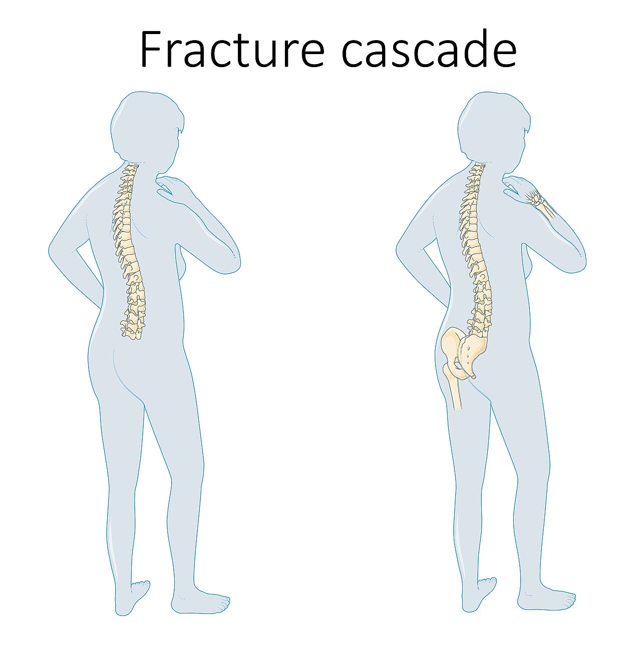
The cardinal movements
The pelvis is not a straight tube. Its inlet is widest transversely and its outlet widest anteroposteriorly, so the fetus must turn as it travels. Engagement brings the biparietal diameter through the inlet. Descent continues throughout, driven by contractions, the pressure of amniotic fluid and, later, maternal pushing. Flexion is forced by resistance from the pelvic floor and reduces the presenting diameter to its smallest. Internal rotation turns the occiput towards anterior, and how far it travels depends on where it started: about 45 degrees from an oblique position such as LOA, about 90 degrees from a transverse position such as LOT, and about 135 degrees from occiput posterior. Extension occurs as the occiput passes under the symphysis and the head is born by extending over the perineum. Restitution and external rotation realign the head with the shoulders, and expulsion follows as the anterior shoulder slips under the symphysis, then the posterior shoulder, then the body.
Knowing this sequence is not academic. When the head is born but restitution does not occur and the chin retracts against the perineum — the turtle sign — you are watching a failed expulsion step, and the manoeuvres in Section 5 are all attempts to complete it.
First stage: latent, active and the honest truth about progress
The first stage runs from the onset of regular contractions to complete dilation. Contemporary practice divides it at 6 cm. The latent phase (0 to about 5 or 6 cm) can legitimately last many hours, particularly in a first labour or an induction, and its length alone is not a reason for caesarean birth. The active phase (6 cm to 10 cm) is when dilation accelerates. Historic Friedman curves demanding 1.2 cm per hour in a nullipara overestimate normal speed; modern data show a much wider range. Arrest of dilation is diagnosed only when the person is at least 6 cm with ruptured membranes and there has been no cervical change for 4 hours or more with adequate contractions, or 6 hours or more with inadequate contractions despite oxytocin.
Nursing care in the first stage is mostly surveillance and support. Vital signs every hour (every 30 minutes if membranes are ruptured or the temperature is rising), temperature every 4 hours intact and every 2 hours ruptured, contraction assessment with fetal heart rate at the interval dictated by risk, position change at least every 30 minutes, and bladder emptying every 2 hours. A full bladder is one of the commonest reversible causes of a stalled labour and of a fundus that will not stay firm afterwards. Vaginal examinations are kept to the minimum necessary once membranes are ruptured, because each one imports organisms.
Group B Streptococcus prophylaxis belongs here. A positive rectovaginal screen at 36 to 37 weeks, GBS bacteriuria in this pregnancy, a previous infant with GBS disease, or unknown status with fever, preterm labour or membranes ruptured 18 hours or more, all call for intrapartum penicillin G 5 million units IV loading then 2.5 to 3 million units every 4 hours. Adequate prophylaxis means at least 4 hours of antibiotic before birth. For a non-anaphylactic penicillin allergy use cefazolin; for severe allergy use clindamycin or vancomycin guided by susceptibility.
Second, third and fourth stages
The second stage runs from complete dilation to birth. It has a passive phase, when the fetus descends without active pushing (“labouring down”, especially useful with an epidural), and an active phase of bearing down. Open-glottis pushing with the urge, in an upright or side-lying position, is preferred to prolonged breath-holding, which raises intrathoracic pressure and reduces placental perfusion. Prolonged second stage is generally defined as more than 3 hours in a nullipara (4 with an epidural) or more than 2 hours in a multipara (3 with an epidural), and even then continued progress may justify continuing. Perineal support, warm compresses and controlled delivery of the head reduce severe lacerations; episiotomy is not performed routinely.
The third stage is birth of the infant to expulsion of the placenta, normally within 5 to 30 minutes. Signs of separation are a firm globular uterus, a rise of the fundus in the abdomen, lengthening of the visible cord and a sudden small gush of blood. A placenta delivering shiny fetal side first is Schultze; maternal side first is Duncan. Active management — a uterotonic (commonly oxytocin 10 units IM, or 10 to 40 units in 500 to 1000 mL of isotonic fluid IV), gentle controlled cord traction with counter-pressure above the symphysis, and fundal massage after delivery of the placenta — roughly halves the risk of haemorrhage. The placenta is inspected for completeness, because retained fragments are a classic delayed cause of bleeding.
The fourth stage is the first 1 to 4 hours after birth, the period of greatest haemorrhage risk. Assess fundus, lochia, bladder, perineum and vital signs every 15 minutes for the first hour, every 30 minutes for the next hour, then hourly. The fundus should be firm, midline and at or just below the umbilicus. A fundus that is boggy, above the umbilicus and displaced to the right almost always means a full bladder — have the patient void or catheterise, then reassess. Lochia rubra should be moderate; saturating a perineal pad in 15 minutes or less, or a steady trickle despite a firm fundus, is haemorrhage until proved otherwise.
| Stage | Boundaries | Typical duration | Nursing priority |
|---|---|---|---|
| First — latent | Onset to about 6 cm | Highly variable; hours | Support, hydration, ambulation, baseline monitoring |
| First — active | 6 cm to 10 cm | Faster, wide normal range | Fetal surveillance, comfort, position change, bladder |
| Second | Complete dilation to birth | Up to 3 h nullipara, 2 h multipara (add 1 h with epidural) | Pushing technique, perineal support, fetal heart after each contraction |
| Third | Birth to placenta | 5 to 30 minutes | Uterotonic, controlled cord traction, inspect placenta |
| Fourth | First 1 to 4 hours postpartum | 1 to 4 hours | Fundus, lochia, bladder, vitals; skin-to-skin and first feed |
Labour progress is judged by cervical and descent change over time against contemporary curves, and the fourth stage — fundus, lochia, bladder — is the hour when the greatest preventable death still happens.
Section 3Reading the Fetal Heart and Restoring Oxygen
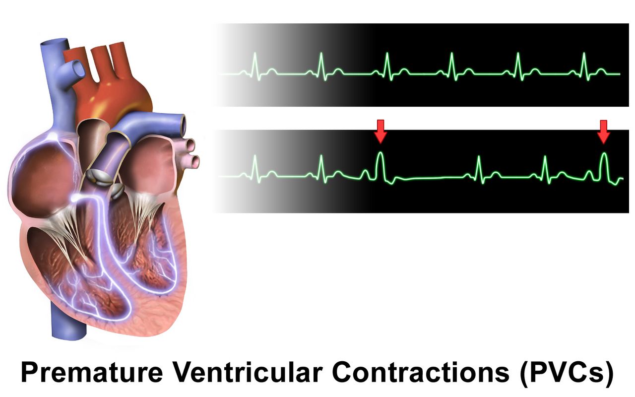
What you are actually monitoring
Fetal oxygenation depends on a chain: maternal blood oxygen, maternal cardiac output, uterine artery flow, the intervillous space, the placenta itself, the umbilical cord and the fetal circulation. Every intrapartum intervention you will ever make targets one of those links. Before applying a monitor, perform Leopold manoeuvres with the bladder empty and a wedge under one hip: they identify the fetal part in the fundus, the location of the back (where you will hear the heart best), the presenting part, and its degree of descent.
For a low-risk labour, intermittent auscultation is an evidence-supported alternative to continuous monitoring: listen for 30 to 60 seconds immediately after a contraction, every 15 to 30 minutes in active first stage and every 5 to 15 minutes in second stage. Continuous electronic monitoring is used when risk factors exist — oxytocin, epidural, meconium, hypertensive disease, diabetes, growth restriction, prematurity, prior caesarean, an abnormal auscultated pattern. Internal monitoring with a fetal scalp electrode and an intrauterine pressure catheter requires ruptured membranes, adequate dilation and an engaged presenting part.
The five terms and the three categories
Baseline is the mean rate over a 10-minute window excluding accelerations, decelerations and marked variability, rounded to increments of 5, requiring at least 2 identifiable minutes. Normal is 110 to 160 bpm. Variability is the beat-to-beat fluctuation of the baseline and is the single most important indicator that the fetal autonomic nervous system is well oxygenated: absent (undetectable), minimal (5 bpm or less), moderate (6 to 25 bpm — the goal), or marked (more than 25 bpm). Accelerations at 32 weeks or more are a rise of at least 15 bpm lasting at least 15 seconds; before 32 weeks, 10 bpm for 10 seconds. They are reassuring.
Decelerations are classified by shape and timing, not depth. Early decelerations are gradual (onset to nadir 30 seconds or more) and mirror the contraction; they reflect head compression and require no intervention. Late decelerations are gradual with the nadir after the peak of the contraction and signal uteroplacental insufficiency — these are the ones that demand action. Variable decelerations are abrupt (onset to nadir under 30 seconds), at least 15 bpm below baseline for at least 15 seconds but under 2 minutes, and reflect cord compression. A prolonged deceleration lasts 2 minutes or more but under 10 minutes; 10 minutes or more is a baseline change.
| Pattern | Cause (VEAL CHOP) | First nursing action |
|---|---|---|
| Variable deceleration | Cord compression | Reposition — lateral, then knee-chest or Trendelenburg if persistent; check for prolapse |
| Early deceleration | Head compression | None; continue to observe and document |
| Acceleration | Oxygenation intact | None; reassuring |
| Late deceleration | Placental insufficiency | Full intrauterine resuscitation and notify the provider |
The NICHD three-tier system converts these findings into a decision. Category I requires all of: baseline 110 to 160, moderate variability, no late or variable decelerations; early decelerations and accelerations may be present or absent. It is normal and needs routine care. Category III is either absent variability accompanied by recurrent late decelerations, recurrent variable decelerations or bradycardia, or a sinusoidal pattern. It predicts abnormal fetal acid-base status and demands immediate intrauterine resuscitation and preparation for expedited birth. Category II is everything in between — the majority of tracings — and is indeterminate: it obliges surveillance, correction of what you can correct, and reassessment, not panic.
Intrauterine resuscitation, in order
When a tracing deteriorates, act in this sequence. First, reposition to a lateral position — it takes seconds, relieves aortocaval compression and often relieves cord compression at the same time. Second, stop or reduce the oxytocin, because the drug that is reducing placental refill is the one thing entirely under your control. Third, give an intravenous fluid bolus of 500 to 1000 mL of an isotonic non-dextrose solution to raise uterine perfusion. Fourth, perform a vaginal examination to check for cord prolapse and rapid descent. Fifth, notify the provider using SBAR. For tachysystole persisting after oxytocin is stopped, a tocolytic such as terbutaline 0.25 mg subcutaneously may be ordered. Amnioinfusion may be ordered for recurrent variable decelerations.
Supplemental oxygen at 10 L/min by non-rebreather mask has long appeared on this list, and many facilities still include it. Current evidence has not demonstrated improved neonatal outcomes from routine oxygen for a category II tracing, so follow your institution’s protocol but do not let reaching for a mask delay the repositioning, the oxytocin and the fluid that actually change uterine blood flow. Digital scalp stimulation producing an acceleration is reassuring and suggests a fetal pH above 7.20.
Moderate variability is the most reassuring finding on a strip, late decelerations mean placental insufficiency, and the first move for almost any worrying pattern is to reposition and stop the oxytocin — not to reach for oxygen.
Section 4Comfort, Analgesia and Anaesthesia
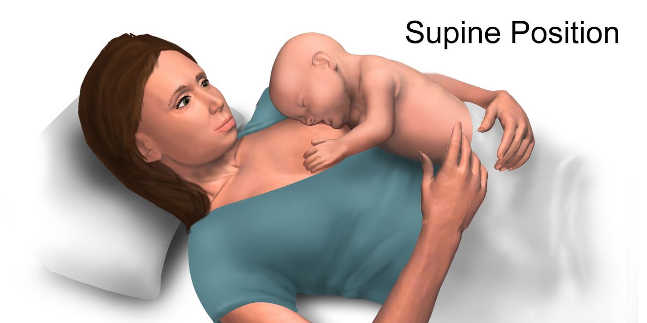
Where labour pain comes from, and what works without a drug
First-stage pain is visceral: cervical stretching and uterine ischaemia referred through T10 to L1, felt in the lower abdomen, back and thighs, and poorly localised. Second-stage pain is somatic: distension of the vagina and perineum carried by the pudendal nerve through S2 to S4, sharp and precisely located. This anatomy explains why a block adequate for the first stage may not cover the second, and why a pudendal block placed late covers perineal repair but not contractions.
Non-pharmacological measures are not a consolation prize. Continuous one-to-one labour support — a partner, a doula, a nurse who stays — reduces caesarean birth, shortens labour and improves satisfaction more reliably than most drugs. Hydrotherapy, warm packs, cold packs, counterpressure over the sacrum for occiput-posterior back pain, the double hip squeeze, position change every 20 to 30 minutes, birth balls, ambulation, rhythmic breathing, focal points, guided imagery, hypnobirthing, acupressure, TENS and intradermal sterile water papules for back pain all have supporting evidence and no fetal effect. Offer them first and keep offering them after an epidural is placed.
Systemic agents and nitrous oxide
Intravenous opioids give partial relief and cross the placenta. Fentanyl 50 to 100 micrograms IV has a short duration and less neonatal depression; butorphanol 1 to 2 mg and nalbuphine 10 mg are mixed agonist-antagonists that cause less nausea but must be avoided in an opioid-dependent patient because they can precipitate withdrawal in the parent and the fetus. The clinical rule is timing: an opioid given within roughly 1 to 4 hours of birth peaks in the newborn at the worst moment, producing respiratory depression and poor feeding. If birth is imminent, hold the dose and tell the provider why.
Two related safety points. A newborn depressed after maternal opioid is managed by effective positive-pressure ventilation, not naloxone; naloxone is no longer part of the initial delivery-room resuscitation sequence, and it must never be given to the newborn of an opioid-dependent parent because it can precipitate seizures. Nitrous oxide in a 50:50 blend with oxygen is self-administered by mask, started about 30 seconds before a contraction, and is safe because a sedated patient drops the mask; only the patient may hold it, and that includes not letting a well-meaning partner help.
Neuraxial analgesia and its complications
An epidural places a catheter in the epidural space for continuous local anaesthetic with an opioid; a spinal injects a single dose into cerebrospinal fluid for caesarean birth; a combined spinal-epidural gives rapid onset plus a catheter. Before placement: informed consent, IV access, a fluid preload or coload of 500 to 1000 mL of isotonic non-dextrose solution, baseline vital signs and fetal heart rate, an empty bladder, and positioning either sitting with the back arched or side-lying curled. Contraindications include patient refusal, coagulopathy or therapeutic anticoagulation, a platelet count below roughly 70 to 100 × 109/L, infection at the site or frank sepsis, uncorrected hypovolaemia and raised intracranial pressure.
Maternal hypotension is the most common complication — a systolic pressure below 100 mmHg or a fall of more than 20 per cent from baseline — and it reduces placental perfusion within seconds, so late decelerations often appear before the patient feels faint. Blood pressure is checked every 5 minutes for the first 15 to 20 minutes after dosing and after every top-up. The sequence when it drops: turn the patient to a lateral position or apply left uterine displacement first, then open the intravenous fluid, then apply oxygen, then notify anaesthesia and give an ordered vasopressor such as ephedrine or phenylephrine.
Other consequences follow from the physiology. Sensory block causes urinary retention — scan or catheterise every 2 hours. Motor block causes fall risk — the patient does not stand unassisted until motor strength and sensation return. Opioid in the mixture causes pruritus and occasionally delayed respiratory depression. A dural puncture produces a post-dural-puncture headache that is positional, worse sitting or standing and relieved lying flat; treat with hydration, analgesia and caffeine, and definitively with an epidural blood patch of 10 to 20 mL of the patient’s own blood. Maternal fever without infection is common and complicates newborn evaluation.
Continuous labour support is the best-evidenced comfort measure in obstetrics, and when an epidural drops the blood pressure the first action is to reposition for left uterine displacement — fluid, oxygen and vasopressors come after.
Section 5Birth, the Newborn’s First Minutes, and the Sequences That Save Lives
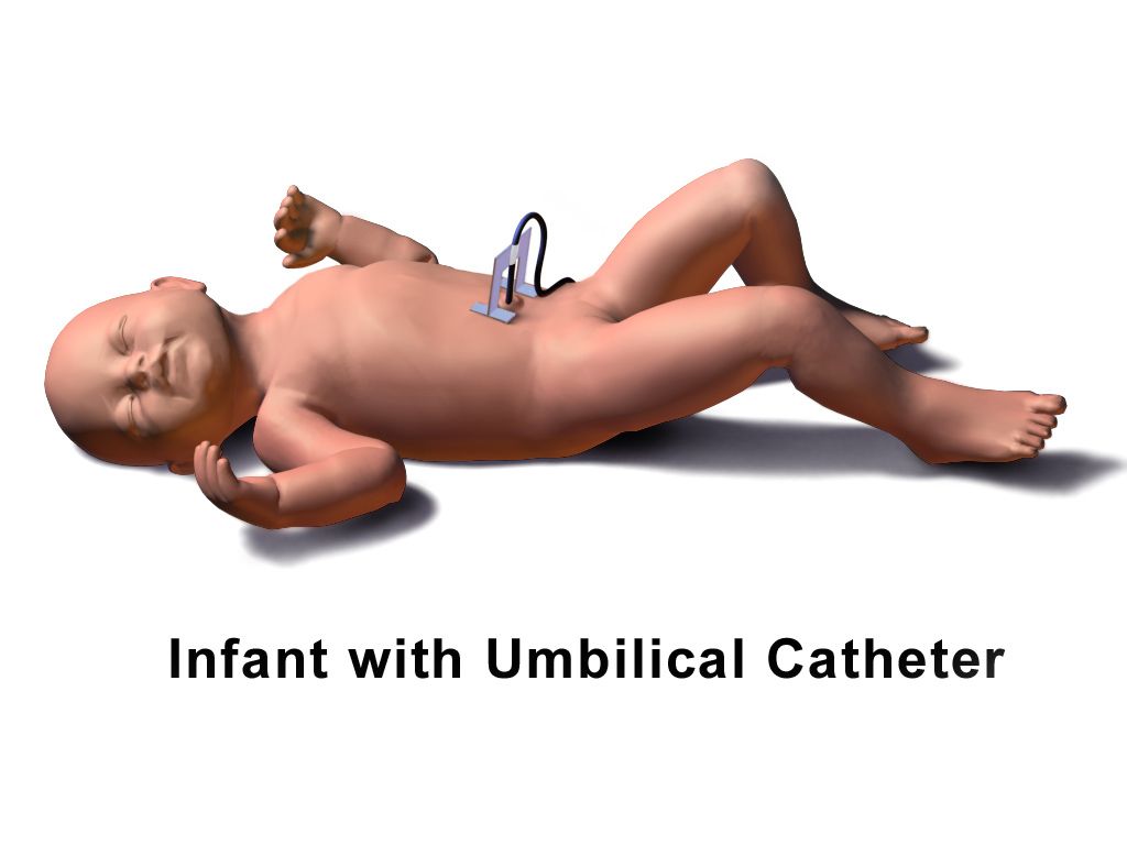
The first ten minutes of a newborn’s life
For a vigorous term newborn, the priorities are warmth, airway and contact. Dry thoroughly, remove the wet linen, place skin-to-skin with a cap, and delay cord clamping 30 to 60 seconds — a transfusion of roughly 20 to 30 mL/kg of placental blood that raises iron stores for months. Assessment at birth is continuous rather than scheduled: watch tone, respiratory effort and heart rate from the first seconds, because no recorded score directs resuscitation and you never wait for one before acting.
Normal newborn axillary temperature is 36.5 to 37.5 °C. If the newborn is not breathing or the heart rate is under 100, begin positive-pressure ventilation within the first minute of life — ventilation is the single most effective intervention. If the heart rate stays under 60 after 30 seconds of ventilation that visibly moves the chest, add compressions in a 3:1 ratio, delivering 90 compressions and 30 breaths each minute.
Postpartum haemorrhage: massage the fundus first
Haemorrhage is defined as cumulative blood loss of 1000 mL or more, or any blood loss accompanied by signs of hypovolaemia, within 24 hours of birth regardless of route. Visual estimation underestimates loss by 30 to 50 per cent, so measure it: weigh blood-soaked items, subtract the dry weight, and count 1 gram as 1 mL. Remember that a healthy pregnant person can lose 1500 mL before the blood pressure falls, because pregnancy adds 40 to 50 per cent to plasma volume — rising pulse, narrowing pulse pressure, restlessness and delayed capillary refill come first.
The causes are the four Ts: Tone (uterine atony, 70 to 80 per cent), Trauma (laceration, haematoma, inversion), Tissue (retained placenta) and Thrombin (coagulopathy). Because atony dominates, the first action for a boggy uterus with heavy bleeding is to massage the fundus, one hand cupping the fundus and the other braced above the symphysis to support the lower segment. Then empty the bladder, call for help, open the intravenous fluids, give uterotonics as ordered and quantify the loss. If the fundus is firm and the bleeding continues, the cause is not tone — look for a laceration or a haematoma, and remember that a steadily worsening perineal or rectal pain with a tense purple swelling is a haematoma.
| Uterotonic | Dose | Do not give if | Watch for |
|---|---|---|---|
| Oxytocin | 10 to 40 units in 500 to 1000 mL isotonic IV; or 10 units IM | Never as an undiluted IV push | Hypotension, tachycardia, water intoxication |
| Methylergonovine | 0.2 mg IM every 2 to 4 hours | Hypertension, preeclampsia, cardiac disease | Check BP before every dose; headache, chest pain |
| Carboprost | 250 micrograms IM every 15 to 90 min, max 8 doses (2 mg) | Asthma | Diarrhoea, vomiting, fever, bronchospasm |
| Misoprostol | 600 to 1000 micrograms oral, sublingual or rectal | Known prostaglandin allergy | Fever, shivering |
| Tranexamic acid | 1 g IV over 10 min, within 3 hours of birth | Active thromboembolic disease | Repeat once after 30 min if bleeding persists |
If pharmacology fails, escalation is mechanical and surgical: bimanual compression, an intrauterine balloon tamponade device, uterine artery embolisation, compression sutures, and hysterectomy as the last resort. Activate the massive transfusion protocol early rather than late.
Magnesium sulfate toxicity, cord prolapse, shoulder dystocia and uterine rupture
Pre-eclampsia is what puts most magnesium infusions in the room, so the criteria matter. It is new-onset blood pressure of 140/90 mmHg or higher on two readings at least 4 hours apart after 20 weeks of gestation, together with proteinuria of 300 mg or more in 24 hours, a protein-to-creatinine ratio of 0.3 or more, or 2+ on dipstick when no quantitative test is available. Proteinuria is not required if any severe feature is present: systolic 160 mmHg or higher or diastolic 110 mmHg or higher, platelets below 100 × 109/L, transaminases at twice the upper limit of normal, severe persistent right upper quadrant or epigastric pain, creatinine above 1.1 mg/dL or doubled from baseline, pulmonary oedema, or new cerebral or visual symptoms. HELLP — Haemolysis, Elevated Liver enzymes, Low Platelets — is the variant that catches people out, because the blood pressure may be only mildly raised or even normal and the patient presents complaining of right upper quadrant pain, nausea and feeling generally unwell. Its criteria are platelets below 100 × 109/L, AST or ALT at least twice normal, and LDH of 600 U/L or more with haemolysis on the blood film. Bedside care is the same for both: magnesium sulfate for seizure prophylaxis, antihypertensives for severe-range pressures, a low-stimulus room with side rails padded and suction and oxygen at hand, strict intake and output, and birth as the only definitive cure.
Magnesium sulfate is given for seizure prophylaxis in preeclampsia and for fetal neuroprotection, typically 4 to 6 g intravenously over 15 to 30 minutes then 1 to 2 g per hour by pump. The therapeutic range is about 4 to 7 mEq/L (4.8 to 8.4 mg/dL). Toxicity climbs in a fixed order: deep tendon reflexes disappear first, at roughly 8 to 12 mg/dL, then respiratory depression from about 12 mg/dL with respiratory arrest by 15 to 20 mg/dL, then cardiac conduction changes and cardiac arrest above roughly 25 mg/dL. That order is the whole reason you assess reflexes hourly — the reflex is the early warning, and by the time the respiratory rate falls you have already missed it. Hold the infusion and notify the provider for absent reflexes, a respiratory rate under 12 per minute, urine output under 30 mL/h (magnesium is cleared renally) or a falling level of consciousness. The first action for suspected toxicity is to stop the infusion, then give the antidote calcium gluconate 1 g (10 mL of a 10 per cent solution) intravenously over about 3 minutes, support ventilation, and draw a serum magnesium level. Expect the newborn to be hypotonic and possibly slow to breathe.
Umbilical cord prolapse follows rupture of membranes with an unengaged presenting part, and is more likely with polyhydramnios, malpresentation, prematurity, multiple gestation or a long cord. It shows as a sudden prolonged deceleration or severe recurrent variables immediately after membranes rupture, or as a pulsating cord felt or seen at the introitus. The first action is to insert a gloved hand and lift the presenting part off the cord, and to keep that hand in place until the infant is delivered — do not remove it, and do not attempt to push the cord back inside. Call for help without leaving the patient. Position knee-chest or Trendelenburg or exaggerated Sims with the hips elevated. Handle the cord as little as possible because touching it causes vasospasm; if it is outside the body, cover it with warm saline-moistened sterile gauze. Stop oxytocin, open fluids, give oxygen, and prepare for immediate caesarean birth.
Shoulder dystocia is the anterior shoulder impacted behind the symphysis after the head is born — the turtle sign. Risks include macrosomia, diabetes in pregnancy, previous dystocia and a prolonged second stage, but half of cases have no risk factor. Call for help and note the time, then work through HELPERR: McRoberts (two people hyperflex the thighs sharply onto the abdomen, flattening the sacral promontory and rotating the symphysis upward) plus suprapubic pressure resolves the majority. Fundal pressure is never used — it drives the shoulder harder into the symphysis and risks uterine rupture. Further steps are internal rotational manoeuvres, delivery of the posterior arm, and the all-fours (Gaskin) position. Complications include brachial plexus injury, clavicular or humeral fracture and hypoxic injury for the newborn, and haemorrhage and third or fourth degree laceration for the birthing parent.
Uterine rupture is rare and catastrophic. Suspect it with a previous classical incision or uterine surgery, a trial of labour after caesarean that is being induced or augmented, excessive oxytocin, grand multiparity or trauma. The most common presenting sign is an abnormal fetal heart rate — a prolonged deceleration or bradycardia, not pain. Other findings are sudden sharp tearing abdominal pain, loss of station as the presenting part retracts, cessation of contractions, palpable fetal parts through the abdominal wall and signs of hypovolaemic shock; external bleeding may be minimal because the haemorrhage is intra-abdominal. Stop the oxytocin, call the emergency team, give oxygen and fluids, and prepare for immediate laparotomy — the interval between rupture and birth is measured in minutes.
| Red flag | What it usually means | First action |
|---|---|---|
| Prolonged deceleration right after membranes rupture | Cord prolapse | Gloved hand elevates the presenting part and stays there; knee-chest |
| Boggy fundus, heavy lochia | Uterine atony | Massage the fundus, then empty the bladder and give uterotonics |
| Fundus firm but bleeding briskly | Laceration or haematoma | Inspect the perineum and cervix; call the provider |
| Absent deep tendon reflexes on magnesium | Impending respiratory depression | Stop the infusion, then calcium gluconate 1 g IV |
| Head born, chin retracts, no restitution | Shoulder dystocia | Call for help, note the time, McRoberts plus suprapubic pressure |
| Sudden bradycardia with tearing pain in a TOLAC | Uterine rupture | Stop oxytocin, activate emergency caesarean |
| Sudden dyspnoea, hypotension, then bleeding from every site | Amniotic fluid embolism | Call the code team, support airway and circulation, anticipate coagulopathy |
In obstetric emergencies the sequence is the treatment: massage the fundus first, stop the magnesium before you reach for calcium gluconate, lift the presenting part off a prolapsed cord and never push it back, and use McRoberts with suprapubic — never fundal — pressure.
The chapter in one breath
Labour is the interaction of five forces: a passenger whose skull moulds and whose position and station you can read from a three-letter abbreviation, a passage whose shape and capacity change with maternal positioning, powers described by frequency, duration, intensity and resting tone, and the position and psyche that quietly determine uterine blood flow. Only progressive cervical change proves labour has begun, and the first act after any rupture of membranes is a full minute of fetal heart rate. The four stages have expected patterns rather than deadlines, and the fourth stage — fundus, lochia, bladder, vital signs on a tightening schedule — carries the greatest preventable mortality. Fetal surveillance rests on five terms and three categories, with moderate variability the most reassuring finding and late decelerations the one that demands action; intrauterine resuscitation begins with repositioning and stopping the oxytocin. Comfort ranges from continuous labour support, the best-evidenced measure of all, to neuraxial analgesia whose commonest complication is hypotension corrected first by left uterine displacement. The newborn’s first minutes are warmth, skin-to-skin contact and delayed cord clamping, with positive-pressure ventilation begun inside the first minute if the infant is not breathing or the heart rate is under 100. And in every emergency — haemorrhage, magnesium toxicity, cord prolapse, shoulder dystocia, uterine rupture — the memorised order of actions is what saves the life.
Words worth knowing
| Term | What it means |
|---|---|
| Attitude | Relationship of fetal parts to one another; complete flexion gives the smallest presenting diameter, about 9.5 cm. |
| Lie | Relationship of the fetal spine to the maternal spine — longitudinal, transverse or oblique. |
| Presentation | The fetal part entering the pelvis first: cephalic, breech or shoulder. |
| Position | Three-letter notation of the fetal reference point within the pelvis, such as LOA (left occiput anterior). |
| Station | Descent in centimetres relative to the ischial spines, from −5 to +5; 0 indicates engagement. |
| Moulding | Overlapping of fetal skull bones at the unfused sutures during descent; resolves within days. |
| Effacement | Thinning and shortening of the cervix, expressed as a percentage. |
| Bishop score | Cervical readiness score out of 13; 8 or more is favourable for induction, 6 or less calls for ripening. |
| Tachysystole | More than five contractions in 10 minutes averaged over 30 minutes. |
| Montevideo units | Sum of contraction amplitudes above baseline over 10 minutes by intrauterine catheter; 200 or more is adequate. |
| Variability | Beat-to-beat fluctuation of the fetal heart baseline; moderate (6 to 25 bpm) indicates intact oxygenation. |
| Late deceleration | Gradual fall in fetal heart rate with the nadir after the contraction peak; uteroplacental insufficiency. |
| Category III tracing | Absent variability with recurrent late or variable decelerations or bradycardia, or a sinusoidal pattern; act immediately. |
| Intrauterine resuscitation | Reposition, stop oxytocin, fluid bolus, check for prolapse, notify — measures that restore fetal oxygen delivery. |
| Cardinal movements | Engagement, descent, flexion, internal rotation, extension, restitution and expulsion. |
| Quantitative blood loss | Measured rather than estimated loss; blood-soaked items are weighed with 1 gram counted as 1 mL. |
| Uterine atony | A soft, poorly contracted uterus; the cause of 70 to 80 per cent of postpartum haemorrhage. |
| McRoberts manoeuvre | Sharp hyperflexion of the thighs onto the abdomen to flatten the sacral promontory in shoulder dystocia. |
| Delayed cord clamping | Waiting 30 to 60 seconds before clamping, transfusing placental blood and improving newborn iron stores. |
Check yourself
INTRAPARTUM COMPLICATIONS AND OBSTETRIC EMERGENCIES
Most labours are ordinary, and then a small number of them are not. The transition is often measured in seconds: membranes rupture and the fetal heart drops and stays down; the head delivers and retracts against the perineum; the placenta separates and the fundus never firms. In these moments the nurse is the person in the room, and the correct first action is frequently not the intuitive one. You do not push a prolapsed cord back in — you lift the presenting part off it. You do not press on the fundus in shoulder dystocia — you flex the hips and press above the pubic bone. You do not reach for the antidote before you stop the magnesium infusion. This chapter builds each emergency the way it is practised: mechanism first, then what you assess, then what you do and in what order, then what goes wrong afterwards. Numbers are given as they are actually written on the order and drawn into the syringe.
By the end of this chapter you can
- Interpret a fetal heart rate tracing using baseline, variability, accelerations and decelerations, and explain the physiological mechanism behind early, variable, late and prolonged decelerations.
- Classify a tracing using the three-tier category system and carry out the intrauterine resuscitation sequence in priority order.
- Describe protracted and arrested labour patterns in non-blaming, descriptive language, and identify the nursing implications of malposition, malpresentation and uterine tachysystole.
- State the first action, the rationale and the contraindicated action for umbilical cord prolapse, shoulder dystocia, uterine rupture, placental abruption and amniotic fluid embolism.
- Administer and monitor magnesium sulfate, recognise the toxicity ladder in the order it appears, and give calcium gluconate correctly.
- Manage immediate postpartum haemorrhage using the four Ts, massaging the fundus first, and match each uterotonic to its dose, route and contraindication.
- Perform the opening steps of neonatal resuscitation, score and interpret the Apgar at 1 and 5 minutes, follow the minute-by-minute preductal saturation targets, and calculate weight-based newborn doses from the kilogram weight, showing the arithmetic and having it independently verified.
- Escalate deterioration using SBAR and apply delegation limits that keep assessment, teaching, evaluation and the unstable patient with the registered nurse.
Section 1Reading the Fetal Heart
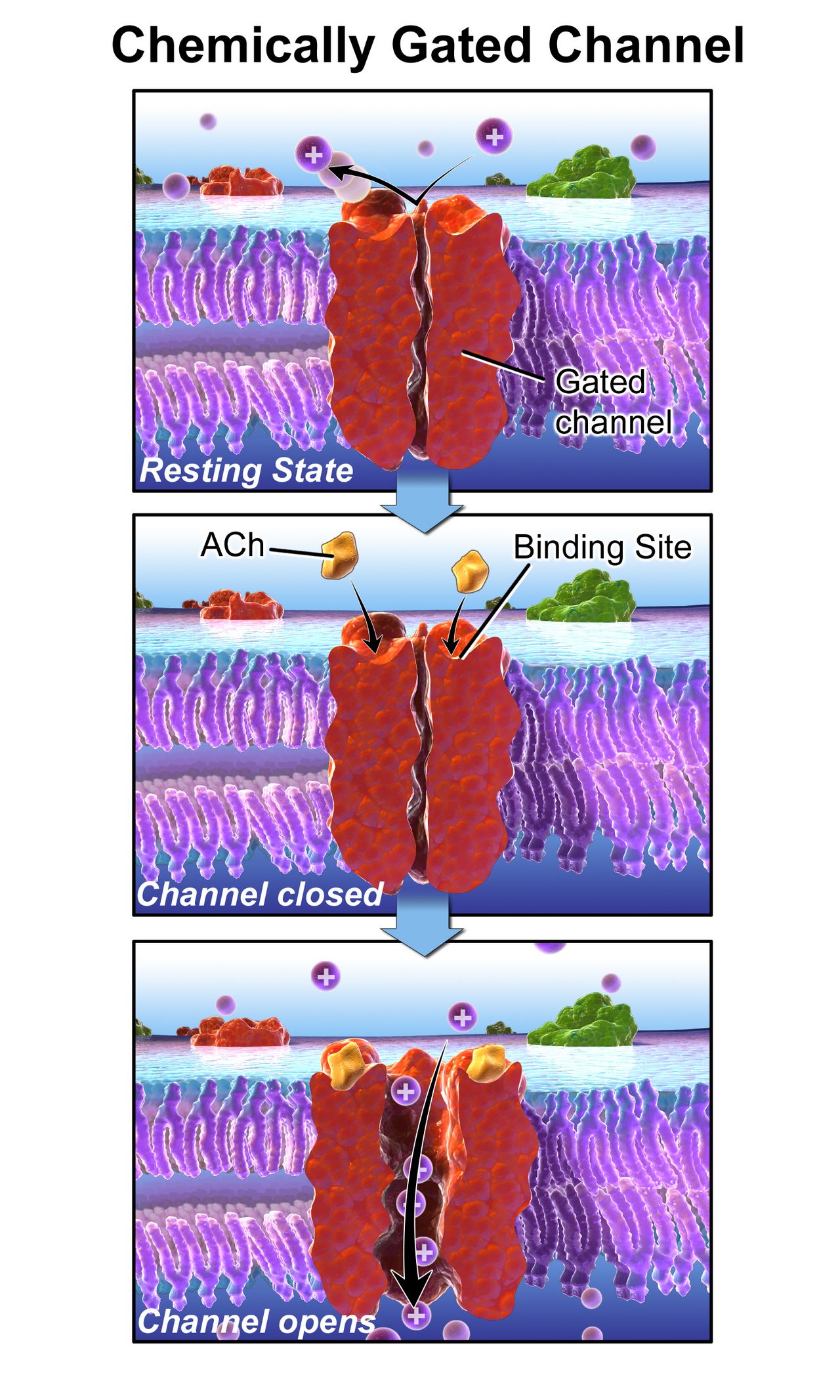
Baseline and variability: the two numbers that matter most
Electronic fetal monitoring does not measure fetal oxygenation. It measures the fetal autonomic nervous system’s response to oxygenation, which is why the tracing is read as a physiological story rather than a number. Every interpretation starts with the baseline: the mean fetal heart rate rounded to the nearest 5 beats per minute over a 10-minute window, excluding accelerations, decelerations and stretches of marked variability, and requiring at least 2 minutes of identifiable baseline in that window. Normal is 110–160 bpm. Above 160 bpm for 10 minutes or longer is tachycardia — think maternal fever and chorioamnionitis first, then dehydration, thyrotoxicosis, beta-agonists such as terbutaline, and early fetal compensation for hypoxia. Below 110 bpm for 10 minutes or longer is bradycardia — think cord compression or occlusion, maternal hypotension after an epidural, rapid descent, uterine rupture, or maternal hypoxia.
Variability is the irregular fluctuation in the baseline, and it is the single most useful feature on the strip. It reflects an intact, well-oxygenated pathway running from cerebral cortex through midbrain to vagus nerve to the fetal heart, so it tells you about the fetal brain, not just the fetal heart. It is graded by the amplitude of the fluctuation from peak to trough: absent (undetectable), minimal (detectable but 5 bpm or less), moderate (6–25 bpm) and marked (more than 25 bpm). Moderate variability is the reassuring finding of the whole tracing — when it is present, the fetus is very unlikely to be acidaemic at that moment, even in the presence of decelerations.
Minimal or absent variability has benign causes as well as ominous ones, and you sort them by asking what changed. A fetal sleep cycle lasts about 20–40 minutes and rarely longer than 80. Opioids, magnesium sulfate and central nervous system depressants flatten variability. Prematurity produces less variability. Fetal tachycardia mechanically reduces it. But absent variability that arrives with recurrent late decelerations, or that persists after a sleep cycle should have ended, is the tracing telling you the fetal central nervous system is being deprived of oxygen, and it is escalated, not observed.
Accelerations and the mechanism of each deceleration
An acceleration is an abrupt increase in the fetal heart rate — at 32 weeks or more, at least 15 bpm above baseline lasting at least 15 seconds from onset to return; before 32 weeks, at least 10 bpm for at least 10 seconds. Accelerations mean an intact somatic nervous system and a non-acidaemic fetus. They can be provoked deliberately: fetal scalp stimulation during a vaginal examination that produces an acceleration predicts a fetal scalp pH of 7.20 or above, and is a legitimate bedside test when the tracing is ambiguous. An acceleration lasting 2 minutes or more but less than 10 minutes is a prolonged acceleration; 10 minutes or more is a change in baseline.
Decelerations are classified by shape and by timing against the contraction, and every classification exists because the shape reveals the mechanism. Two words do the work. Gradual means the onset to the nadir takes 30 seconds or longer, and gradual implies a reflex or chemoreceptor pathway. Abrupt means onset to nadir takes less than 30 seconds, and abrupt implies mechanical interruption of cord blood flow.
Early decelerations are gradual, mirror the contraction, and the nadir sits under the peak. The mechanism is fetal head compression during descent, which raises intracranial pressure, triggers a vagal response and slows the heart. They are benign, need no intervention, and typically appear in active labour at 4–7 cm and again in the second stage. Variable decelerations are abrupt, drop at least 15 bpm below baseline, last at least 15 seconds but less than 2 minutes, and vary in shape and in their relationship to contractions. The mechanism is umbilical cord compression: the thin-walled umbilical vein is squeezed first, venous return falls, the fetus accelerates briefly, then the arteries are compressed, systemic resistance rises, baroreceptors fire and the rate plunges. Occasional variables are common and tolerated; recurrent deep variables with slow return, loss of variability or a rising baseline are not. Late decelerations are gradual and shifted — the onset, the nadir and the recovery each come after the corresponding point of the contraction. The mechanism is uteroplacental insufficiency: the contraction transiently reduces intervillous perfusion, fetal oxygen tension falls, chemoreceptors respond, and in a fetus with poor reserve the response repeats with every contraction. Late decelerations are never normal. A prolonged deceleration falls at least 15 bpm below baseline for 2 minutes or more but less than 10 minutes; beyond 10 minutes it is a new baseline. A sinusoidal pattern — a smooth, undulating sine wave of 3–5 cycles per minute persisting 20 minutes or more — suggests severe fetal anaemia and is an emergency.
| Deceleration | Shape and timing | Mechanism | Response |
|---|---|---|---|
| Early | Gradual; nadir under the peak of the contraction | Head compression, vagal reflex | None; document |
| Variable | Abrupt; any relationship to contractions | Umbilical cord compression | Reposition; consider amnioinfusion if recurrent |
| Late | Gradual; onset, nadir and recovery all after the contraction | Uteroplacental insufficiency | Full intrauterine resuscitation; never ignore |
| Prolonged | 2 minutes or more, less than 10 minutes | Cord occlusion, hypotension, tachysystole, rupture, prolapse | Vaginal exam to exclude prolapse; full resuscitation |
| Sinusoidal | Smooth sine wave, 3–5 cycles per minute, 20 minutes or more | Severe fetal anaemia (vasa praevia, feto-maternal haemorrhage, isoimmunisation) | Category III; prepare for immediate birth |
Three categories and the intrauterine resuscitation sequence
The three-tier system converts the description into a decision. Category I requires all of the following: baseline 110–160 bpm, moderate variability, no late or variable decelerations; early decelerations and accelerations may be present or absent. Category I is normal and predicts normal fetal acid–base status. Category III is either absent variability accompanied by recurrent late decelerations, recurrent variable decelerations or bradycardia, or a sinusoidal pattern. Category III predicts abnormal acid–base status and demands prompt evaluation and, if it does not resolve, expedited birth. Category II is everything in between — the great majority of tracings in labour. Category II is not a diagnosis; it is an instruction to look harder, intervene, and re-evaluate.
Intrauterine resuscitation aims at three physiological targets: relieve cord compression, restore uteroplacental perfusion, and reduce uterine activity. The sequence, in the order a nurse actually performs it, is: (1) stop the oxytocin if it is infusing — you remove the cause before you treat the effect; (2) reposition the birthing parent to a lateral position (either side) or knee-chest, which lifts the uterus off the inferior vena cava and often shifts the cord; (3) perform a vaginal examination to exclude cord prolapse and to assess rapid descent; (4) give an intravenous fluid bolus of 500–1000 mL of an isotonic crystalloid such as lactated Ringer’s if the person is not fluid-restricted, which is the specific treatment for post-epidural hypotension; (5) treat hypotension with the ordered vasopressor if fluids are insufficient; (6) consider a tocolytic — terbutaline 0.25 mg subcutaneously — for tachysystole; (7) notify the provider and prepare for expedited birth. Supplemental oxygen at 10 L/min by non-rebreather mask remains in many unit protocols, but current evidence has not demonstrated improved neonatal outcomes from routine maternal oxygen for a category II tracing; follow your facility protocol and know that it is not the intervention that fixes the problem. Amnioinfusion — warmed isotonic fluid instilled through an intrauterine pressure catheter — is the targeted treatment for recurrent variable decelerations caused by oligohydramnios.
Document what you saw, what you did and what happened next, using the standard terminology rather than words such as “good strip” or “fetal distress”. Interpretation of the fetal heart rate tracing is an assessment: it is a registered nurse function and it is not delegated to assistive personnel. Unlicensed personnel may apply the monitor belts and reposition a stable patient; they may not tell you what the strip means.
Moderate variability is the strongest bedside evidence that a fetus is currently well oxygenated, and when a tracing deteriorates the first move is to remove the cause — stop the oxytocin and reposition — before anything else.
Section 2When the Labour Pattern Deviates
Describing the pattern, not blaming the patient
The older phrase “failure to progress” is retired for good reason. It assigns the problem to the labouring person, it is imprecise, and it has been associated with caesarean births performed before labour was given a fair chance. Describe what you observe: a protracted pattern (progress slower than expected) or an arrest pattern (no progress over a defined interval despite adequate uterine activity). Contemporary labour curves place the onset of the active phase at about 6 cm, not 4 cm, and the latent phase is now understood to be far longer and far more variable than Friedman’s original curve implied. Arrest of dilatation is defined at 6 cm or more with ruptured membranes and no cervical change for 4 hours or more with adequate contractions, or 6 hours or more with inadequate contractions. In the second stage, arrest is considered after 3 hours of pushing in a nulliparous person and 2 hours in a multiparous person, with an additional hour allowed for regional anaesthesia.
Adequacy of contractions is quantified, not guessed. With an intrauterine pressure catheter, Montevideo units are calculated by summing the peak-minus-baseline pressures of every contraction in a 10-minute window; 200 MVUs or more is adequate. Without a catheter, palpate: a contraction you can indent easily is mild, one that indents with difficulty is moderate, and one that feels like a forehead is strong. A hypotonic pattern gives infrequent, weak, poorly coordinated contractions with a soft resting uterus and slow progress, and it responds to augmentation, amniotomy, position change and emptying the bladder. A hypertonic pattern gives frequent, painful, ineffective contractions with elevated resting tone, most often in the latent phase; it responds to therapeutic rest, hydration and analgesia, and augmenting it makes it worse.
Non-pharmacological measures are genuine interventions. Upright and mobile positions, a birthing ball, hands and knees, side-lying with a peanut ball between the knees for a person with an epidural, hydration, an empty bladder every 2 hours, and continuous labour support all shorten labour and reduce operative birth. Say “your labour is taking a slower path” rather than anything that implies the person is failing at it.
Malposition, malpresentation and the diameters they present
Occiput posterior position is the commonest malposition. The fetal occiput lies against the sacrum, the poorly flexed head presents a larger diameter, and the labour is characterised by severe continuous low back pain, early urge to push, a prolonged second stage and a higher rate of operative birth. Nursing management is positional and effective: hands and knees, lunges, open-knee-chest, side-lying on the side opposite the fetal spine, and counter-pressure to the sacrum. Most rotate spontaneously.
Breech presentation occurs in about 3–4 per cent of term births. Frank breech has hips flexed and knees extended; complete breech has both flexed; footling has one or both feet presenting and carries the greatest risk of cord prolapse because the presenting part does not fill the pelvic inlet. External cephalic version may be offered at 36–37 weeks. Face presentation may deliver vaginally if the chin is anterior (mentum anterior) but cannot if the chin is posterior. Brow presentation presents the largest diameter of all and usually requires caesarean unless it converts. A transverse lie cannot deliver vaginally. Compound presentation, most often a hand alongside the head, raises the risk of cord prolapse and of laceration.
Two nursing observations matter across all of these. First, in any malpresentation, the moment membranes rupture is the moment of highest risk for cord prolapse — auscultate or observe the fetal heart rate immediately after every rupture, spontaneous or artificial, for a full minute. Second, an unengaged presenting part in the presence of ruptured membranes means the patient is not ambulated without a provider order.
Induction, augmentation and tachysystole
Cervical readiness is scored with the Bishop score across dilatation, effacement, station, consistency and position; a score of 8 or more predicts a vaginal birth as likely as spontaneous labour, and 6 or less indicates that cervical ripening should precede oxytocin. Ripening agents include vaginal misoprostol 25 mcg every 3–6 hours, dinoprostone gel or insert, and mechanical methods such as a transcervical balloon catheter. Misoprostol is contraindicated in anyone with a previous caesarean or other uterine scar because it markedly increases the risk of uterine rupture.
Oxytocin is a high-alert medication. It is mixed as a dilute solution (commonly 30 units in 500 mL of isotonic fluid), delivered by infusion pump as a secondary line piggybacked into the primary line at the port closest to the patient so it can be stopped instantly, and titrated against uterine response rather than to a target rate. Typical protocols start at 0.5–2 milliunits per minute and increase by 1–2 milliunits per minute no more often than every 30–60 minutes. The goal is contractions every 2–3 minutes lasting 40–90 seconds, of moderate to strong intensity (40–70 mmHg by catheter), with a resting tone below 20 mmHg and a resting interval of at least 60 seconds. Because oxytocin is structurally similar to antidiuretic hormone, prolonged high-dose infusion in large fluid volumes can cause water intoxication — watch for headache, nausea, confusion and a falling serum sodium.
Tachysystole is more than 5 contractions in 10 minutes averaged over a 30-minute window. It matters because the placenta is perfused between contractions, not during them; take away the resting interval and you take away fetal oxygenation. Management depends on the tracing. With a category I tracing, reposition, give a fluid bolus and reduce the oxytocin rate. With a category II or III tracing, stop the oxytocin outright, reposition, give a bolus, and if the pattern persists give terbutaline 0.25 mg subcutaneously. Never treat tachysystole by increasing the oxytocin because “the contractions are not doing anything”.
Preterm birth, group B streptococcus and trial of labour after caesarean
When preterm labour cannot be stopped, the intrapartum priorities are fetal lung maturity, neuroprotection and infection prophylaxis. Antenatal corticosteroids — betamethasone 12 mg intramuscularly, two doses 24 hours apart, or dexamethasone 6 mg intramuscularly every 12 hours for four doses — reduce respiratory distress syndrome, intraventricular haemorrhage and necrotising enterocolitis. Magnesium sulfate given before 32 weeks reduces the risk of cerebral palsy. Tocolytics such as nifedipine or indomethacin are used to buy the 48 hours that steroids need, not to prolong the pregnancy indefinitely.
Group B streptococcus prophylaxis is given intrapartum, not antenatally, because the aim is to reduce the bacterial load in the birth canal at the time the newborn passes through it. Penicillin G is first line: 5 million units intravenously as a loading dose, then 2.5–3 million units every 4 hours until birth. Prophylaxis is considered adequate only if the first dose was given at least 4 hours before birth — a fact that shapes the newborn’s observation plan. Ampicillin 2 g then 1 g every 4 hours is an alternative; cefazolin is used for low-risk penicillin allergy, clindamycin for high-risk allergy with a susceptible isolate, and vancomycin when susceptibility is unknown.
A trial of labour after caesarean is reasonable after one or two previous low transverse incisions and requires a facility able to perform an immediate caesarean. It is contraindicated after a classical (vertical) uterine incision, previous uterine rupture, or extensive transfundal surgery. Continuous electronic monitoring is standard, prostaglandin ripening with misoprostol is not used, and oxytocin is titrated with particular caution. The nurse’s single most important surveillance task is described in the next section.
Describe the labour pattern rather than blaming the person producing it, quantify uterine activity before augmenting it, and remember that the placenta is perfused between contractions — which makes the resting interval, not the contraction, the number to protect.
Section 3Sudden Intrapartum Emergencies
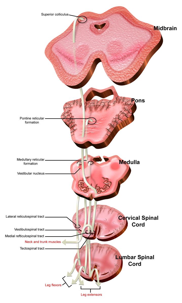
Umbilical cord prolapse
The cord slips below the presenting part and is compressed between it and the bony pelvis. Blood flow to the fetus stops, and the fetal heart rate shows a sudden prolonged deceleration or severe recurrent variable decelerations, classically within moments of membranes rupturing. Risk factors are anything that prevents the presenting part from filling the pelvic inlet: footling breech, transverse lie, unengaged presenting part, polyhydramnios, prematurity, multiple gestation, a long cord, and artificial rupture of membranes performed at a high station. The cord may be visible at the introitus, palpable in the vagina, or entirely occult with the diagnosis made only from the tracing.
The first action is to call for help and, with a gloved hand in the vagina, elevate the presenting part off the cord — and then to keep that hand exactly where it is, through transfer, through skin preparation, through spinal placement, until the baby is delivered. Simultaneously position the patient in knee-chest or steep Trendelenburg or exaggerated Sims to let gravity assist, stop the oxytocin, give oxygen at 10 L/min by non-rebreather, open the intravenous fluids, and prepare for immediate caesarean birth. A tocolytic such as terbutaline 0.25 mg subcutaneously may be ordered to reduce uterine activity, and filling the bladder with 500–700 mL of sterile saline through an indwelling catheter is an alternative method of elevating the presenting part when a delay is anticipated.
Two actions are wrong and both are intuitive. Do not attempt to push the cord back into the uterus — it does not stay, and manipulation causes vasospasm that worsens the very ischaemia you are trying to relieve. Do not repeatedly handle or palpate the cord to check for pulsation for the same reason. If a loop lies outside the introitus, cover it lightly with a warm saline-moistened gauze and leave it alone.
Shoulder dystocia
The head delivers and the anterior shoulder impacts behind the pubic symphysis. The sign is the turtle sign — the head emerges and retracts tightly against the perineum — along with failure of restitution and failure of the shoulders to deliver with normal gentle traction. Risk factors include fetal macrosomia, diabetes in pregnancy, a previous shoulder dystocia, post-term pregnancy, obesity, prolonged second stage and operative vaginal birth — but roughly half of cases occur with no risk factor at all, which is why every nurse rehearses the sequence.
Call for help and note the time; the clock is the documentation. Then work through HELPERR: Help; Evaluate for episiotomy (which makes room for the operator’s hand, not for the shoulder); Legs — McRoberts manoeuvre, hyperflexing the hips sharply onto the abdomen, which straightens the sacrum and rotates the symphysis cephalad; Pressure — suprapubic pressure, applied by a second person with the heel of the hand just above the pubic bone and directed downward and laterally toward the fetal chest to adduct the shoulder; Enter for internal rotational manoeuvres (Rubin II, Woods corkscrew); Remove the posterior arm; Roll the patient onto all fours (Gaskin manoeuvre). McRoberts plus suprapubic pressure alone resolves more than half of cases, and delivery is aimed for within about 5 minutes.
Fundal pressure is absolutely contraindicated. Pushing on the fundus drives the impacted shoulder harder against the symphysis, and it has caused uterine rupture. Excessive lateral traction on the fetal head causes brachial plexus injury. Complications for the newborn include brachial plexus injury with Erb palsy, clavicular or humeral fracture and hypoxic injury; for the birthing parent, postpartum haemorrhage and third- or fourth-degree perineal laceration. Document who was present, the sequence and duration of each manoeuvre, the time of head and body delivery, and cord gases if obtained.
Uterine rupture, abruption and the bleeding you cannot see
Uterine rupture is a complete separation of the uterine wall. The most common early sign is not pain — it is an abnormal fetal heart rate, usually a prolonged deceleration or sudden bradycardia. Other findings are a sudden tearing or sharp abdominal pain, cessation or loss of the contraction pattern, loss of station as the presenting part retracts upward on examination, palpable fetal parts through the abdominal wall, signs of hypovolaemic shock, and vaginal bleeding that may be minimal because most of the haemorrhage is intra-abdominal. Management is immediate laparotomy: stop the oxytocin, give oxygen, open two large-bore intravenous lines, activate the massive transfusion protocol, notify the operating theatre and the neonatal team, and prepare the patient. Fetal survival falls sharply beyond roughly 10–18 minutes of sustained bradycardia, and hysterectomy may be required.
Placental abruption is premature separation of a normally implanted placenta. The picture is painful dark red bleeding with a firm, board-like, exquisitely tender uterus, elevated resting tone, frequent low-amplitude contractions and a non-reassuring tracing. A concealed abruption bleeds behind the placenta, so the visible loss badly understates the true loss — trust the maternal heart rate, the fundal height rising over time and the fetal tracing more than the pad. Disseminated intravascular coagulation is a real risk. By contrast, placenta praevia presents with painless bright red bleeding and a soft, non-tender uterus, and the absolute rule is that no vaginal examination is performed in a patient with undiagnosed third-trimester bleeding until praevia has been excluded by ultrasound. Vasa praevia produces painless bright red bleeding at the moment of membrane rupture followed within minutes by fetal bradycardia or a sinusoidal pattern; the blood is fetal, the fetal blood volume is only about 80–85 mL per kilogram, and the response is immediate caesarean birth.
Amniotic fluid embolism is rare, unpredictable and catastrophic. Amniotic debris entering the maternal circulation triggers an anaphylactoid response: abrupt hypoxia and respiratory distress, sudden hypotension and cardiovascular collapse, often a seizure, and then disseminated intravascular coagulation with torrential bleeding. Management is supportive and immediate — high-quality cardiopulmonary resuscitation with continuous manual left uterine displacement rather than a tilted board, 100 per cent oxygen and early intubation, aggressive volume and vasopressor support, and massive transfusion. If there is no return of spontaneous circulation, perimortem caesarean birth is begun at 4 minutes with the aim of delivering by 5 minutes, because emptying the uterus restores venous return and improves the chance of resuscitating both patients.
| Emergency | Recognising it | FIRST action | Never do this |
|---|---|---|---|
| Cord prolapse | Prolonged deceleration right after rupture of membranes; cord palpated or seen | Gloved hand lifts the presenting part off the cord and stays there; knee-chest or Trendelenburg | Push the cord back in or repeatedly handle it |
| Shoulder dystocia | Turtle sign; shoulders will not deliver | Call for help, note the time, McRoberts plus suprapubic pressure | Fundal pressure; hard traction on the head |
| Uterine rupture | Sudden fetal bradycardia, tearing pain, loss of station, shock | Stop oxytocin, activate emergency caesarean, resuscitate volume | Wait to see whether the tracing recovers |
| Abruption | Painful bleeding, rigid tender uterus, high resting tone | Lateral position, oxygen, two large-bore lines, type and crossmatch, coagulation studies | Judge severity by visible blood alone |
| Undiagnosed third-trimester bleeding | Painless bright red bleeding, soft uterus | Ultrasound before any examination | Digital or speculum vaginal examination |
| Amniotic fluid embolism | Abrupt hypoxia, collapse, then coagulopathy | CPR with manual left uterine displacement; call the code and the theatre | Delay perimortem caesarean beyond 4 minutes without ROSC |
In every intrapartum emergency the correct first action relieves the mechanical or perfusion problem — lift the presenting part, flex the hips, empty the uterus — and the intuitive action of touching the thing that looks wrong is usually the one that causes harm.
Section 4Hypertensive Crisis and Magnesium Sulfate
Severe features, acute severe hypertension and eclampsia
Pre-eclampsia becomes pre-eclampsia with severe features when any one of the following appears: systolic blood pressure 160 mmHg or above or diastolic 110 mmHg or above on two occasions at least 4 hours apart (or once, if severe hypertension is confirmed on repeat within minutes and treatment is being started); platelet count below 100 000 per microlitre; serum creatinine above 1.1 mg/dL or doubled; transaminases at least twice the upper limit of normal; pulmonary oedema; or new cerebral or visual symptoms. Persistent headache unrelieved by paracetamol, visual scotomata or blurring, and epigastric or right upper quadrant pain are the three symptoms that change the plan, and the epigastric pain is stretching of the hepatic capsule, not indigestion.
Acute-onset severe hypertension — a sustained systolic of 160 mmHg or more or a diastolic of 110 mmHg or more — is a hypertensive emergency because of the risk of haemorrhagic stroke, and it should be treated within 30 to 60 minutes, ideally within 30. Three first-line regimens exist. Intravenous labetalol 20 mg over 2 minutes, then 40 mg after 10 minutes, then 80 mg after another 10 minutes, to a cumulative maximum around 300 mg — avoided in asthma, bradycardia below 60, and decompensated heart failure. Intravenous hydralazine 5–10 mg over 2 minutes, repeated at 10 mg after 20 minutes if the threshold is still exceeded, to a cumulative maximum of about 20–30 mg before moving to labetalol — expect reflex tachycardia, headache and occasional maternal hypotension. Oral immediate-release nifedipine 10 mg, repeated at 20 mg after 20 minutes, which is the option when intravenous access is not yet established. Recheck the blood pressure at 10-minute intervals during treatment and do not aim for normotension — a target of roughly 140–150 over 90–100 mmHg preserves uteroplacental perfusion.
Eclampsia is a generalised tonic-clonic seizure in a person with a hypertensive disorder of pregnancy. The first action during the seizure is protect the airway and prevent injury: stay with the patient, call for help, turn the patient to the side, lower the bed, pad the rails, remove hazards, and note the time. Do not restrain the limbs and do not insert anything into the mouth. After the seizure ends, work airway first and in this order: suction the airway, give oxygen at 10 L/min by non-rebreather, secure or confirm intravenous access, give the ordered magnesium sulfate bolus through it, draw laboratory studies, and only then evaluate the fetus. Transient fetal bradycardia with reduced variability after an eclamptic seizure is expected and usually recovers as the parent is stabilised — rushing to caesarean during or immediately after a seizure endangers an unstable patient. Birth follows stabilisation; it does not replace it. Magnesium sulfate is continued for at least 24 hours after birth or after the last seizure.
Magnesium sulfate: the drug, the monitoring and the toxicity ladder
Magnesium sulfate prevents and treats eclamptic seizures by antagonising calcium at the neuromuscular junction and raising the cerebral seizure threshold. It is not an antihypertensive — a common and dangerous misunderstanding. The standard regimen is a loading dose of 4–6 g intravenously over 15–30 minutes, followed by a maintenance infusion of 1–2 g per hour. It is a high-alert medication: it goes on a volumetric pump, on its own clearly labelled secondary line, with an independent double-check by a second nurse of the concentration, the pump settings and the dose.
The therapeutic serum range is roughly 4–7 mEq/L (about 5–8 mg/dL). Toxicity then declares itself in a predictable order, and knowing the order is what makes the monitoring meaningful: deep tendon reflexes disappear before respiratory depression appears. The patellar reflex is therefore the early-warning system, and it is checked hourly. If the epidural blocks the patellar reflex, use the biceps or brachioradialis reflex instead.
| Serum magnesium (approximate) | What you find | What it means |
|---|---|---|
| 1.5–2.5 mg/dL | Normal non-pregnant range | Sub-therapeutic for seizure prophylaxis |
| 5–8 mg/dL (4–7 mEq/L) | Flushing, warmth, mild drowsiness, slurred speech, feeling “heavy” | Therapeutic — expected, reassure the patient |
| 8–12 mg/dL | Loss of deep tendon reflexes | Stop the infusion and notify — this is the warning |
| 12–18 mg/dL | Respiratory rate below 12, respiratory depression | Toxicity; antidote and airway support |
| 18–24 mg/dL | Altered conduction, widened QRS, profound hypotension | Severe toxicity |
| Above 25–30 mg/dL | Cardiac arrest | Full resuscitation |
Monitoring parameters are checked hourly and the infusion is held if any one of them fails: respiratory rate below 12 per minute, absent deep tendon reflexes, urine output below 30 mL per hour (or below 100 mL in 4 hours), a serum level above the therapeutic range, or a sudden fall in level of consciousness. Urine output matters because magnesium is cleared entirely by the kidney — a patient with oliguria accumulates the drug even on an unchanged infusion rate. Also monitor continuous pulse oximetry, cardiac and fetal heart rate, lung sounds for pulmonary oedema, and strict intake and output.
The antidote is calcium gluconate 1 g, which is 10 mL of a 10 per cent solution, given intravenously over about 3 minutes. The sequence when toxicity is recognised is: (1) stop the magnesium infusion; (2) call for help and notify the provider; (3) give calcium gluconate as ordered; (4) support ventilation with oxygen, bag-mask or intubation as needed; (5) draw a magnesium level; (6) continuous monitoring. Stopping the infusion comes before the antidote — giving calcium while magnesium continues to run is treating a tap that is still open. Keep the calcium gluconate, a bag-valve-mask and suction at the bedside of every patient on magnesium.
The newborn is also a magnesium patient. Babies born to a parent on infusion may show hypotonia, lethargy, poor suck and respiratory depression at birth. Anticipate the need for respiratory support and have the neonatal team present; naloxone is not the answer here and is no longer part of neonatal resuscitation at all.
HELLP syndrome and the coagulation problem
HELLP is Haemolysis, Elevated Liver enzymes and Low Platelets, and it can occur with only modest hypertension, which is exactly why it is missed. The laboratory definition uses lactate dehydrogenase of 600 units/L or more or bilirubin of 1.2 mg/dL or more with schistocytes on the smear, transaminases at least twice the upper limit of normal, and a platelet count below 100 000 per microlitre. The classic presenting complaint is right upper quadrant or epigastric pain, often with nausea, vomiting and general malaise that the patient may describe as feeling like influenza.
The nursing implications are bleeding implications. Platelets below 100 000 mean minimising venipunctures, using the smallest needle, applying pressure for longer, avoiding intramuscular injections where possible, using a soft toothbrush, and watching for petechiae, gum bleeding, bruising and haematuria. Regional anaesthesia is generally withheld below a platelet count in the 70 000–80 000 range because of the risk of epidural haematoma — the exact threshold is set by anaesthesia and by institutional policy. Subcapsular hepatic haematoma is a rare but lethal complication; sudden severe right upper quadrant pain with shoulder pain and shock suggests rupture and no one palpates that abdomen deeply. Definitive treatment of HELLP, like that of pre-eclampsia with severe features, is birth.
Magnesium sulfate prevents seizures rather than lowering blood pressure, its toxicity always removes deep tendon reflexes before it depresses respirations, and the response to toxicity begins by stopping the infusion and then giving calcium gluconate 1 g over about 3 minutes.
Section 5The Fourth Stage and Two Patients at Once
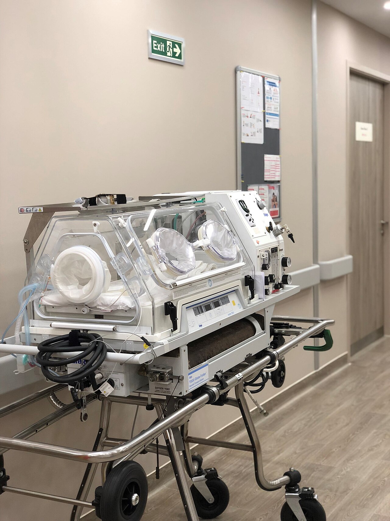
Postpartum haemorrhage: massage the fundus first
Postpartum haemorrhage is a cumulative blood loss of 1000 mL or more, or blood loss accompanied by signs or symptoms of hypovolaemia, within 24 hours of birth regardless of route. Loss is quantified, not estimated: blood is measured in graduated under-buttock drapes and soaked materials are weighed, with 1 gram of weight above dry weight counted as 1 mL of blood. Visual estimation understates loss by 30–50 per cent, and the healthy young obstetric patient compensates so well that the blood pressure may not fall until 25–30 per cent of blood volume is gone. Tachycardia, narrowing pulse pressure, restlessness, air hunger and a rising shock index (heart rate divided by systolic blood pressure; 0.9 or above is concerning) all appear first.
Causes are organised as the four Ts. Tone — uterine atony — causes 70–80 per cent of cases and is more likely after an over-distended uterus, prolonged or very rapid labour, prolonged oxytocin exposure, grand multiparity, chorioamnionitis or magnesium sulfate. Trauma covers lacerations, haematoma and uterine inversion. Tissue covers retained placental fragments and abnormally adherent placenta. Thrombin covers coagulopathy, including disseminated intravascular coagulation after abruption or amniotic fluid embolism.
The first action for heavy postpartum bleeding is to palpate and massage the fundus, because tone is the most likely cause and fundal massage is both the assessment and the treatment. Only after the uterus is being massaged do you call for help, empty the bladder (a full bladder displaces the uterus — a fundus that is boggy and deviated to the right is a bladder problem until proven otherwise), establish or check large-bore intravenous access, begin or increase oxytocin, quantify the loss, and send a full blood count, coagulation screen, fibrinogen and crossmatch. If the fundus is firm and midline and the patient is still bleeding steadily bright red, tone is not the cause — look for a laceration and call the provider to inspect the birth canal. If the patient reports severe unrelenting perineal, vaginal or rectal pressure and pain with a firm fundus and rising pulse, suspect a concealed haematoma.
| Uterotonic | Dose and route | Watch for | Contraindication |
|---|---|---|---|
| Oxytocin | 10–40 units in 500–1000 mL isotonic fluid IV infusion, or 10 units IM | Hypotension if pushed rapidly undiluted; water intoxication | None absolute; never give undiluted rapid IV push |
| Methylergonovine | 0.2 mg IM every 2–4 hours (oral for maintenance) | Sharp rise in blood pressure; cramping, nausea | Hypertension, pre-eclampsia — check the blood pressure before every dose |
| Carboprost tromethamine | 250 mcg IM or intramyometrial every 15–90 minutes, maximum 8 doses (2 mg) | Diarrhoea, nausea, vomiting, fever, flushing | Asthma; caution in cardiac, renal or hepatic disease |
| Misoprostol | 600–1000 mcg orally, sublingually or rectally, single dose | Shivering, pyrexia, diarrhoea | Few; heat-stable and useful when refrigeration is unavailable |
| Tranexamic acid | 1 g IV over 10 minutes, within 3 hours of birth; may repeat once after 30 minutes | Give slowly — rapid infusion causes hypotension | Known thromboembolic disease; give within the 3-hour window |
If bleeding continues, escalation is stepwise and rapid: bimanual uterine compression, intrauterine balloon tamponade, a vacuum-induced haemorrhage-control device, uterine artery embolisation, compression sutures, arterial ligation, and hysterectomy as the definitive last step. Activate the massive transfusion protocol early, transfusing red cells, plasma and platelets in roughly a 1:1:1 ratio, keep the patient warm, and remember that hypothermia and acidosis worsen coagulopathy. One special case: never pull on a cord against an unseparated placenta, because uterine inversion — sudden severe pain, profound shock out of proportion to visible loss, and a fundus that cannot be found — is managed by stopping uterotonics, replacing the uterus, and only then restarting them.
The newborn who does not transition, and weight-based dosing
At every birth, three questions are asked in the first seconds: is the baby term, does the baby have good tone, and is the baby breathing or crying? Three yeses mean skin-to-skin contact with the parent, delayed cord clamping for 30–60 seconds, drying and routine care. Any no means the baby goes to the radiant warmer for the initial steps — warm, dry, stimulate, position the airway in the sniffing position, clear secretions only if they are obstructing — with heart rate and respirations assessed within the first 60 seconds, the “Golden Minute”.
If the heart rate is below 100 beats per minute or the baby is apnoeic or gasping, begin positive-pressure ventilation at 40–60 breaths per minute, starting with 21 per cent oxygen for babies of 35 weeks or more and 21–30 per cent for those below 35 weeks, with the pulse oximeter on the right hand or wrist to read preductal saturation. Ventilation is the single most important step in neonatal resuscitation; almost every newborn who needs help needs air moved into the lungs and nothing more. If the chest does not rise, run MR SOPA: Mask adjust, Reposition the head, Suction mouth then nose, Open the mouth, Pressure increase, Alternative airway. If the heart rate remains below 60 after 30 seconds of ventilation that visibly moves the chest, start chest compressions with the two-thumb encircling technique at a ratio of 3 compressions to 1 breath, giving 90 compressions and 30 breaths — 120 events — each minute, with oxygen increased to 100 per cent. Targeted preductal saturations climb slowly and deliberately: 60–65 per cent at 1 minute, 65–70 at 2, 70–75 at 3, 75–80 at 4, 80–85 at 5 and 85–95 at 10 minutes. The Apgar score is assigned at 1 and 5 minutes, and every 5 minutes to 20 minutes if it remains below 7, but it is a description of the transition, not the trigger for it — you never wait for an Apgar score to start resuscitating.
If the heart rate is still below 60 after 60 seconds of coordinated compressions and ventilation through a secure airway, epinephrine is given, preferably through an umbilical venous catheter. The intravenous dose is 0.02 mg/kg (acceptable range 0.01–0.03 mg/kg) of the 0.1 mg/mL concentration, repeated every 3–5 minutes; the endotracheal dose, used only while access is being obtained, is higher at 0.05–0.1 mg/kg. For suspected hypovolaemia — pallor, weak pulses, poor response, a history of abruption, vasa praevia or cord accident — give a volume expander of 10 mL/kg of normal saline or O-negative blood over 5–10 minutes. Work the arithmetic every time, out loud, with a second nurse:
The Apgar score has five components, each scored 0, 1 or 2 for a total out of 10, and the mnemonic APGAR names them: Appearance (colour), Pulse (heart rate), Grimace (reflex irritability), Activity (muscle tone) and Respiration (respiratory effort). Heart rate scores 0 if absent, 1 if below 100 and 2 if 100 or above. Respiratory effort scores 0 if absent, 1 for slow, irregular or a weak cry, and 2 for a good lusty cry. Tone scores 0 for flaccid, 1 for some flexion, 2 for active motion. Reflex irritability scores 0 for no response, 1 for a grimace, 2 for a cry, cough or sneeze. Colour scores 0 for blue or pale, 1 for a pink body with blue hands and feet — acrocyanosis, which is normal in the first hours — and 2 for completely pink. A total of 7 to 10 is reassuring, 4 to 6 is moderately depressed, and 0 to 3 is severely depressed. Note again what the score is not: it is scored after the Golden Minute has already been spent resuscitating, and it never determines whether resuscitation starts.
Escalation, delegation and the debrief
Escalate in a structure the receiving clinician can act on. SBAR for an intrapartum haemorrhage sounds like this: Situation — “This is Priya, registered nurse in room 4. Ms Okafor is 20 minutes postpartum from a vaginal birth and I have quantified 900 mL of blood loss and it is continuing.” Background — “Gravida 4, 41 weeks, oxytocin augmentation for 14 hours, birth weight 4.3 kg, shoulder dystocia resolved with McRoberts and suprapubic pressure.” Assessment — “The fundus is boggy two fingerbreadths above the umbilicus and deviated right. Heart rate 122, blood pressure 96 over 54, she is pale and anxious. I have massaged the fundus, emptied 400 mL from the bladder and increased the oxytocin.” Recommendation — “I need you at the bedside now, I am activating the haemorrhage protocol, and I would like an order for a second uterotonic. Her blood pressure history is normal, so methylergonovine is appropriate.”
Red flags that trigger that call without waiting: any category III tracing; a prolonged deceleration lasting more than 2 minutes; sudden fetal bradycardia after rupture of membranes; loss of station with abdominal pain in a person labouring after caesarean; a saturated pad in less than 15 minutes or a clot larger than a lemon; systolic 160 or diastolic 110 mmHg confirmed on repeat; a new headache, visual change or epigastric pain in a hypertensive patient; absent deep tendon reflexes or a respiratory rate below 12 on magnesium; and a newborn heart rate below 100 after 30 seconds of effective ventilation.
Delegation limits do not soften in an emergency. Assessment, teaching, evaluation, fetal heart rate interpretation, titration of oxytocin or magnesium, intravenous push medication, blood product administration and the care of an unstable patient stay with the registered nurse. Assistive personnel may take vital signs on stable patients, provide comfort measures and positioning, assist with ambulation of a stable patient, weigh pads for quantification (with the nurse interpreting the result), obtain supplies and the emergency trolley, and call for extra hands. Delegating a task never delegates the accountability for the outcome. Afterwards, hold a team debrief, because obstetric emergencies are traumatic for staff as well as families, and explain to the family in plain, non-blaming language what happened and why — a parent who believes their body failed carries that belief into the next pregnancy.
For heavy postpartum bleeding the fundus is massaged first because it is both the assessment and the treatment, and for the newborn in the next cot ventilation is the intervention that matters — with the Apgar scored at 1 and 5 minutes as a description of the transition rather than the trigger for it, and every newborn dose calculated from the kilogram weight with the arithmetic shown and verified by a second nurse.
The chapter in one breath
Intrapartum emergencies are won by sequence. The fetal heart rate tracing is read as baseline, variability, accelerations and decelerations, and the shape of a deceleration names its mechanism — early from head compression, variable from cord compression, late from uteroplacental insufficiency — while moderate variability remains the best bedside evidence of adequate oxygenation. When a tracing worsens, remove the cause first: stop the oxytocin, reposition laterally, examine to exclude a prolapsed cord, bolus fluid, and prepare for birth. Labour that deviates from the expected pattern is described, not blamed, and uterine activity is quantified before it is augmented, because the placenta is perfused between contractions. In the acute emergencies, the intuitive act is usually the wrong one: lift the presenting part off a prolapsed cord rather than pushing the cord back; use McRoberts and suprapubic pressure for shoulder dystocia and never fundal pressure; suspect rupture from a sudden bradycardia and loss of station. Magnesium sulfate prevents seizures rather than lowering pressure, abolishes deep tendon reflexes before it depresses respirations, and its toxicity is answered by stopping the infusion and then giving calcium gluconate 1 g over about 3 minutes. For postpartum haemorrhage, massage the fundus first, work the four Ts, and match each uterotonic to its contraindication. And because every birth is two resuscitations, the newborn is met with the initial steps, ventilation before compressions, preductal saturation targets that climb slowly through the first 10 minutes, and an Apgar at 1 and 5 minutes that describes the transition without ever delaying it.
Words worth knowing
| Term | What it means |
|---|---|
| Baseline fetal heart rate | Mean rate over a 10-minute window rounded to the nearest 5 bpm, excluding accelerations, decelerations and marked variability; normal 110–160 bpm. |
| Variability | Irregular fluctuation of the baseline; absent, minimal (5 bpm or less), moderate (6–25 bpm) or marked (more than 25 bpm). Moderate is reassuring. |
| Early deceleration | Gradual fall mirroring the contraction, caused by fetal head compression and a vagal reflex; benign. |
| Variable deceleration | Abrupt fall of 15 bpm or more lasting 15 seconds to 2 minutes, caused by umbilical cord compression. |
| Late deceleration | Gradual fall whose onset, nadir and recovery all follow the contraction, caused by uteroplacental insufficiency; never normal. |
| Category III tracing | Absent variability with recurrent late or variable decelerations or bradycardia, or a sinusoidal pattern; predicts abnormal fetal acid–base status. |
| Intrauterine resuscitation | Sequence of stopping oxytocin, repositioning, examining, fluid bolus, treating hypotension and considering tocolysis, to restore fetal oxygenation. |
| Tachysystole | More than five contractions in 10 minutes averaged over 30 minutes; removes the resting interval during which the placenta is perfused. |
| Montevideo units | Sum of peak-minus-baseline intrauterine pressures over 10 minutes; 200 or more indicates adequate uterine activity. |
| Bishop score | Cervical readiness score from dilatation, effacement, station, consistency and position; 6 or less favours ripening before induction. |
| Umbilical cord prolapse | Descent of the cord below the presenting part; managed by elevating the presenting part and knee-chest or Trendelenburg positioning, never by replacing the cord. |
| Shoulder dystocia | Impaction of the anterior shoulder behind the pubic symphysis after delivery of the head; managed by McRoberts and suprapubic pressure, never fundal pressure. |
| Uterine rupture | Complete separation of the uterine wall; earliest sign is usually an abnormal fetal heart rate, with loss of station and shock following. |
| Amniotic fluid embolism | Anaphylactoid response to amniotic debris causing abrupt hypoxia, collapse and disseminated intravascular coagulation; perimortem caesarean begins at 4 minutes without return of circulation. |
| Magnesium sulfate toxicity | Progressive loss of deep tendon reflexes, then respiratory depression, then cardiac arrest; treated by stopping the infusion and giving calcium gluconate 1 g over about 3 minutes. |
| HELLP syndrome | Haemolysis, elevated liver enzymes and low platelets, often with right upper quadrant pain and only modest hypertension. |
| Postpartum haemorrhage | Cumulative blood loss of 1000 mL or more, or any loss with signs of hypovolaemia, within 24 hours of birth; first action is fundal massage. |
| Four Ts | Tone, Trauma, Tissue and Thrombin — the causes of postpartum haemorrhage in order of frequency. |
| Quantified blood loss | Measured rather than estimated loss, using graduated drapes and weighed materials at 1 gram equals 1 mL. |
| Apgar score | Appearance, Pulse, Grimace, Activity and Respiration, each scored 0 to 2, assigned at 1 and 5 minutes; it describes the transition and never determines whether resuscitation begins. |
Check yourself
The Postpartum Period
The six weeks that follow birth are the most physiologically turbulent stretch of the entire childbearing year, and they are the stretch most often treated as an afterthought. In the space of a few minutes a uterus that weighed roughly 1000 g begins shrinking toward 60–80 g, a circulation carrying an extra 1.5 L of plasma unloads itself, clotting factors stay elevated for weeks, and a person who has not slept is handed a newborn and a stack of teaching. Most of what happens is normal. The nurse’s work is to know normal so precisely that the abnormal announces itself early — a fundus one fingerbreadth too high and displaced to the right, a pulse of 108 in someone who should be bradycardic, a headache that paracetamol will not touch on day four, a mood that has not lifted by day fourteen. Postpartum haemorrhage, hypertensive crisis, sepsis, thromboembolism and perinatal mood disorders are the five leading contributors to maternal death, and almost all of them are recognisable at the bedside before they are catastrophic.
By the end of this chapter you can
- Trace the mechanism of uterine involution, predict fundal height by postpartum day, and distinguish normal lochia progression from the patterns that signal haemorrhage, retained tissue or infection.
- Interpret the cardiovascular, haematological, renal and endocrine adaptations of the puerperium, including physiological bradycardia, puerperal diuresis and the leucocytosis that makes a white cell count useless on its own.
- Perform a structured BUBBLE-HE assessment and state the vital-sign thresholds that trigger escalation using the Maternal Early Warning Criteria.
- Sequence the response to postpartum haemorrhage correctly — massage the fundus first — and select uterotonics by dose, route and contraindication using the four Ts framework.
- Recognise magnesium sulfate toxicity in the order the signs actually appear, stop the infusion first, and give calcium gluconate as the antidote.
- Screen for puerperal infection, venous thromboembolism and postpartum preeclampsia, and construct an SBAR escalation for each.
- Support lactation physiologically, differentiate postpartum blues from postpartum depression and postpartum psychosis, and act on a positive screen.
- Deliver POST-BIRTH warning-sign teaching, contraceptive counselling and fourth-trimester follow-up, and identify what may and may not be delegated to unlicensed assistive personnel.
Section 1Involution: How the Reproductive Tract Comes Home
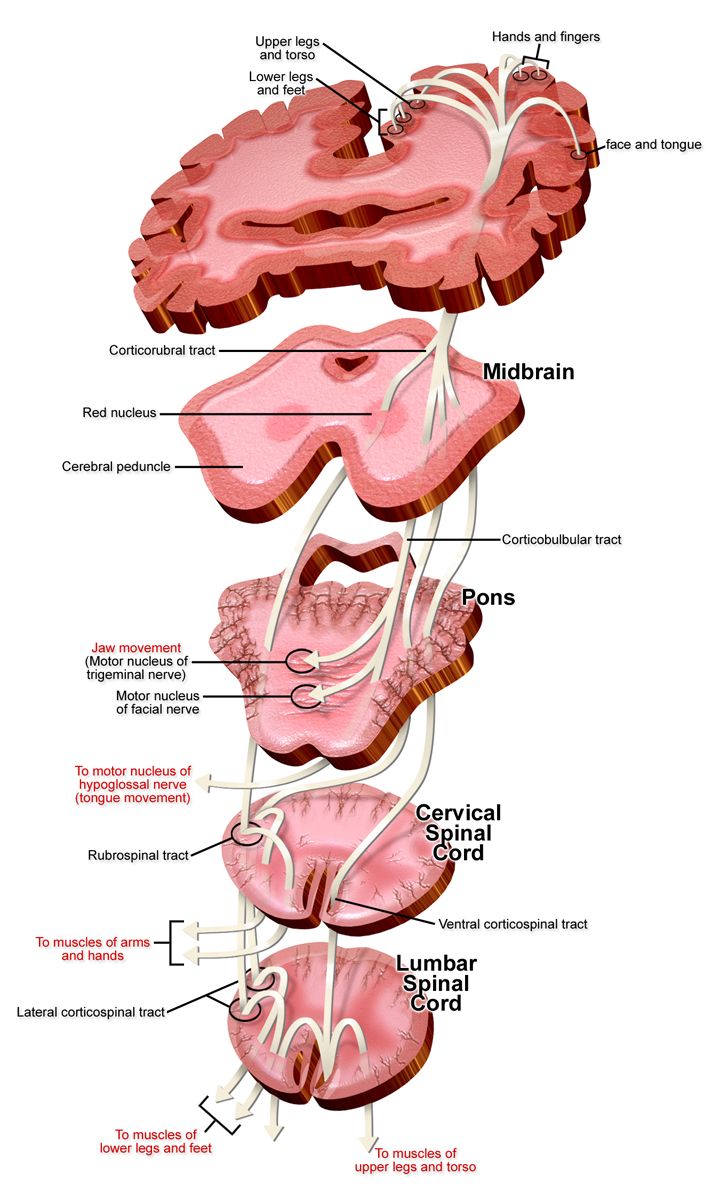
The mechanism: ligature, autolysis, regeneration
Involution is not the uterus deflating. Three separate processes run at once. The first is mechanical: after the placenta separates, the interlacing bundles of myometrium contract and retract around the spiral arterioles that fed the placental bed. These crossing muscle fibres are the living ligature, and they are the single reason a person does not exsanguinate from a placental site the size of a saucer with an arterial supply of roughly 500–800 mL per minute at term. Platelets and fibrin then seal what the muscle has already clamped. When the myometrium is well contracted the fundus is felt as a firm, midline, grapefruit-like mass. When it relaxes, the ligature opens and bleeding is immediate and heavy. This is why uterine atony causes 70–80 per cent of primary postpartum haemorrhage, and why the first thing a nurse does about postpartum bleeding is put a hand on the fundus.
The second process is autolysis. Deprived of oestrogen and progesterone, the hypertrophied myocytes digest their own excess cytoplasm through proteolytic enzymes. Cell number changes very little; cell size collapses. The uterus falls from about 1000 g immediately after birth to roughly 500 g at one week, 300–350 g at two weeks and 60–80 g by six weeks — near, though rarely identical to, its pre-pregnancy weight. The third process is regeneration of the endometrium. The superficial decidua sloughs as lochia; the basal layer regenerates the endometrial lining over most of the cavity by about day 16, while the placental site itself takes six weeks to re-epithelialise from the margins inward. That slow, exfoliative healing of the placental site — rather than scarring — is what allows the next pregnancy to implant normally, and it explains why bleeding can restart around day 7–14 when the eschar over the site separates.
Clinically, fundal height tracks this timetable. Immediately after birth the fundus sits midway between the symphysis pubis and the umbilicus. Within about twelve hours it rises to the level of the umbilicus, then descends approximately 1 cm — one fingerbreadth — each day. By day six it should be about midway between symphysis and umbilicus, and by day ten to fourteen it has dropped behind the symphysis and is no longer palpable abdominally. Document position in fingerbreadths relative to the umbilicus (U/1 means one below, 1/U means one above), consistency (firm or boggy) and midline position. Afterpains — the cramping of these contractions — are mild in a first-time parent whose uterus stays tonically contracted, and considerably stronger in multiparas, in over-distended uteri (twins, polyhydramnios, macrosomia) and during breastfeeding or chestfeeding, when suckling releases endogenous oxytocin.
Reading lochia
Lochia is the shedding of decidua, blood, serous exudate, leucocytes and bacteria. It moves through three predictable stages, and the direction of travel matters more than the exact day. Lochia rubra is dark red and lasts days 1–3, occasionally to day 4; small clots are acceptable but clots larger than a plum, or clots the size of an egg or bigger, are reportable. Lochia serosa is pinkish-brown and serosanguineous, running roughly from day 4 to day 10. Lochia alba is creamy white or yellowish, from about day 10 to as long as six weeks. Total volume across the puerperium is on the order of 240–270 mL. Lochia should smell fleshy or musty, like menstrual flow. A foul or fetid odour means infection until proven otherwise. Progression that goes backwards — serosa returning to rubra — suggests retained placental fragments, subinvolution or simply too much activity too soon, and always earns an assessment rather than reassurance.
Quantify rather than eyeball. Visual estimation of blood loss underestimates true loss by 30–50 per cent, and the error grows as the loss grows. Quantitative blood loss (QBL) means weighing blood-soaked materials and subtracting the known dry weight: 1 gram equals 1 millilitre. A pad that weighs 340 g when its dry weight was 40 g holds 300 mL. Where weighing is not in place, describe pad saturation over a stated time interval rather than using the word “normal”.
| Descriptor | Stain on a perineal pad in one hour | Approximate volume | Action |
|---|---|---|---|
| Scant | Less than 2.5 cm (1 inch) | Under 10 mL | Routine |
| Light / small | Less than 10 cm (4 inches) | 10–25 mL | Routine |
| Moderate | Less than 15 cm (6 inches) | 25–50 mL | Routine, reassess |
| Heavy | Pad saturated within one hour | 50–80 mL | Assess fundus and bladder, notify |
| Excessive | Pad saturated in 15 minutes or less; blood pooling under the buttocks; steady trickle | Haemorrhage | Emergency — begin the haemorrhage sequence |
Cervix, vagina, perineum and the comfort plan
The cervix is soft, oedematous and bruised immediately after birth, admitting two fingers for the first few days. By the end of the first week it admits a fingertip; by six weeks it is closed. The external os never regains its pre-pregnancy round shape after a vaginal birth — it heals as a transverse slit, a permanent anatomical record of parity that will be visible on every future speculum examination. The vagina is smooth, gaping and oedematous at first; rugae return by about three to four weeks. In someone who is lactating, ovarian oestrogen stays suppressed, so the vaginal epithelium remains thin and poorly lubricated for months. Warn about dyspareunia and recommend a water-based lubricant — this is not a trivial issue, and people rarely raise it unprompted.
Assess the perineum in the Sims’ (lateral) position with a good light, using REEDA: Redness, Ecchymosis, Oedema, Discharge or drainage, and Approximation of the wound edges. Note the degree of any laceration or episiotomy (first degree: skin and vaginal mucosa; second: perineal muscle; third: extending into the anal sphincter; fourth: through the rectal mucosa). Third- and fourth-degree injuries change management — no rectal suppositories, no enemas, stool softeners as standing orders, and a higher index of suspicion for infection and for later incontinence. Haemorrhoids are common and worsened by second-stage pushing.
The comfort plan is time-dependent and students routinely reverse it. Apply ice or a cold pack for the first 24 hours, 20 minutes on and at least 10 minutes off, to limit oedema and haematoma formation through vasoconstriction. After 24 hours, switch to heat — a warm sitz bath for 20 minutes two or three times daily — to promote circulation, healing and comfort. Witch-hazel pads, a peri-bottle of warm water used front-to-back with every void, topical anaesthetic spray and a scheduled oral analgesic regimen complete the plan. Teach the patient to change the pad with every void and to always wipe front to back.
A firm, midline fundus descending about one centimetre a day with lochia progressing rubra to serosa to alba is the signature of normal involution — any deviation in height, tone, position, colour, volume or odour is a finding to be assessed, not observed.
Section 2Systemic Adaptation and the Structured Assessment
Cardiovascular, haematological and fluid adaptation
Pregnancy expands plasma volume by 40–50 per cent, adding roughly 1200–1500 mL. That expansion is the body’s pre-payment for birth: it is why a healthy person can lose 500 mL at a vaginal birth or 1000 mL at a caesarean and remain haemodynamically unremarkable. It is also why the vital signs lie. Because the reserve is spent silently, a postpartum patient may lose 1000–1500 mL — a quarter of blood volume — with nothing more than mild tachycardia, and then decompensate abruptly. Never let a normal blood pressure reassure you about bleeding. The shock index (heart rate divided by systolic blood pressure) is more sensitive: a value of 0.9 or higher in a postpartum patient warrants urgent evaluation even when each individual number looks acceptable.
Immediately after birth, the emptied uterus autotransfuses 500–750 mL into the systemic circulation and vena caval compression is relieved, so cardiac output rises 60–80 per cent above pre-labour values in the first ten to fifteen minutes. Stroke volume and cardiac output fall steadily thereafter, reaching near-baseline by about two weeks and fully by six to twelve weeks. The immediate surge triggers a vagally mediated puerperal bradycardia of 40–60 beats per minute that can persist six to ten days; it is normal and benign, though a rate under 50 still meets the Maternal Early Warning Criteria below and earns a bedside look rather than a shrug. The corollary matters far more: tachycardia above 100 in a postpartum patient is never normal and means haemorrhage, hypovolaemia, infection, pain, anxiety or thromboembolism until proven otherwise.
Excess extracellular fluid leaves by two routes. Puerperal diuresis begins within twelve hours and can reach 3000 mL per day between days two and five; profuse diaphoresis, especially at night, does the rest. Expect a further 2–3 kg of weight loss on top of the 5–6 kg lost at birth. Haematologically, pregnancy is a prothrombotic state that does not switch off at delivery: fibrinogen and factors VII, VIII, IX and X stay elevated for four to six weeks, and the risk of venous thromboembolism is highest in the first three weeks postpartum, remaining above baseline to roughly twelve weeks. Leucocytosis of 20,000–30,000/mm3 is a normal response to labour, so a raised white cell count alone cannot diagnose infection; a rising count with fever, tachycardia and localising signs can. Haemoglobin and haematocrit are unreliable in the first 24 hours because plasma has not yet redistributed; by day three to seven the haematocrit rises as diuresis concentrates the blood. As a rule of thumb, each 500 mL of blood loss lowers haemoglobin by about 1–1.5 g/dL and haematocrit by 3–4 percentage points.
The other systems
The urinary tract is the postpartum system most likely to cause trouble quietly. Bladder tone is reduced by progesterone, by the pressure of the presenting part, by regional anaesthesia and by perineal oedema and pain, and sensation of fullness may be absent. Diuresis then fills that insensate bladder fast. A distended bladder pushes the uterus upward and laterally — classically up and to the right — preventing contraction and causing bleeding. Expect a spontaneous void within six hours of birth or of urinary catheter removal; measure the first void and expect at least 150 mL. A post-void residual over 150 mL, repeated small voids of under 150 mL, or a palpable bladder after voiding indicates retention and needs catheterisation and provider notification. Mild proteinuria (1+) and glycosuria can persist for a few days; dilated ureters and renal pelves take up to six to eight weeks to return to normal, which sustains the risk of pyelonephritis.
The gastrointestinal tract is hungry and slow: appetite returns sharply, while progesterone-related hypomotility, dehydration, perineal pain, opioids and fear of the first bowel movement together produce constipation. Aim for a bowel movement within two to three days with fluids, fibre, ambulation and a stool softener such as docusate. Musculoskeletally, relaxin-softened joints and the widened symphysis take six to eight weeks to stabilise, and diastasis recti narrows over the same period; teach that lifting anything heavier than the baby is unwise early on and that ligamentous laxity, not clumsiness, explains the altered balance. The skin shows fading of melasma and linea nigra over months; striae fade from red-purple to silver but do not disappear. Endocrine reversal is dramatic: oestrogen, progesterone and human placental lactogen fall precipitously within hours, prolactin rises in the lactating parent, and thyroid function may swing enough to cause postpartum thyroiditis at three to six months. In someone not lactating, ovulation returns at a mean of about 45 days and can occur as early as day 27; in a fully lactating parent it is delayed but never guaranteed.
BUBBLE-HE and the thresholds that trigger escalation
The standard postpartum assessment is remembered as BUBBLE-HE, and it should be performed with vital signs on the recommended schedule: every 15 minutes for the first hour, every 30 minutes for the second hour, hourly for the next four hours, then every four to eight hours once stable. Ask the patient to empty the bladder before a fundal assessment, position her supine with knees flexed, place the lower hand just above the symphysis to support the lower uterine segment, and palpate with the upper hand. If the fundus is boggy, massage in a circular motion with the flat of the hand until firm, then express clots — never massage a firm fundus, which fatigues the myometrium and causes bleeding, and never push on the fundus without splinting below, which risks uterine inversion.
| Letter | What you assess | Expected finding |
|---|---|---|
| B — Breasts | Softness, filling, engorgement, nipple integrity, latch | Soft days 1–2, filling day 2–3, engorged day 3–5; nipples intact |
| U — Uterus | Fundal height, tone, position | Firm, midline, descending about 1 cm/day |
| B — Bladder | Void within 6 h, volume, distension, retention | Voiding at least 150 mL, non-palpable bladder |
| B — Bowel | Bowel sounds, flatus, first stool, softener use | Active sounds; stool by day 2–3 |
| L — Lochia | Colour, amount, odour, clots | Rubra to serosa to alba; fleshy odour; no large clots |
| E — Episiotomy / perineum / incision | REEDA, haemorrhoids, caesarean incision | Edges approximated, mild oedema, no drainage |
| H — Homans / lower extremities | Unilateral pain, swelling, warmth, erythema, calf tenderness | Symmetrical, non-tender (Homans’ sign is unreliable — do not rely on it) |
| E — Emotional status & bonding | Affect, sleep, support, interaction with the newborn, mood screening | Engaged, en face contact, appropriate variability |
| Maternal Early Warning Criteria — any single one triggers prompt bedside evaluation |
|---|
| Systolic blood pressure under 90 or over 160 mmHg |
| Diastolic blood pressure over 100 mmHg |
| Heart rate under 50 or over 120 beats per minute |
| Respiratory rate under 10 or over 30 breaths per minute |
| Oxygen saturation under 95 per cent on room air |
| Oliguria: less than 35 mL per hour for two or more hours |
| Agitation, confusion or unresponsiveness |
| A patient with hypertension reporting a non-remitting headache or shortness of breath |
Temperature deserves its own rule. A temperature up to 38.0 °C (100.4 °F) in the first 24 hours is usually dehydration and the muscular work of labour, and is treated with oral fluids and reassessment. A temperature of 38.0 °C or higher on any two of the first ten postpartum days, excluding the first 24 hours, defines puerperal morbidity and must be investigated. Delegation is constrained here: unlicensed assistive personnel may take and record vital signs on a stable patient, assist with ambulation and perineal care, apply ice packs, record intake and output and help position for feeding. Fundal assessment, lochia evaluation, interpretation of any value, all patient teaching, evaluation of response to interventions and the care of any unstable patient remain with the registered nurse and cannot be delegated.
Postpartum vital signs are read against postpartum norms — bradycardia to 40–60 and leucocytosis to 30,000 are expected, while tachycardia over 100, oliguria under 35 mL/h or a saturation under 95 per cent are early warnings that demand a bedside evaluation.
Section 3Postpartum Haemorrhage: the Sequence That Saves
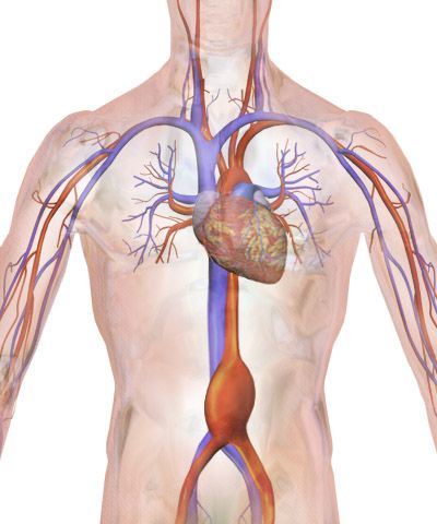
Definitions, quantification and why the numbers deceive
Postpartum haemorrhage (PPH) is defined as cumulative blood loss of 1000 mL or more, or blood loss accompanied by signs or symptoms of hypovolaemia, within 24 hours of birth, regardless of the mode of birth. The older route-specific thresholds (500 mL vaginal, 1000 mL caesarean) survive as triggers for heightened vigilance rather than as diagnoses. Primary or early PPH occurs within the first 24 hours; secondary or late PPH occurs from 24 hours to twelve weeks and is most often caused by retained placental fragments, subinvolution of the placental site or endometritis.
The deception is physiological. Because of pregnancy’s volume expansion, the classic shock cascade is delayed and then compressed. A loss of 10–15 per cent of blood volume may produce no change at all. At 20–25 per cent the patient may show only tachycardia, narrowed pulse pressure, restlessness and cool, pale skin with delayed capillary refill. Hypotension often does not appear until 30–40 per cent has gone, at which point the patient is in decompensated shock and the window is closing. Restlessness, air hunger and a sense of impending doom are cerebral hypoperfusion, not anxiety. Believe them.
Risk factors cluster around a uterus that is over-distended, exhausted or scarred: multiple gestation, polyhydramnios, macrosomia, grand multiparity, prolonged or very rapid labour, oxytocin augmentation, chorioamnionitis, magnesium sulfate exposure, general anaesthesia, operative vaginal birth, previous PPH, placenta praevia or accreta spectrum, and coagulopathy. A formal haemorrhage risk assessment is performed on admission, on admission to the postpartum unit and whenever the clinical picture changes. Crucially, a substantial proportion of haemorrhages occur in patients with no risk factors at all, which is why every birthing unit maintains a haemorrhage cart, a massive transfusion protocol and staged emergency orders.
The four Ts and what you do first
| Cause | Share | Bedside clue | Directed action |
|---|---|---|---|
| Tone — uterine atony | 70–80% | Boggy, soft, difficult-to-locate fundus; heavy dark bleeding; clots | Massage, empty bladder, uterotonics, tamponade |
| Trauma — laceration, haematoma, inversion, rupture | 15–20% | Firm fundus with continuous bright red trickle; severe rectal or perineal pain; bulging discoloured mass | Inspect and repair; haematoma drainage; surgical management |
| Tissue — retained placenta or fragments | up to 10% | Uterus fails to stay contracted; placenta incomplete on inspection; late bleeding day 7–14 | Manual removal, ultrasound, uterine evacuation |
| Thrombin — coagulopathy | 1–2% | Oozing from puncture sites, gums, incision; blood that will not clot; known abruption, sepsis, amniotic fluid embolism, HELLP | Product replacement, treat the underlying cause |
The sequence is the intervention. When bleeding is heavy, the first nursing action is to massage the fundus — it is fastest, requires no order, no equipment and no drug, and it corrects the cause in three-quarters of cases while you are calling for help. Do not leave the bedside to page anyone, do not start with vital signs, and do not begin with oxygen. The full sequence runs as follows, with several steps happening simultaneously once help arrives:
| Order | Action | Rationale |
|---|---|---|
| 1 | Massage the fundus while splinting the lower segment; express clots once firm | Restores the living ligature; corrects 70–80% of cases immediately |
| 2 | Call for help — activate the obstetric haemorrhage protocol | This is a team event; one nurse cannot massage, medicate, document and transfuse |
| 3 | Empty the bladder (void or indwelling catheter) | A distended bladder mechanically prevents contraction; the indwelling catheter then measures urine output as a perfusion marker |
| 4 | Two large-bore IVs (16–18 gauge); isotonic crystalloid wide open; warm the fluids | Access before collapse; hypothermia worsens coagulopathy |
| 5 | Increase or start oxytocin infusion, then add second-line uterotonics by protocol | Pharmacological contraction of the myometrium |
| 6 | Oxygen 10–15 L/min by non-rebreather; flat or modified Trendelenburg; keep warm | Maximises oxygen delivery on a falling haemoglobin |
| 7 | Continuous vital signs, pulse oximetry, strict intake and output, QBL by weight | Trends detect decompensation; QBL replaces guessing |
| 8 | Type and crossmatch, CBC, fibrinogen, coagulation studies; transfuse 1:1:1 if massive | Fibrinogen under 200 mg/dL in obstetric bleeding predicts severe haemorrhage |
| 9 | Escalate: bimanual compression, intrauterine balloon tamponade, tranexamic acid, uterine artery embolisation, B-Lynch suture, hysterectomy | Stepwise mechanical and surgical control |
Uterotonics: the right drug for the right patient
Every uterotonic has a patient it must not be given to, and those contraindications are examined at the bedside, not in the pharmacy. Check the blood pressure before methylergonovine. Ask about asthma before carboprost. Read the drug back with the dose and the route before giving it.
| Drug | Dose and route | Do not give if | Nursing notes |
|---|---|---|---|
| Oxytocin (first line) | 10–40 units in 500–1000 mL isotonic solution IV, titrated; or 10 units IM | Hypersensitivity | Never give undiluted rapid IV push — causes hypotension and dysrhythmia. Watch for water intoxication with high-dose prolonged infusion. |
| Methylergonovine | 0.2 mg IM every 2–4 hours; 0.2 mg orally every 6–8 hours | Hypertension, preeclampsia, cardiac disease | Check BP before every dose and hold per protocol if systolic is above 140. Never IV push — risk of hypertensive crisis and stroke. |
| Carboprost tromethamine (15-methyl prostaglandin F2-alpha) | 250 mcg IM or intramyometrial every 15–90 minutes; maximum 8 doses (2 mg total) | Asthma; active cardiac, pulmonary, hepatic or renal disease | Causes bronchospasm. Diarrhoea, nausea, vomiting and fever are expected — premedicate with an antiemetic and an antidiarrhoeal. |
| Misoprostol | 600–1000 mcg orally, sublingually or rectally (single dose) | Known prostaglandin allergy | Heat-stable, no refrigeration — the backbone of low-resource management. Shivering and pyrexia are common. |
| Tranexamic acid (adjunct, not a uterotonic) | 1 g IV over 10 minutes, ideally within 3 hours of birth; may repeat once after 30 minutes if bleeding continues | Known thromboembolic disease (relative) | Antifibrinolytic. Giving it faster than 10 minutes causes hypotension. Benefit falls sharply after 3 hours. |
When the fundus is firm: trauma, tissue, inversion and coagulopathy
A firm, well-contracted fundus with ongoing bright red bleeding is not atony — it is a laceration until someone with a light and a speculum proves otherwise. Cervical, vaginal and periurethral tears bleed briskly and continuously; the correct nursing action is to notify the provider immediately and prepare for inspection and repair, not to massage harder.
A haematoma presents differently again: bleeding is concealed, so there is little on the pad, but the patient reports severe, unrelenting perineal, vaginal or rectal pain and pressure out of all proportion to what an episiotomy should cause, and analgesia does not touch it. Look for a tense, bulging, purple-blue mass, unilateral perineal swelling, difficulty voiding, and unexplained tachycardia with falling haemoglobin. Small haematomas are managed with ice and observation; larger ones require incision, evacuation and ligation. Any patient whose perineal pain escalates rather than settles is being assessed for a haematoma.
Uterine inversion is rare, sudden and lethal if the sequence is wrong. Suspect it when the fundus cannot be palpated abdominally, a dark red-grey mass appears at or protruding from the introitus, the patient reports sudden severe pain, and shock develops that is out of proportion to visible blood loss. The management order is unusual and must be memorised: stop all uterotonics (a contracting uterus cannot be replaced), call for help and anaesthesia, do not remove the placenta if it is still attached, do not massage the inverted fundus, establish two IVs and begin fluid resuscitation, anticipate a uterine relaxant such as terbutaline or nitroglycerin, and only after the provider has replaced the uterus do you restart uterotonics to hold it in place. Similarly, in uterine rupture — suspected intrapartum with a sudden tearing pain, loss of fetal station and an abnormal fetal heart rate, and postpartum with shock, abdominal rigidity and haemodynamic collapse — the definitive answer is immediate surgical exploration, with the nurse’s role being fluid resuscitation, blood products and preparation for laparotomy.
In postpartum haemorrhage the first action is fundal massage — assess tone before you reach for a drug, empty the bladder before you blame the uterus, and match the uterotonic to the patient’s blood pressure and airway history before you give it.
Section 4Ongoing Danger: Hypertension, Magnesium, Infection and Clots
Postpartum hypertension and magnesium sulfate safety
Hypertensive disorders do not end with the placenta. Around half of eclamptic seizures related to term pregnancy occur after birth, and postpartum preeclampsia may first appear from 48 hours up to six weeks after delivery, most commonly in the first week — often in a patient whose pregnancy was entirely normotensive. Teach every patient the presenting symptoms before discharge: a headache that does not respond to paracetamol or ibuprofen, visual changes (scotomata, blurring, photophobia), right upper quadrant or epigastric pain, sudden facial or hand swelling, shortness of breath, or a seizure. Blood pressure of 140/90 mmHg or higher on two occasions at least four hours apart is hypertension; 160/110 mmHg or higher is severe range and must be treated within 30 to 60 minutes with intravenous labetalol 20 mg, intravenous hydralazine 5–10 mg, or immediate-release oral nifedipine 10 mg, repeated and escalated per protocol.
Magnesium sulfate is not an antihypertensive. It is an anticonvulsant and a central nervous system depressant given for seizure prophylaxis, and it is typically continued for 24 hours after birth, which means postpartum nurses run it. A usual regimen is a loading dose of 4–6 g intravenously over 15–30 minutes followed by a maintenance infusion of 1–2 g per hour on a pump, always as a secondary line so it can be stopped independently. The therapeutic range is 4–7 mEq/L (about 4.8–8.4 mg/dL). Because magnesium is cleared entirely by the kidney, urine output is a drug-safety parameter, not merely a fluid parameter: output below 30 mL per hour means the drug is accumulating.
The toxicity signs appear in a fixed order, and knowing that order is what prevents an arrest. Deep tendon reflexes disappear before respiratory depression. That is the whole teaching point: the patellar reflex is the early-warning system, checked hourly, and its loss buys you time before the diaphragm is affected. If reflexes are absent, respirations are under 12 per minute, urine output is under 30 mL per hour, or the patient is slurring, flushed and profoundly sleepy, the first action is to stop the infusion — immediately, without an order, because stopping a drug that is poisoning the patient is within nursing scope. Then apply oxygen, call for help, obtain a magnesium level, and administer the antidote: calcium gluconate 1 g (10 mL of a 10 per cent solution) intravenously over about 3 minutes. Calcium gluconate must be physically present at the bedside whenever magnesium is running.
| Serum magnesium | What you see | What you do |
|---|---|---|
| 1.5–2.5 mEq/L | Normal, no effect | — |
| 4–7 mEq/L | Therapeutic: flushing, warmth, mild drowsiness, sluggish reflexes | Continue; hourly reflexes, respirations, output, level of consciousness |
| 8–10 mEq/L | Loss of deep tendon reflexes | Stop the infusion, notify, obtain level |
| 10–13 mEq/L | Respiratory depression, respirations under 12 | Stop infusion, oxygen, calcium gluconate 1 g IV over 3 minutes, rapid response |
| 15–25 mEq/L | Altered cardiac conduction, cardiac arrest | Resuscitation, calcium, intensive care |
Puerperal infection: from endometritis to sepsis
Endometritis is the commonest puerperal infection and is markedly more frequent after caesarean birth, especially after prolonged rupture of membranes, prolonged labour, multiple vaginal examinations, internal monitoring, manual removal of the placenta or chorioamnionitis. It typically declares itself on postpartum day 2–3 with fever of 38.0 °C or higher, tachycardia (the pulse commonly rises about 10 beats per minute for each 0.5 °C), uterine or parametrial tenderness, subinvolution, chills, malaise and foul-smelling lochia. Treatment is intravenous broad-spectrum antibiotics, classically clindamycin with gentamicin, continued until the patient has been afebrile for 24 to 48 hours. Position the patient in semi-Fowler’s to promote lochial drainage, push oral fluids to 3–4 L per day unless contraindicated, and reinforce hand hygiene and front-to-back perineal care.
Other sources are the wound (caesarean incision or perineal repair — look for erythema, induration, warmth, purulent drainage and dehiscence), the urinary tract (dysuria, frequency, suprapubic or flank pain, often after catheterisation or regional anaesthesia), the breast (see the next section) and, rarely, septic pelvic thrombophlebitis, which is suspected when fever persists despite adequate antibiotics. Sepsis in the postpartum patient can be catastrophically fast and is masked by normal postpartum physiology: leucocytosis is expected, mild tachycardia is common, and a young healthy patient compensates until she does not. Treat a temperature of 38.0 °C or above — or below 36.0 °C, because hypothermia is the more sinister of the two — plus a heart rate above 110 plus any of confusion, hypotension or oliguria as a sepsis alert. Obtain cultures, give antibiotics within one hour, give a fluid bolus and escalate.
Venous thromboembolism, anaemia and immunisation before discharge
The postpartum period carries the highest thrombotic risk of a woman’s life — roughly a twenty- to eighty-fold increase in the first six weeks, concentrated in the first three. Virchow’s triad is fully satisfied: hypercoagulability from persistently elevated clotting factors, venous stasis from immobility and pelvic vein compression, and endothelial injury from birth or surgery. Superimposed risks include caesarean birth, obesity, age over 35, smoking, preeclampsia, haemorrhage, infection, multiparity and thrombophilia. Deep vein thrombosis shows as unilateral leg pain, swelling, warmth, erythema and calf tenderness — and Homans’ sign is neither sensitive nor specific, so it is no longer used to rule anything in or out. Pulmonary embolism presents with sudden dyspnoea, pleuritic chest pain, tachypnoea, tachycardia, anxiety, cough or haemoptysis, and hypoxia; it is a rapid-response call, with the nurse elevating the head of the bed, applying high-flow oxygen and staying with the patient. Prevention is unglamorous and effective: early ambulation, hydration, sequential compression devices, and pharmacological prophylaxis such as enoxaparin 40 mg subcutaneously daily for higher-risk patients, with therapeutic dosing calculated as 1 mg/kg subcutaneously every 12 hours.
Before discharge, close three loops. First, Rh status: an RhD-negative person who gives birth to an RhD-positive infant and who is not already alloimmunised receives Rho(D) immune globulin 300 mcg intramuscularly within 72 hours of birth; the standard 300 mcg dose neutralises about 15 mL of fetal red cells (30 mL of fetal whole blood), and a Kleihauer-Betke or flow cytometry test determines whether additional vials are needed after a large fetomaternal haemorrhage. Second, immunity and immunisation: a rubella non-immune patient receives MMR before discharge with counselling to avoid pregnancy for 28 days — the vaccine is a live virus but is compatible with breastfeeding; Tdap is given postpartum if it was not given during pregnancy. Third, anaemia: check haemoglobin at least 24 hours after any significant loss, prescribe oral iron with vitamin C between meals, warn about dark stools and constipation, and teach that fatigue, dizziness on standing, palpitations and breathlessness are reasons to call rather than reasons to push through.
The postpartum killers — haemorrhage, hypertensive crisis, sepsis and thromboembolism — announce themselves through reflexes, respiratory rate, urine output, temperature, heart rate and a headache that will not go away, and every one of those is a nursing observation before it is a laboratory result.
Section 5Feeding, Mood and the Fourth Trimester
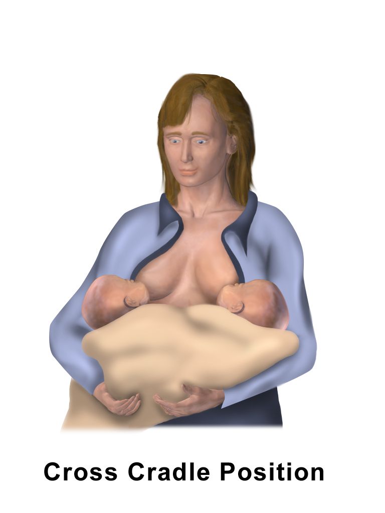
Lactation physiology and the problems that actually present
Milk production runs on a simple loop. Prolactin from the anterior pituitary makes milk; oxytocin from the posterior pituitary ejects it (the let-down reflex, often felt as tingling and accompanied by afterpains). Both are released by suckling, so supply is driven by demand and by drainage. Retained milk raises feedback inhibitor of lactation and shuts production down locally, which is why the answer to almost every low-supply question is more frequent, more effective removal of milk. Placental delivery causes the progesterone withdrawal that permits lactogenesis II — secretory activation, the moment the milk “comes in” — at roughly 30 to 72 hours after birth. Before that, the newborn receives colostrum: thick, yellow, low in volume (about 2–20 mL per feed on day one, which matches a newborn stomach the size of a cherry), extraordinarily high in secretory IgA and protein, and laxative enough to clear meconium and reduce bilirubin. Transitional milk follows for about two weeks, then mature milk, with watery foremilk giving way to fat-rich hindmilk within a feed.
Expect 8 to 12 feeds in 24 hours, driven by the infant’s cues — rooting, hand-to-mouth, stirring, lip-smacking — with crying as a late and inefficient cue. Assess the latch rather than the clock: mouth wide open, lips flanged out, chin indenting the breast, a substantial portion of the areola taken in with more below than above, audible swallowing, and no pinching pain after the first few sucks. Persistent nipple pain means a shallow latch or a positioning problem — break the suction with a clean finger and start again rather than enduring it. Judge adequacy by output: by day 4 to 5 expect at least six wet nappies and three to four yellow, seedy stools a day. Newborns normally lose up to 7 to 10 per cent of birth weight, with the nadir at day 3 to 4 and return to birth weight by day 10 to 14.
Engorgement peaks at day 3 to 5 as vascular and lymphatic congestion accompanies secretory activation. Both breasts are firm, hot, tight and painful, and a low-grade temperature can occur. Manage it by feeding frequently, applying cold between feeds to reduce oedema, using brief warmth or reverse-pressure softening just before a feed so the infant can latch onto a softened areola, and expressing only enough to relieve pressure. Mastitis is different and unilateral: a wedge-shaped area of redness, heat and tenderness with fever of 38.5 °C or higher and flu-like aching. The key teaching, and the one most often got wrong, is that breastfeeding continues on the affected side — the milk is safe for the infant and drainage is treatment. Add rest, fluids, analgesia and a ten- to fourteen-day course of a penicillinase-resistant antibiotic such as dicloxacillin 500 mg four times daily or cephalexin 500 mg four times daily; an untreated mastitis can become a breast abscess. For a parent who is not lactating, suppression means a well-fitting supportive bra, cold packs, avoiding any nipple stimulation or expression, and analgesia; symptoms settle in 24 to 72 hours.
Medications at the couplet’s bedside
The postpartum nurse gives medications to two patients with a roughly twentyfold difference in body mass, and the newborn’s doses are never improvised from adult practice. Routine newborn prophylaxis illustrates the discipline: vitamin K1 (phytonadione) 0.5 to 1 mg intramuscularly into the vastus lateralis within 6 hours of birth to prevent vitamin K deficiency bleeding, and erythromycin 0.5 per cent ophthalmic ointment as a 1 cm ribbon to each lower conjunctival sac within an hour or two of birth. Both are fixed newborn doses, verified against the order and the infant’s weight before they are drawn up.
For the birthing parent, analgesia is chosen with lactation in mind: scheduled ibuprofen 600 mg every 6 hours with paracetamol 650–1000 mg every 6 hours (maximum 3000–4000 mg per 24 hours) controls most postpartum pain, while codeine and tramadol are contraindicated during breastfeeding because ultra-rapid CYP2D6 metabolisers can transfer dangerous morphine concentrations into milk and cause infant respiratory depression. Ibuprofen is safe with breastfeeding and treats afterpains and perineal pain well; time the dose about thirty minutes before a feed when afterpains are severe.
Psychological adaptation and mood
Rubin’s phases still describe the arc usefully. In taking-in (the first 24 to 48 hours) the birthing parent is dependent, focused on her own needs, hungry, sleepy and given to retelling the birth story — which is integration work, not chatter. In taking-hold (roughly day 2 to day 10) she becomes independent, task-focused, eager to master care and highly receptive to teaching. This is the teaching window; instruction delivered during taking-in is rarely retained. Letting-go follows over weeks as the family reorganises around the new member and grieves the relationships and identity that existed before. Watch also for bonding behaviours — en face gaze, fingertip-to-palm progression of touch, calling the infant by name, engrossment in a partner — and for sibling and cultural patterns, which vary widely and are not deficits.
Distinguish three conditions that are not on a single continuum. Postpartum blues affect 50 to 80 per cent of birthing parents, begin around day 3 to 5, peak at day 5, and resolve spontaneously by day 10 to 14. They present as tearfulness, mood lability, irritability, anxiety and fatigue, with intact function and no thoughts of harm; treatment is rest, nutrition, reassurance and practical support. Postpartum depression affects roughly one in seven, can begin any time within the first year, and lasts beyond two weeks with pervasive low mood, anhedonia, guilt, disturbed sleep even when the baby sleeps, poor concentration, appetite change and difficulty bonding. Screen with the Edinburgh Postnatal Depression Scale; a score of 10 or more warrants follow-up, 13 or more is a strong positive, and any positive response to item 10 — thoughts of self-harm — requires immediate assessment for safety before the visit ends. Treatment includes psychotherapy, sertraline or another lactation-compatible SSRI, and support. Postpartum psychosis occurs in about 1 to 2 per 1000, typically within the first two weeks and often within 72 hours, and is a psychiatric emergency: delusions frequently concerning the infant, hallucinations, confusion, disorganisation, and grossly impaired reality testing, with substantial risk of suicide and of harm to the infant. Do not leave the parent alone with the baby, ensure continuous supervision, and arrange urgent psychiatric admission. Bipolar disorder is the strongest risk factor, and every patient with that history should be flagged antenatally.
Going home: teaching, contraception and follow-up
Discharge teaching is the last safety net, and it works better as a short list of alarms than as a lecture. The POST-BIRTH warning signs give the patient and her family concrete thresholds. Call emergency services for pain in the chest, obstructed breathing or shortness of breath, seizures, or thoughts of hurting oneself or the baby. Call the provider for bleeding that soaks more than one pad in an hour or clots the size of an egg or larger, an incision that is not healing, a red or swollen leg that is painful or warm, a temperature of 38.0 °C (100.4 °F) or higher, or a headache that does not improve or that comes with visual changes. Teach the patient and a support person together, teach back rather than ask “any questions?”, and give the numbers to call in writing.
Fertility returns before periods do. Ovulation can precede the first menses, so contraception is discussed before discharge. Progestin-only methods (the progestin-only pill, the implant, depot medroxyprogesterone, the levonorgestrel intrauterine device) and copper intrauterine devices are compatible with breastfeeding and can be started immediately postpartum, including immediate post-placental IUD insertion. Combined hormonal contraceptives are avoided in the first 21 days postpartum for everyone because of thromboembolic risk, and until 42 days if additional VTE risk factors are present. The lactational amenorrhoea method is about 98 per cent effective only when all three conditions hold simultaneously: exclusive breastfeeding day and night, amenorrhoea, and an infant under six months old. Advise resuming intercourse when lochia has stopped and the perineum is comfortable, and warn about vaginal dryness during lactation. Finally, follow the fourth-trimester model: contact within the first three weeks — within 72 hours for hypertensive disorders — and a comprehensive visit no later than 12 weeks that reviews mood, bleeding, blood pressure, feeding, contraception, chronic disease and the plan for future pregnancies.
The fourth trimester is nursing work: protect the feeding relationship, give newborn prophylaxis and parental analgesia with lactation in mind, screen the mood rather than assume it, and send the family home with specific alarms rather than general reassurance.
The chapter in one breath
The puerperium reverses nine months of adaptation in six weeks. The uterus involutes through myometrial retraction, autolysis and regeneration — falling from about 1000 g to 60–80 g, descending roughly a centimetre a day, and shedding lochia rubra, serosa and alba. The plasma volume that protected the patient in labour now unloads through diuresis and diaphoresis, producing a benign bradycardia of 40–60 and a leucocytosis to 30,000 that make ordinary vital-sign rules useless; tachycardia over 100, oliguria under 35 mL per hour or a saturation under 95 per cent are the real warnings. Assessment is structured (BUBBLE-HE), quantified (weigh blood, 1 g equals 1 mL) and never delegated. In haemorrhage the sequence is the treatment — massage the fundus first, call for help, empty the bladder, then choose a uterotonic that the patient’s blood pressure and airway will tolerate. A firm fundus with bright red bleeding is a laceration; concealed pain is a haematoma; no palpable fundus with disproportionate shock is inversion, and uterotonics stop. Magnesium toxicity takes reflexes before respirations, so hourly reflexes buy the time that calcium gluconate then uses. Hypertension, sepsis and thromboembolism stalk the weeks after discharge, and mood must be screened, not assumed. Newborn prophylaxis and parental analgesia are given with lactation in mind, and every family goes home with named alarms.
Words worth knowing
| Term | What it means |
|---|---|
| Involution | Return of the uterus toward its pre-pregnancy size and position through retraction, autolysis and endometrial regeneration; complete by about six weeks. |
| Subinvolution | Delayed or arrested involution — a boggy, higher-than-expected uterus with persistent or recurrent rubra, usually from retained fragments or infection. |
| Living ligature | The interlacing myometrial fibres that mechanically clamp the spiral arterioles of the placental site when the uterus contracts. |
| Lochia | Postpartum uterine discharge of blood, decidua, leucocytes and exudate, progressing rubra (days 1–3) to serosa (days 4–10) to alba (day 10 to six weeks). |
| Afterpains | Cramping from intermittent uterine contractions, stronger in multiparas, over-distended uteri and during breastfeeding because suckling releases oxytocin. |
| Quantitative blood loss (QBL) | Measured rather than estimated blood loss, obtained by weighing soaked materials and subtracting dry weight; 1 gram equals 1 millilitre. |
| Postpartum haemorrhage | Cumulative blood loss of 1000 mL or more, or any loss with signs of hypovolaemia, within 24 hours of birth regardless of mode of birth. |
| Uterine atony | Failure of the myometrium to contract after birth; the cause of 70–80 per cent of primary postpartum haemorrhage. |
| Four Ts | Tone, Trauma, Tissue, Thrombin — the diagnostic framework for postpartum haemorrhage. |
| Shock index | Heart rate divided by systolic blood pressure; a value of 0.9 or higher in a postpartum patient suggests significant haemorrhage before hypotension appears. |
| Uterine inversion | The fundus turning inside out into the uterine cavity or vagina; managed by stopping uterotonics, not removing the placenta, and replacing the uterus before restarting uterotonics. |
| Puerperal bradycardia | Normal heart rate of 40–60 beats per minute for the first six to ten days, caused by the sudden rise in stroke volume after birth. |
| Puerperal morbidity | Temperature of 38.0 °C or higher on any two of the first ten postpartum days, excluding the first 24 hours — the classic definition of postpartum infection. |
| Endometritis | Infection of the uterine lining, usually on day 2–3, with fever, uterine tenderness, subinvolution and foul lochia; more common after caesarean birth. |
| Lactogenesis II | Secretory activation — the copious milk production that begins 30 to 72 hours after birth once progesterone falls with placental delivery. |
| REEDA | Perineal wound assessment: Redness, Ecchymosis, Oedema, Discharge, Approximation. |
| BUBBLE-HE | The structured postpartum assessment: Breasts, Uterus, Bladder, Bowel, Lochia, Episiotomy/perineum, Homans/lower extremities, Emotional status. |
| Taking-hold | Rubin’s second phase, roughly day 2 to day 10, when the birthing parent becomes independent and task-focused — the optimal window for teaching. |
| Lactational amenorrhoea method | Contraception that is about 98 per cent effective only when exclusive breastfeeding, amenorrhoea and an infant under six months are all present. |
Check yourself
Postpartum Complications
The hours and weeks after birth are the most dangerous stretch of the childbearing year, and they are the stretch we watch least closely. A birthing parent who walked into the unit healthy can bleed a litre in ten minutes with a blood pressure that has not yet moved, develop severe-range hypertension five days after discharge, throw a pulmonary embolus at three weeks, or be quietly overwhelmed by intrusive thoughts at six. Most of these deaths are judged preventable, and what usually fails is not knowledge of the disease but the speed and order of the response. This chapter is therefore built around sequences: what you assess, what you do first, what you do next, and what you say when you escalate. Massage the fundus before you reach for a drug. Stop the magnesium before you draw up the antidote. Ask about the thoughts before the family leaves the room.
By the end of this chapter you can
- Define early and late postpartum haemorrhage using cumulative quantitative blood loss, and explain why pregnancy’s hypervolaemia delays hypotension.
- Perform the first-response sequence for uterine atony in order — massage the fundus first — and justify each subsequent step.
- Compare the uterotonics (oxytocin, methylergonovine, carboprost, misoprostol) and tranexamic acid by dose, route, timing and absolute contraindication.
- Distinguish the four Ts at the bedside, including genital tract haematoma, retained placental fragments, disseminated intravascular coagulation and uterine inversion.
- Recognise endometritis, mastitis and maternal sepsis, and calculate weight-based antibiotic and fluid doses, showing the arithmetic and the independent double check.
- Manage venous thromboembolism, postpartum preeclampsia and magnesium sulfate toxicity, naming the antidote calcium gluconate and the order in which toxicity appears.
- Differentiate postpartum blues, perinatal depression and anxiety and postpartum psychosis, and act on a positive self-harm item.
- Escalate any of the above using SBAR, and apply delegation limits so that assessment, teaching, evaluation and the unstable patient stay with the registered nurse.
- Teach the POST-BIRTH warning signs to a family in plain, non-blaming language before discharge.
Section 1Early Postpartum Haemorrhage and the First Ten Minutes
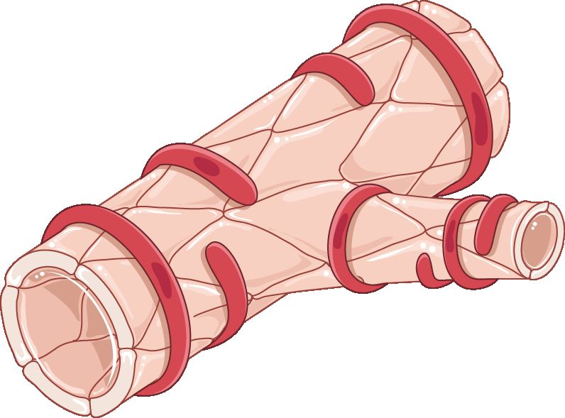
Why the numbers deceive you
Postpartum haemorrhage is defined as cumulative blood loss of 1000 mL or more, or blood loss of any volume accompanied by signs or symptoms of hypovolaemia, within 24 hours of birth — regardless of whether the birth was vaginal or caesarean. That second clause matters more than the number. A small person who has lost 700 mL and is suddenly restless, yawning and tachycardic is haemorrhaging; a large person who has lost 1100 mL and is chatting comfortably may not yet be. Loss of more than 500 mL after a vaginal birth, even if it does not meet the definition, should still prompt you to look for a cause. Haemorrhage occurring after the first 24 hours and up to 12 weeks postpartum is called late or secondary haemorrhage and has a different cause list.
Visual estimation of blood loss is unreliable, and the error runs in the dangerous direction: clinicians underestimate large losses and overestimate small ones. Quantitative blood loss (QBL) replaces the guess with a scale. Weigh the blood-soaked pads, drapes, chux and lap sponges, subtract the known dry weight, and convert — one gram equals approximately one millilitre. Use a calibrated under-buttocks drape at vaginal birth and read the graduated pouch, subtracting amniotic fluid volume collected before the placenta delivers. In theatre, measure canister volume and subtract irrigation. QBL is cumulative and is recorded at defined intervals, not once at the end.
The deeper trap is physiological. Pregnancy expands plasma volume by roughly 40 to 50 per cent, which is nature’s own transfusion in anticipation of birth. A previously healthy birthing person can therefore lose 1000 to 1500 mL before the systolic blood pressure falls at all. Blood pressure is a late sign. Tachycardia, narrowing pulse pressure, rising respiratory rate, cool clammy skin, delayed capillary refill and above all a change in mental state — anxiety, air hunger, “something is wrong” — arrive first. Trust the trend and the person, not the single number on the monitor.
Two tools sharpen the eye. The shock index is heart rate divided by systolic blood pressure; a normal postpartum value is 0.5 to 0.7, a value of 0.9 or above should trigger a second look and 1.4 or above signals severe compromise. The maternal early warning criteria flag any of the following for immediate bedside evaluation by a clinician who can escalate: systolic pressure below 90 or above 160 mmHg, diastolic above 100, heart rate below 50 or above 120, respiratory rate below 10 or above 30, oxygen saturation below 95 per cent on room air, urine output under 35 mL per hour for two hours or more, or altered mental status, agitation, unrelieved headache or breathlessness.
The four Ts, and why tone comes first
Haemostasis at the placental site is mechanical, not chemical. The myometrium is arranged in interlacing bundles that surround the spiral arterioles; when those bundles contract, they strangle the vessels shut. Clinicians call them the living ligatures. Clotting factors help, but a uterus that will not contract cannot be clotted into safety. This is why the whole first-line management of haemorrhage is aimed at making the uterus squeeze.
Causes sort into four Ts. Tone — uterine atony — accounts for roughly 70 to 80 per cent of cases. Trauma (lacerations, haematoma, rupture, inversion) accounts for most of the remainder, around 20 per cent. Tissue means retained placenta or membranes and includes the placenta accreta spectrum. Thrombin means a coagulation problem, whether inherited, acquired in pregnancy, or consumed by the haemorrhage itself. In practice you assess tone first because it is both the most likely cause and the one you can treat with your hands in the first five seconds.
Risk factors for atony cluster around an overstretched, exhausted or chemically relaxed uterus: multiple gestation, polyhydramnios, a large baby, high parity, a long labour or a very rapid one, prolonged oxytocin exposure during labour, chorioamnionitis, magnesium sulfate infusion, general anaesthesia with halogenated agents, and retained placental tissue. Risk assessment is repeated at three defined points — at prenatal booking, again on admission in labour, and again immediately after birth — because the category changes as labour unfolds. But remember the humbling statistic: a substantial proportion of haemorrhages occur in people categorised as low risk. Every birth gets a fundal check.
One reversible cause hides in plain sight. A full bladder sits directly beneath the lower uterine segment, pushes the uterus up and to one side, and mechanically prevents it from contracting down. A fundus that is boggy, high and deviated laterally — classically to the right — is a bladder problem until you have emptied the bladder. Have the patient void; if they cannot, catheterise. Document the volume.
What you do first
The single most common examination error in postpartum haemorrhage is reaching for a syringe before reaching for the fundus. Massage is faster than any drug, needs no order, and works on the cause of four out of five haemorrhages. Place one hand flat above the symphysis pubis to support and anchor the lower uterine segment — this is not optional, because massaging an unsupported uterus can invert it — and with the other hand cup the fundus and massage firmly in a circular motion until it firms to the consistency of a tennis ball. Expel clots only once the uterus is firm; expressing clots from a boggy uterus simply pushes the fundus further up and can invert it.
Then the sequence unfolds in parallel, not one at a time, because the team you called in step two does several of these at once.
| Order | Action | Why this, why now |
|---|---|---|
| 1 | Massage the fundus with lower-segment support | Mechanical contraction is the definitive haemostat; works in seconds without an order |
| 2 | Call for help — activate the haemorrhage protocol | One nurse cannot massage, give drugs, quantify loss and document; this is a team event |
| 3 | Increase or start the oxytocin infusion | First-line uterotonic, already hanging in most units |
| 4 | Empty the bladder (void or catheterise) | A distended bladder mechanically prevents involution |
| 5 | Two large-bore IV lines (18 gauge or larger); warmed isotonic crystalloid | Volume and a route for blood products; small cannulae will not transfuse fast |
| 6 | Oxygen 10 to 15 L per minute by non-rebreather mask (below 10 L the reservoir bag collapses); keep the patient flat and warm | Maximises oxygen carriage on the red cells that remain; hypothermia worsens coagulopathy |
| 7 | Bloods: full blood count, type and crossmatch, coagulation screen, fibrinogen, lactate | Guides transfusion; fibrinogen predicts severity |
| 8 | Second-line uterotonic if bleeding continues; tranexamic acid | Escalate by drug class rather than repeating a failed one |
| 9 | Quantify blood loss continuously; vital signs every 5 minutes; indwelling catheter for hourly output | Urine output under 30 mL per hour signals inadequate perfusion |
| 10 | Tamponade device, then surgical management | Escalate before the patient becomes coagulopathic, not after |
Notice what is not first. Not the blood pressure cuff, not the call to pharmacy, not fetching the emergency box. Hands on the fundus, voice calling for help. Everything else follows.
Uterotonics, tranexamic acid and what comes after the drugs
Each uterotonic has a contraindication that can kill the patient you are trying to save, and each of those contraindications describes a patient you will meet often. Learn the pairs: methylergonovine with hypertension, carboprost with asthma.
| Drug | Dose and route | Watch for | Do not give if |
|---|---|---|---|
| Oxytocin | 10 to 40 units in 500 to 1000 mL isotonic crystalloid IV infusion, titrated; or 10 units IM | Hypotension and tachycardia if pushed fast; water intoxication with large volumes | Never give undiluted as a rapid IV bolus |
| Methylergonovine | 0.2 mg IM, may repeat every 2 to 4 hours; oral 0.2 mg every 6 to 8 hours for maintenance | Sudden severe hypertension, headache, chest pain | Hypertension, preeclampsia, cardiac disease; avoid the IV route |
| Carboprost tromethamine | 250 micrograms IM or intramyometrial, repeat every 15 to 90 minutes, maximum 8 doses (2 mg total) | Diarrhoea, vomiting, fever, flushing — pre-medicate with an antiemetic and antidiarrhoeal | Asthma (bronchospasm); caution in hepatic, renal or cardiac disease |
| Misoprostol | 600 to 1000 micrograms sublingual, oral or rectal, single dose | Shivering and pyrexia — expected, not infection | Few absolute contraindications; useful where refrigeration is unavailable |
| Tranexamic acid | 1 g IV over 10 minutes, ideally within 3 hours of birth; may repeat 1 g if bleeding persists after 30 minutes or restarts within 24 hours | Hypotension if infused faster than 1 mL per minute; seizures at high dose | Known thromboembolic disease; benefit falls sharply after 3 hours |
Tranexamic acid is an antifibrinolytic, not a uterotonic. It does not make the uterus contract; it stops the clot that forms from being dissolved. Its mortality benefit is time-dependent, which is why it belongs in the early bundle rather than the salvage phase. Give it in addition to uterotonics, never instead of them.
When drugs fail, the next tier is mechanical. Intrauterine balloon tamponade — a fluid-filled balloon inflated inside the uterine cavity, commonly to 300 to 500 mL — presses the endometrial surface against the myometrium. A newer vacuum-induced device does the opposite, applying low-level suction so the uterus collapses inward on itself. Both buy time. While a device is in place, keep an indwelling urinary catheter, watch for concealed bleeding above the balloon (the fundal height rising with a firm uterus and a falling haematocrit), and record drainage-port output as part of QBL. Surgical options follow: uterine compression sutures such as the B-Lynch, uterine or internal iliac artery ligation, uterine artery embolisation by interventional radiology, and finally hysterectomy.
Massive haemorrhage becomes a coagulopathy if you replace only red cells and crystalloid. Massive transfusion protocols deliver packed red cells, fresh frozen plasma and platelets in a balanced ratio, often 1:1:1. Fibrinogen deserves special attention in obstetrics: pregnancy raises it to roughly 350 to 650 mg/dL, so a “normal-looking” level of 250 is actually low for a pregnant patient, and a level below 200 mg/dL predicts severe haemorrhage and calls for cryoprecipitate or fibrinogen concentrate. Keep the patient warm and correct acidosis and hypocalcaemia — the lethal triad of hypothermia, acidosis and coagulopathy is what turns a survivable bleed into an unsurvivable one.
In postpartum haemorrhage the first action is a firm fundal massage with the lower uterine segment supported, because uterine tone — not a drug and not the blood pressure reading — is what stops the bleeding.
Section 2Trauma, Tissue, Thrombin and Uterine Inversion
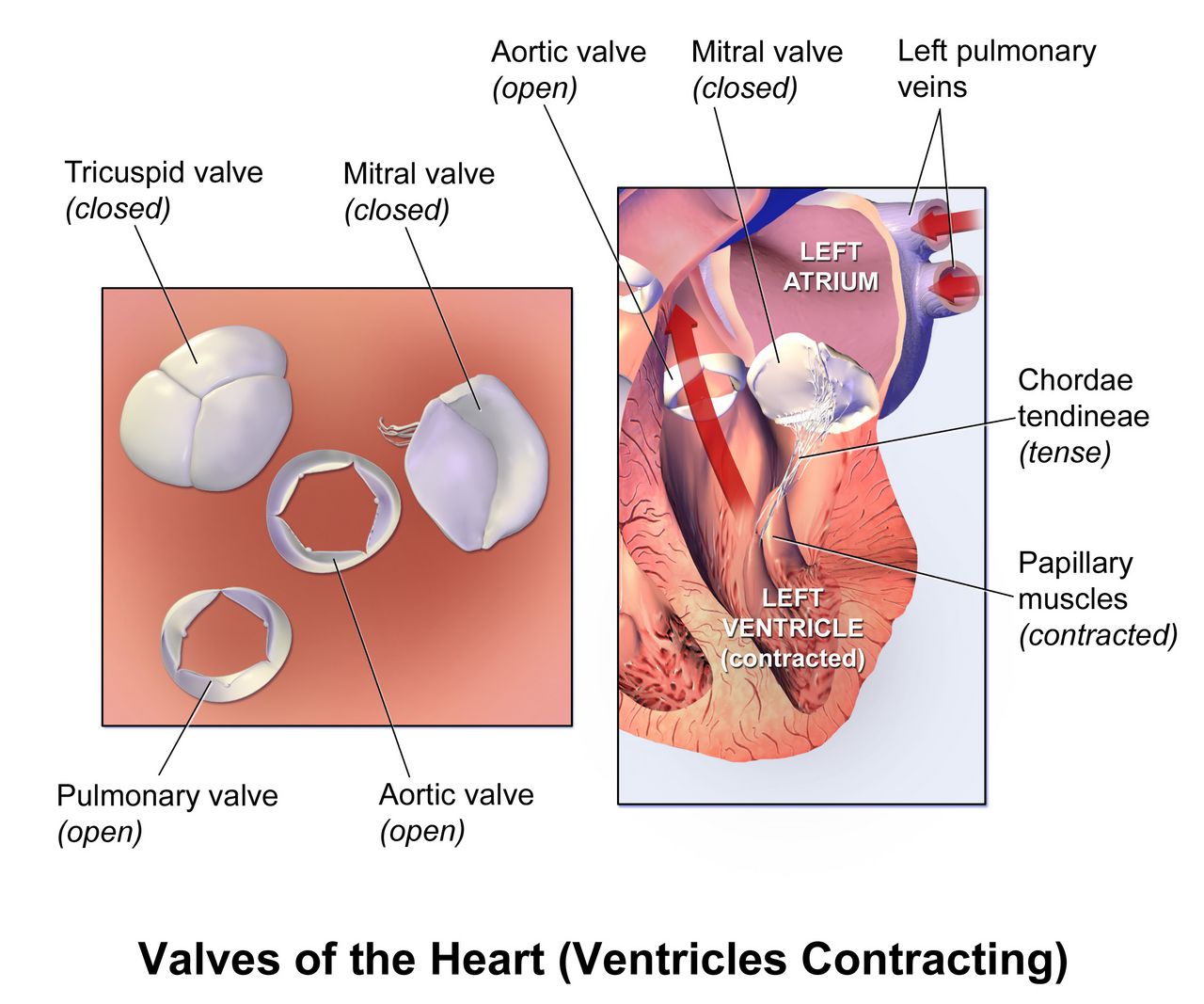
Trauma: lacerations and the haematoma you cannot see
The clinical discriminator is simple enough to teach in one line: a boggy fundus is atony; a firm fundus with continued bleeding is trauma. If you have massaged the uterus to the firmness of a tennis ball, emptied the bladder, given oxytocin, and blood is still running — often a steady bright red trickle rather than dark clots — the source is a tear. Someone must inspect the cervix, vagina and perineum under good light with adequate analgesia. That is a provider task, but you set it up, position the patient, and keep quantifying.
Genital tract haematoma is the version of trauma that bleeds where you cannot see it. Blood collects in the vulvar, vaginal or retroperitoneal space while the perineal pad stays deceptively dry. The presentation is pain — severe, unrelenting perineal, vaginal or rectal pressure that is out of proportion to the repair and is not relieved by ice or the usual analgesia. On inspection there may be a tense, bluish-purple, unilateral bulging mass; on the vaginal side there may be nothing visible at all. Meanwhile the pulse climbs, the haematocrit falls and the fundus stays firm. A patient who says “I feel like I need to have a bowel movement” an hour after a repair is describing a haematoma until proven otherwise.
Small, non-expanding haematomas of roughly 3 to 5 cm are managed conservatively with ice for the first 12 hours, analgesia, careful marking of the borders and serial reassessment. Larger or expanding collections require incision, evacuation of clot, ligation of the bleeding vessel and often a drain, with a urinary catheter because oedema obstructs the urethra. Retroperitoneal haematoma is rare, is usually associated with caesarean or uterine artery injury, and presents as shock with no external bleeding whatsoever; it is diagnosed by imaging and managed surgically or by embolisation.
Tissue: retained fragments, accreta and late haemorrhage
The placenta is normally delivered within 5 to 30 minutes of the baby. Beyond 30 minutes it is called retained, and the cavity cannot contract around the adherent tissue. Every placenta is inspected for completeness — missing cotyledons or a torn vessel running to the edge of the membranes suggests a succenturiate lobe still inside. Management is manual removal under analgesia or ultrasound-guided curettage, with antibiotic cover, and the patient needs vigilant fundal and bleeding checks afterwards.
The placenta accreta spectrum — accreta (adherent to myometrium), increta (invading myometrium) and percreta (through the serosa, sometimes into bladder) — is the modern face of catastrophic obstetric haemorrhage, and it tracks the caesarean rate. Suspect it when a prior caesarean is combined with a placenta praevia. Where it is diagnosed antenatally the birth is planned in a centre with blood banking, interventional radiology and surgical backup, and the plan is usually caesarean hysterectomy with the placenta left in situ. An accreta discovered at the time of birth, in a unit that has not prepared for it, is a genuine emergency: do not tug on the cord, activate massive transfusion, and get help.
Retained fragments are also the leading cause of late (secondary) postpartum haemorrhage, occurring between 24 hours and 12 weeks. The typical story is a patient who returns on day 7 to 14 with heavier bleeding than she had at day 3, sometimes with clots and cramping. Related is subinvolution: the fundus is higher and softer than the day count predicts, and lochia rubra persists past the usual 3 to 4 days or returns after becoming serosa. Normal involution descends the fundus about 1 cm (one fingerbreadth) per day, from the level of the umbilicus in the first 12 hours to non-palpable abdominally by day 10 to 14. Evaluation is by ultrasound; treatment is uterotonics such as oral methylergonovine 0.2 mg every 6 to 8 hours, antibiotics if infection is present, and curettage if tissue is retained.
| Finding | Suggests | Your next move |
|---|---|---|
| Boggy fundus, dark blood and clots, fundus above umbilicus | Atony | Massage with lower-segment support, then uterotonic |
| Firm fundus, steady bright red trickle | Laceration | Notify for inspection and repair; keep quantifying loss |
| Firm fundus, minimal external loss, severe unilateral perineal or rectal pain, rising pulse | Haematoma | Inspect, mark borders, notify urgently, prepare for evacuation |
| Boggy fundus deviated to one side, high | Distended bladder | Void or catheterise, then reassess tone |
| Fundus not palpable, mass at introitus, profound shock, severe pain | Uterine inversion | Stop uterotonics, call for help, do not remove the placenta |
| Persistent oozing from IV sites and gums, blood not clotting | Coagulopathy or DIC | Send fibrinogen and coagulation screen, activate massive transfusion |
Thrombin: coagulopathy, DIC and amniotic fluid embolism
Some patients arrive with a bleeding tendency. Von Willebrand disease is the commonest inherited bleeding disorder in people who menstruate; factor levels rise physiologically during pregnancy and then fall back within one to two weeks of birth, which is exactly why these patients characteristically bleed late rather than immediately. Immune thrombocytopenia, liver disease, and therapeutic anticoagulation are the other common set-ups.
More often, coagulopathy is acquired. Disseminated intravascular coagulation in obstetrics follows placental abruption, severe preeclampsia and HELLP syndrome, intrauterine fetal demise, sepsis, amniotic fluid embolism, and any haemorrhage large enough to consume its own clotting factors. Clinically you see bleeding from every breached surface at once: oozing from cannula sites and the gums, petechiae, bruising at the blood-pressure cuff, haematuria, and blood in the drain that will not clot. Laboratory findings are a falling platelet count, prolonged prothrombin and activated partial thromboplastin times, rising D-dimer and a falling fibrinogen. Treatment is to stop the bleeding source and replace what has been consumed — plasma, platelets, cryoprecipitate — while supporting perfusion.
Amniotic fluid embolism is rare, unpredictable and unpreventable. It presents as sudden cardiorespiratory collapse during labour, at birth, or within about 30 minutes after, classically as a triad of abrupt hypoxia, hypotension and coagulopathy, often with seizure. There is no test that makes the diagnosis in real time and no specific treatment. Management is high-quality resuscitation: airway and 100 per cent oxygen, chest compressions with manual left uterine displacement if still undelivered, early massive transfusion for the coagulopathy, and immediate obstetric and critical care presence. Your role is to recognise it in seconds and call the code.
Uterine inversion: the emergency where you stop the uterotonic
Uterine inversion — the fundus turning inside out and descending through the cervix — is rare, occurring in perhaps 1 in 2000 to 1 in 20 000 births, and it is the one obstetric haemorrhage where the reflex to give a uterotonic is exactly wrong. It is usually caused by excessive cord traction on an uncontracted uterus or by fundal pressure, which is one reason cord traction is only ever applied with counter-pressure on the lower segment and only with a contracted uterus.
The presentation is unmistakable once you have seen it: sudden severe lower abdominal pain, a dark red-purple mass appearing at or protruding from the introitus, a fundus that cannot be palpated abdominally or feels like a dimple or crater, and shock that is out of proportion to the visible blood loss because vagal stimulation from traction on the peritoneum adds neurogenic shock to the haemorrhagic shock.
The sequence is: call for help and anaesthesia immediately; stop all uterotonics, because a contracting cervical ring makes replacement impossible; do not attempt to remove the placenta if it is still attached, because separating it increases blood loss before the uterus is back; establish large-bore access and start volume resuscitation; anticipate a uterine relaxant such as terbutaline, nitroglycerin, magnesium sulfate or a halogenated inhalational agent; and support the provider’s manual replacement, in which the fundus is pushed back through the cervix. Only after the uterus is replaced do you restart uterotonics, vigorously, to keep it there — and then remove the placenta. If manual replacement fails, laparotomy follows.
When the fundus is firm and the patient is still bleeding, still hurting or still tachycardic, the problem is trauma, retained tissue, coagulopathy or inversion — and in inversion you stop the uterotonic, leave the placenta alone and get the uterus back first.
Section 3Puerperal Infection, Mastitis and Maternal Sepsis
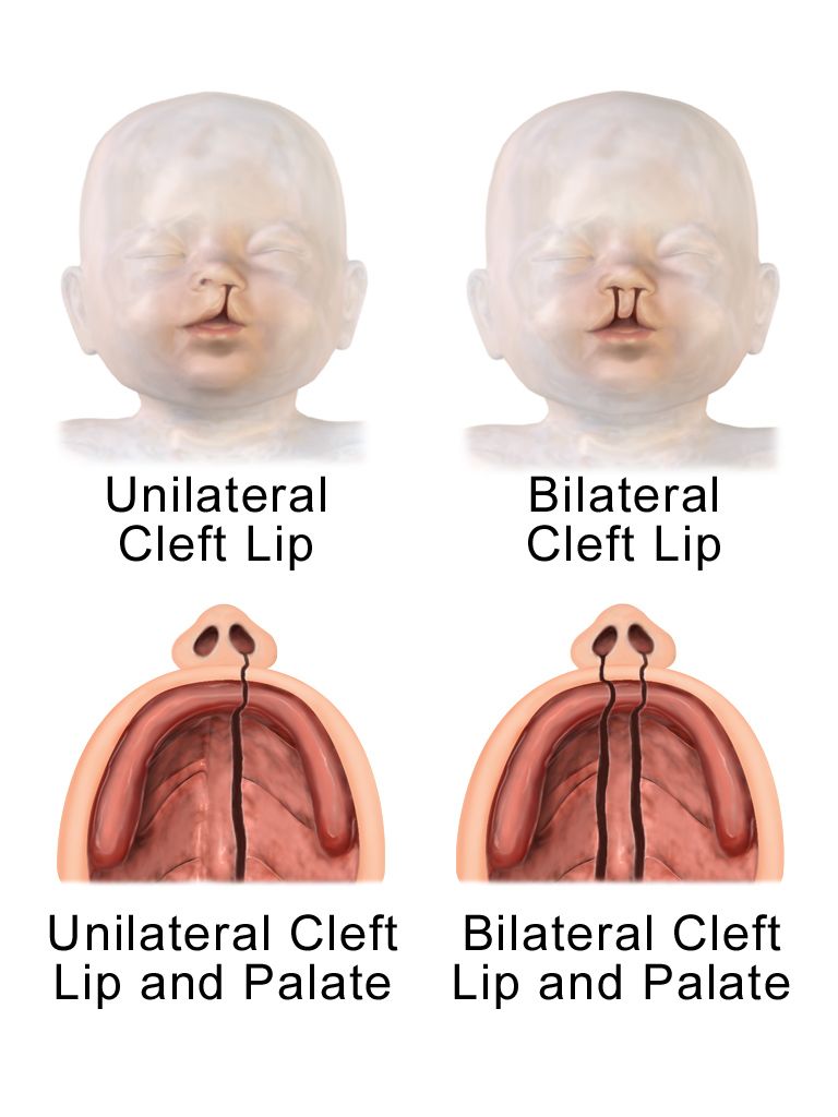
Defining puerperal infection and recognising endometritis
Puerperal infection is classically defined as an oral temperature of 38.0°C (100.4°F) or higher on any two of the first ten days postpartum, excluding the first 24 hours, with temperatures taken at least four times a day. The exclusion of the first day matters: a transient low-grade temperature in the first 24 hours is common and usually reflects dehydration and the work of labour, and it responds to fluids. A temperature on day two or later does not get explained away.
Endometritis is the commonest puerperal infection and is polymicrobial, arising from ascending genital tract flora. Risk rises steeply with caesarean birth, particularly an unscheduled caesarean after labour and ruptured membranes, and with prolonged rupture of membranes, multiple vaginal examinations, internal monitoring, manual removal of the placenta, chorioamnionitis in labour, prolonged labour, retained tissue, anaemia and diabetes. Prophylactic antibiotics given within 60 minutes before caesarean incision substantially reduce it.
Assessment findings appear on day two to day five: fever, chills, tachycardia that tracks the temperature, lower abdominal or uterine tenderness that is exquisite on fundal palpation, lochia that is foul-smelling or profuse (though endometritis caused by group A streptococcus may have scant, odourless lochia), malaise and anorexia. Treatment is intravenous broad-spectrum antibiotics — the classic regimen is clindamycin 900 mg IV every 8 hours plus gentamicin, dosed by weight — continued until the patient has been afebrile and asymptomatic for 24 to 48 hours, at which point oral antibiotics are usually unnecessary. Nursing care adds fluids, antipyretics, Fowler position to promote lochial drainage, meticulous perineal hygiene, and continued support for feeding, because most of these antibiotics are compatible with lactation.
If the fever persists after 48 to 72 hours of appropriate antibiotics, the differential widens: abscess, retained products, wound infection, septic pelvic thrombophlebitis (a diagnosis of exclusion in a patient who looks surprisingly well between fever spikes), or a resistant organism. Persistent fever on adequate therapy is an escalation trigger, not a reason to wait another day.
Wound, perineal and urinary infection
Caesarean wound infection usually presents on day four to seven with erythema extending beyond the incision line, induration, warmth, tenderness, purulent or serosanguineous drainage and fever. Management is drainage and opening of the wound where needed, antibiotics, and packing with dressing changes; teach the family to report increasing pain, spreading redness, new drainage or wound edges separating. Perineal and episiotomy infections present with disproportionate pain, oedema, erythema at the repair, and sometimes wound breakdown; sitz baths, analgesia and antibiotics are the mainstay.
The rare but lethal variant is necrotising fasciitis. The clue is pain far out of proportion to the visible wound, rapid progression over hours, dusky or bronze skin discoloration, bullae, crepitus, anaesthesia over the affected skin, and systemic toxicity. This is a surgical emergency requiring immediate wide debridement; antibiotics alone will not save the patient. If a postpartum wound looks mild but the patient is in agony and looks unwell, escalate immediately rather than reassess in an hour.
Urinary tract infection is common because of birth trauma to the bladder, epidural-related retention, catheterisation and postpartum diuresis. Cystitis presents with frequency, urgency, dysuria and suprapubic tenderness; pyelonephritis adds high fever, rigors, flank or costovertebral angle tenderness, nausea and vomiting, and requires intravenous antibiotics and hydration. Prevention is unglamorous and effective: encourage voiding within 4 to 6 hours of birth, scan or palpate for retention, remove catheters as early as possible, and encourage fluids.
Mastitis and the feeding relationship
Lactational mastitis typically appears in the second to fourth week postpartum and is usually unilateral. The lactating parent describes a hard, hot, red, wedge-shaped area of the breast, aching muscles, chills and a fever often above 38.5°C — many say it feels like influenza arriving in an hour. Risk factors are milk stasis from missed or shortened feeds, a poor latch, oversupply, pressure from a tight bra or a bag strap, and nipple trauma providing a portal of entry, most often for Staphylococcus aureus.
The single most important teaching point is that feeding or expressing must continue. Milk from an infected breast is safe for a healthy term infant, and stopping abruptly worsens the stasis that caused the problem and raises the risk of abscess. Current guidance is to feed on demand rather than to try to drain the breast: extra pumping and deliberate over-emptying drive hyperlactation and worsen the inflammation. Apply cold — ice packs between and after feeds — and do not apply heat before feeds or use deep massage, both of which increase oedema and inflammation; gentle lymphatic stroking towards the axilla is the only manual treatment. Alongside that, the plan is rest, fluids, analgesia and an anti-inflammatory such as ibuprofen, and, if there is no improvement within 24 to 48 hours or the parent is systemically unwell, a 10 to 14 day antibiotic course — dicloxacillin 500 mg orally four times daily or cephalexin 500 mg four times daily, with trimethoprim-sulfamethoxazole or clindamycin where meticillin-resistant S. aureus is suspected. Symptoms should improve within 48 to 72 hours; if they do not, or a fluctuant mass is palpable, ultrasound looks for an abscess requiring needle or surgical drainage.
Language matters here as much as pharmacology. A parent with mastitis is frequently exhausted, in pain and being told by well-meaning relatives to stop feeding. Nothing in that story is a failure. Describe what is happening mechanically — milk is not moving, and the breast is inflamed — and offer the fix. Never label an infant “a difficult feeder” or a parent “non-compliant”; describe the latch, the transfer and the plan.
Maternal sepsis: the hour that counts, and doing the arithmetic
Sepsis is life-threatening organ dysfunction caused by a dysregulated response to infection, and in obstetrics it is doubly dangerous because the young, physiologically compensated patient looks well until she does not. Standard adult screening tools underperform in pregnancy and the puerperium because tachycardia, tachypnoea and a modestly raised white cell count are all normal. Use obstetric-specific triggers: temperature above 38.0°C or below 36.0°C, heart rate above 120, respiratory rate above 30 (or above 25 with any other trigger), systolic pressure below 90, oxygen saturation below 95 per cent, and any altered mental state. Lactate of 2 mmol/L or more raises concern; 4 mmol/L or more is severe.
The bundle is time-defined. Draw blood cultures before antibiotics but do not let culture collection delay them; administer broad-spectrum antibiotics within one hour of recognition; measure lactate and repeat it if elevated; give a crystalloid fluid bolus of 30 mL per kilogram for hypotension or lactate of 4 mmol/L or more; and start vasopressors if the mean arterial pressure remains below 65 mmHg after fluid. Also find and remove the source — retained tissue, an abscess, an infected wound.
Two of those steps are weight-based, and weight-based dosing is where arithmetic errors become patient harm. Show your work every time.
A postpartum temperature of 38.0°C or higher after the first 24 hours is infection until proven otherwise, and once sepsis is suspected the clock is one hour to antibiotics with every weight-based dose calculated, written and independently checked.
Section 4Thromboembolism, Postpartum Hypertension and the Failing Heart
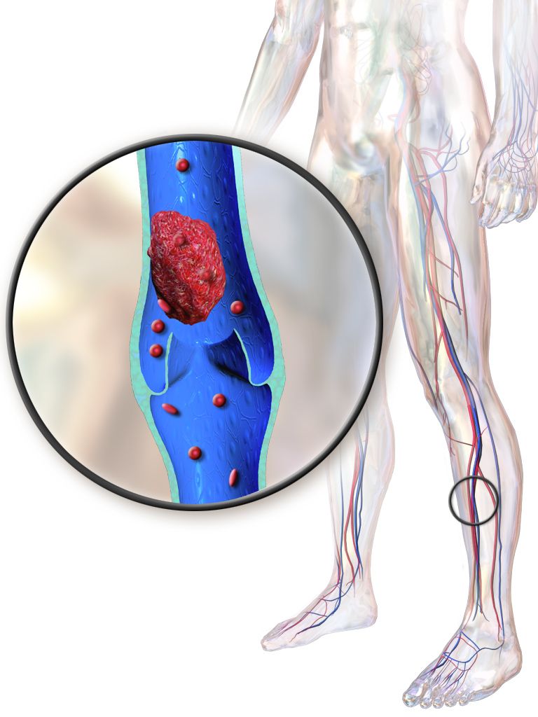
Venous thromboembolism: highest risk, lowest vigilance
Pregnancy satisfies all three limbs of Virchow’s triad simultaneously: venous stasis from the gravid uterus and reduced venous tone, endothelial injury at birth or caesarean, and a physiologically hypercoagulable state that evolved to protect against haemorrhage. That protective adaptation does not switch off at birth. Thrombotic risk is roughly four to five times baseline in pregnancy but is at its highest in the first three weeks after birth — by some estimates twenty-fold or more — and remains raised for six to twelve weeks. This is precisely the window when the patient is at home, sleep-deprived, sitting still to feed, and least likely to be assessed.
Deep vein thrombosis presents with unilateral leg pain, swelling, warmth, erythema and sometimes a palpable cord; a calf circumference difference of more than 2 cm between legs is meaningful. Left-sided DVT predominates in pregnancy. Do not use the Homans sign — dorsiflexing the foot to elicit calf pain — it is neither sensitive nor specific and may dislodge clot. Diagnosis is by compression ultrasonography. Superficial thrombophlebitis is tender and erythematous along a visible vein and is managed with warmth, elevation, compression and anti-inflammatories.
Pulmonary embolism is the killer. Sudden dyspnoea, pleuritic chest pain, tachypnoea, tachycardia, apprehension or a sense of impending doom, cough, haemoptysis and hypoxaemia. In a postpartum patient, sudden breathlessness is never “just anxiety” until an embolus has been excluded. The first actions are: stay with the patient, sit her upright, apply high-flow oxygen, call the rapid response team, obtain vital signs including oxygen saturation, and establish or confirm intravenous access — then anticoagulation, imaging and, for massive embolism with haemodynamic collapse, thrombolysis or embolectomy. Notice again that the first action is oxygenation and calling for help, not fetching the heparin.
Anticoagulation and the arithmetic that goes with it
Low-molecular-weight heparin is the agent of choice in pregnancy and lactation. It does not cross the placenta, it is not meaningfully excreted in milk, it has predictable pharmacokinetics and it does not require routine monitoring. Therapeutic enoxaparin is 1 mg per kilogram subcutaneously every 12 hours; prophylactic dosing is typically 40 mg subcutaneously once daily, or 30 mg every 12 hours in some protocols. For an 82 kg patient the therapeutic dose is 82 × 1 = 82 mg subcutaneously every 12 hours — a calculation that should be written out and checked, not estimated from a vial size.
Unfractionated heparin is used when rapid reversibility matters, when renal function is poor, or around the time of neuraxial anaesthesia, because its short half-life and complete reversal with protamine sulfate (approximately 1 mg per 100 units of heparin) make timing easier; protamine only partially reverses low-molecular-weight heparin. Warfarin is teratogenic and is avoided in pregnancy but is acceptable during lactation, and it is bridged with heparin until the international normalised ratio is therapeutic. Direct oral anticoagulants are generally avoided while breastfeeding or chestfeeding because of limited safety data.
Nursing care wraps around the drug. Teach subcutaneous injection into the abdominal fat pad at least 5 cm from the umbilicus, alternating sides, without expelling the air bubble and without rubbing the site. Teach the bleeding precautions — soft toothbrush, electric razor, no non-steroidal anti-inflammatory drugs unless prescribed, report bruising, nosebleeds, blood in urine or stool, and heavier lochia. Reinforce mobility and hydration, apply graduated compression or sequential devices, and time anticoagulant doses carefully in anyone with an epidural catheter, because spinal haematoma is the feared complication of getting that interval wrong.
Postpartum preeclampsia, eclampsia and magnesium sulfate toxicity
Hypertensive disease does not end with the placenta. Postpartum preeclampsia can present up to six weeks after birth, most commonly between 48 hours and seven days, and in a meaningful number of patients it is new — there was no hypertension in pregnancy at all. Severe features are systolic pressure of 160 mmHg or more or diastolic of 110 mmHg or more on two occasions, a headache unrelieved by analgesia, visual disturbance, right upper quadrant or epigastric pain, platelets below 100 000 per microlitre, creatinine above 1.1 mg/dL or doubled from baseline, transaminases at twice the upper limit of normal, or pulmonary oedema. HELLP syndrome is the laboratory-defined severe variant, and its three criteria are in its name: haemolysis (schistocytes on the smear, lactate dehydrogenase of 600 units/L or more, rising indirect bilirubin), elevated liver enzymes at twice the upper limit of normal or higher, and low platelets below 100 000 per microlitre. HELLP can occur with only mildly raised, or occasionally normal, blood pressure, which is why right upper quadrant or epigastric pain in a postpartum patient earns a full panel rather than an antacid. A patient telephoning on day five with a headache that will not go away and “spots” in her vision needs a blood pressure measured today, not an appointment next week.
Acute severe-range hypertension must be treated within 30 to 60 minutes of confirmation. First-line agents are intravenous labetalol 20 mg over 2 minutes, escalating to 40 mg then 80 mg at 10-minute intervals to a cumulative maximum of 300 mg; intravenous hydralazine 5 to 10 mg over 2 minutes, repeated every 20 minutes; or immediate-release oral nifedipine 10 mg. Avoid labetalol in asthma and in bradycardia. Magnesium sulfate is given for seizure prophylaxis in severe features and for treatment of eclampsia; it is not an antihypertensive and does not lower blood pressure.
Magnesium sulfate is loaded at 4 to 6 g intravenously over 15 to 30 minutes and maintained at 1 to 2 g per hour, always on a pump, always as a secondary line. Therapeutic serum levels are approximately 4 to 7 mEq/L, which is 5 to 8 mg/dL (multiply mEq/L by 1.2 to get mg/dL). Because magnesium is cleared renally, oliguria means accumulation, and urine output is a toxicity parameter, not just a fluid parameter. Assess before and hourly during the infusion: deep tendon reflexes, respiratory rate, urine output, level of consciousness, blood pressure, and continuous oximetry.
Toxicity appears in a fixed order, and the order is the whole lesson. Deep tendon reflexes disappear first, at roughly 8 to 12 mg/dL, before the respiratory rate falls at around 12 to 15 mg/dL, before ECG changes, before cardiac arrest above roughly 25 to 30 mg/dL. The absent patellar reflex is therefore your early warning, which is why you check reflexes hourly on a patient who otherwise looks perfectly comfortable. If reflexes are absent, the respiratory rate is below 12 per minute, urine output is under 30 mL per hour, or the patient is newly drowsy or slurring: stop the infusion first, then call for help, then give the antidote — calcium gluconate 1 g intravenously, which is 10 mL of a 10 per cent solution, administered slowly over about 3 minutes. Keep calcium gluconate and a bag-valve mask at the bedside of every patient on magnesium.
| Serum magnesium (mg/dL) | What you see | Action |
|---|---|---|
| 5 to 8 | Therapeutic; flushing, warmth, feeling hot, mild drowsiness and nausea may be normal at this level. New slurred speech is not — it is early central nervous system depression | Continue, reassure, keep monitoring hourly; if speech becomes slurred, stop the infusion and draw a level |
| 8 to 12 | Deep tendon reflexes diminished then absent; new drowsiness, slurred speech | Stop infusion, notify provider, draw a level |
| 12 to 15 | Respiratory rate below 12, respiratory depression | Stop infusion, oxygen and support ventilation, calcium gluconate 1 g IV over 3 minutes |
| Above 15 to 25 | ECG changes, hypotension, altered consciousness | Emergency response, calcium gluconate, intensive care |
| Above 25 to 30 | Cardiac arrest | Resuscitation with calcium gluconate given during the code |
Peripartum cardiomyopathy and the heart that fails quietly
Peripartum cardiomyopathy is systolic heart failure with an ejection fraction below 45 per cent, arising in the last month of pregnancy or within five months of birth, without another identifiable cause. It is easy to miss because every symptom is also a normal postpartum complaint: fatigue, breathlessness, ankle swelling, a cough. What distinguishes it is the pattern — breathlessness lying flat (orthopnoea), waking at night gasping (paroxysmal nocturnal dyspnoea), a persistent dry cough, palpitations, and oedema that is worsening rather than resolving in the second week when postpartum diuresis should have cleared it. Risk is higher with hypertensive disease of pregnancy, multiple gestation, advanced maternal age and Black race, the last reflecting structural inequities in access and care rather than biology.
Assessment includes crackles, a third heart sound, jugular venous distension, tachycardia, hypoxaemia and weight gain; diagnosis is by echocardiography with elevated natriuretic peptides. Management is standard heart-failure therapy adapted for lactation: diuretics for congestion, beta-blockade, afterload reduction with hydralazine and nitrates if still pregnant, and angiotensin-converting enzyme inhibitors postpartum using lactation-compatible agents such as enalapril or captopril. Anticoagulation is considered when the ejection fraction is severely reduced, because of thrombus risk. Counselling about future pregnancy is essential, since recurrence risk is significant and highest when function has not normalised.
All three of the conditions in this section share one nursing failure mode: the symptom is attributed to normal recovery. Breathlessness, a headache that will not lift, a swollen calf, oedema that is getting worse — each has a benign explanation that is usually right and occasionally lethal. Assess, do not assume, and escalate with structure.
Magnesium toxicity announces itself by the loss of deep tendon reflexes before respiratory depression, so hourly reflex checks are the monitor — and if toxicity appears you stop the infusion first and then give calcium gluconate 1 g intravenously.
Section 5Perinatal Mood, Psychosis and the Fourth Trimester
Blues, depression, anxiety — telling them apart
Postpartum blues affects roughly half to four-fifths of birthing parents. It begins around day three to five as oestrogen and progesterone fall precipitously, peaks around day five, and resolves without treatment by day ten to fourteen. The picture is tearfulness that arrives without an obvious trigger, mood lability, irritability, anxiety and poor sleep — but crucially the parent still functions, still cares for the infant, and still enjoys moments of the day. Treatment is sleep, food, practical help, honest reassurance that this is common and self-limiting, and a clear instruction about when to call: if it lasts beyond two weeks, if it deepens, or if the parent cannot care for herself or the baby.
Perinatal depression affects about one in seven and may begin in pregnancy or at any point in the first year, most commonly around four to six weeks postpartum. It is distinguished from the blues by duration beyond two weeks, by severity, and by functional impairment. Listen for persistent low mood or numbness, loss of interest, guilt and worthlessness (“the baby would be better off with someone else”), inability to sleep even when the baby sleeps, appetite change, poor concentration, feeling detached from the infant, and thoughts of death or self-harm. Depression in the non-birthing parent is also real, affecting roughly one in ten partners, and is worth asking about.
Anxiety disorders are at least as common as depression and are under-recognised because vigilance is culturally expected of new parents. Perinatal generalised anxiety, panic disorder, and obsessive-compulsive presentations with intrusive thoughts are all seen. Intrusive thoughts — sudden, vivid, unwanted images of harm coming to the baby — are horrifying to the parent, who typically hides them for fear of losing the child. The clinical distinction is critical and reassuring: in obsessive-compulsive presentations the thoughts are ego-dystonic, meaning the parent is repelled by them, has no wish to act, and avoids the trigger. In psychosis the thoughts are ego-syntonic, driven by delusion, and carry real risk. Post-traumatic stress after a frightening birth or an intensive-care admission is also common and responds to trauma-focused therapy.
Screening as a clinical act
Screening is recommended at least once in the perinatal period and in practice at several points: at the first prenatal visit, later in pregnancy, before discharge, and at the comprehensive postpartum visit; paediatric practices additionally screen the parent at the 1, 2, 4 and 6-month well-child visits, because that is where the family actually appears. The Edinburgh Postnatal Depression Scale is the most widely used instrument — ten items, each scored 0 to 3, maximum 30, with a score of 10 to 13 or above prompting further evaluation. It deliberately excludes somatic items such as fatigue and sleep disturbance, which are universal after birth and would otherwise inflate every score. The Patient Health Questionnaire-9 is a common alternative.
The item that must never be totalled away is item 10 of the Edinburgh scale, which asks about thoughts of self-harm. Any response other than “never” requires immediate evaluation, regardless of the total score. A parent can score 6 overall and still be in danger. Ask directly, in plain words, without euphemism: “Have you had thoughts of hurting yourself, or thoughts that your family would be better off without you? Have you had thoughts of harming the baby?” Asking does not plant the idea. Not asking leaves it unspoken.
Screening is an assessment, and assessment is registered-nurse work. Administering a form can be supported by others, but interpreting the score, acting on item 10, teaching the family and evaluating the response cannot be delegated. Neither can the initial assessment of any postpartum patient, the assessment of an unstable patient, the titration of oxytocin or magnesium, blood product administration, or discharge teaching. Assistive personnel can take vital signs on a stable patient, assist with ambulation and hygiene, apply ice packs, weigh pads for you to quantify and interpret, and support feeding position — and they must be told exactly what to report back and how quickly.
Treatment is effective and should be offered without delay. Psychotherapy — cognitive behavioural and interpersonal therapy in particular — is first-line for mild to moderate illness. Selective serotonin reuptake inhibitors are used for moderate to severe illness, with sertraline commonly chosen during lactation because milk transfer is low; if a parent did well on a particular agent before, restarting that agent is usually preferable to switching. Two neuroactive steroid treatments specific to postpartum depression now exist: zuranolone, taken orally 50 mg once daily for 14 days, and brexanolone, given as a 60-hour intravenous infusion under a monitoring programme because of the risk of excessive sedation and sudden loss of consciousness. Both carry sedation warnings that affect infant care and driving, and both require a conversation about who is holding the baby.
Postpartum psychosis is an emergency
Postpartum psychosis occurs in approximately 1 to 2 births per 1000 and typically begins early — often within the first two weeks, frequently between days three and ten. The strongest risk factor is bipolar disorder, personal or family, and a previous episode of postpartum psychosis carries a high recurrence risk. The presentation is dramatic and fluctuating: insomnia with no sense of needing sleep, agitation, rapid mood swings, confusion and disorganised thinking that waxes and wanes hour to hour, hallucinations, and delusions that very often centre on the infant — that the baby is possessed, defective, not hers, or in danger from which only she can save him. The risk of infanticide is estimated at around 4 per cent and of suicide around 5 per cent, which is why this is treated as an emergency without exception.
What you do first: do not leave the parent alone, and do not leave the parent alone with the infant. Stay, assign continuous observation, remove obvious means of harm from the room, and call for immediate psychiatric evaluation. Notify the provider, involve the family in the safety plan, arrange for another responsible adult to care for the infant, and expect inpatient admission with mood stabilisation, antipsychotics and sometimes electroconvulsive therapy, which is highly effective here. Speak calmly, do not argue with the delusion and do not agree with it; orient to what is real and safe, and keep the environment quiet and low-stimulus.
Frame this correctly for the family. Postpartum psychosis is a medical illness of the brain in the days after birth. It is not a character flaw, not a parenting failure, and not evidence that this person cannot ever parent. Most recover fully with treatment. The family will remember the words you use in the first hour for the rest of their lives.
The fourth trimester: discharge, follow-up and the whole family
Maternal mortality in high-income systems is now weighted towards the postpartum period, and mental health conditions including suicide and overdose, together with cardiovascular conditions, are leading causes of death in the year after birth. Most of these deaths occur after discharge. That makes discharge teaching a clinical intervention, not paperwork. Contemporary guidance replaces the single six-week visit with ongoing contact: a check within the first three weeks, additional contacts as needed, and a comprehensive visit no later than twelve weeks that covers mood, bleeding, blood pressure, feeding, pain, sexuality, contraception, chronic disease follow-up and future pregnancy planning.
Teach the warning signs by symptom, in plain language, and give them in writing to more than one adult in the household, because a parent who is bleeding, febrile or depressed is not the best keeper of her own instructions. The POST-BIRTH warning signs give the list an order that families remember: Pain in the chest, Obstructed breathing or shortness of breath, Seizures, Thoughts of hurting yourself or the baby, Bleeding that soaks more than one pad an hour or clots the size of an egg or larger, Incision that is not healing, Red or swollen leg that is painful or warm, Temperature of 38.0°C (100.4°F) or higher, and a Headache that will not go away or comes with vision changes.
| Warning sign to teach | What it may mean | What the family does |
|---|---|---|
| Chest pain, trouble breathing, or a seizure | Pulmonary embolism, cardiomyopathy, eclampsia | Call emergency services immediately |
| Thoughts of harming yourself or the baby | Perinatal depression or psychosis | Call emergency services or the crisis line immediately; do not stay alone |
| Soaking more than one pad an hour, or clots the size of an egg or larger | Late postpartum haemorrhage | Call emergency services immediately |
| Temperature of 38.0°C (100.4°F) or higher | Endometritis, wound infection, mastitis, sepsis | Call the provider the same day |
| An incision or tear that is red, draining, opening or increasingly painful | Wound infection | Call the provider the same day |
| A red, swollen, warm or painful leg | Deep vein thrombosis | Call the provider the same day; do not massage the leg |
| A headache that will not go away, or a bad headache with vision changes | Postpartum preeclampsia | Get the blood pressure checked the same day |
Finally, hold the language. Describe the labour pattern rather than saying anyone failed to progress. Describe the latch and the milk transfer rather than calling an infant a difficult feeder. Describe barriers — transport, childcare, cost, fear, exhaustion — rather than calling a parent non-compliant. Use “birthing parent” and “chestfeeding” where they fit and “mother” and “breastfeeding” where they fit, and follow the family’s lead. For families experiencing loss, offer choices rather than assumptions about seeing and holding, use the baby’s name, provide memory-making, and remember that grief and postpartum physiology occur in the same body at the same time — the fundal checks, the bleeding, the milk coming in and the mood screening all still apply.
Perinatal mental health is assessed with a scored tool and acted on immediately — any positive self-harm item is an emergency regardless of the total, and postpartum psychosis means the parent is never left alone with the infant.
The chapter in one breath
Postpartum complications kill quickly and quietly, and almost every one of them turns on a sequence rather than a fact. Haemorrhage is defined by 1000 mL of quantified loss or by any loss with signs of hypovolaemia, and because pregnancy expands plasma volume by nearly half, the pulse, the respiratory rate and the patient’s own words change long before the blood pressure does. Tone causes most of it, so the first action is a firm fundal massage with the lower segment supported, then help, oxytocin, an empty bladder, two large-bore lines, and tranexamic acid within three hours — escalating through methylergonovine (never in hypertension) and carboprost (never in asthma) to tamponade and surgery. A firm fundus that keeps bleeding means a laceration, a concealed haematoma announced by disproportionate pain, retained tissue, coagulopathy, or an inversion in which you stop the uterotonics and leave the placenta alone. Fever after the first day is infection until disproved, and sepsis gives you one hour to antibiotics with every weight-based dose written out and checked. Thromboembolism peaks in the first three weeks; postpartum preeclampsia can arrive after discharge; magnesium toxicity takes the reflexes before it takes the breathing, and the antidote is calcium gluconate after you stop the infusion. And in the fourth trimester, a scored mood screen and a positive self-harm item outrank every other task in the room.
Words worth knowing
| Term | What it means |
|---|---|
| Postpartum haemorrhage | Cumulative blood loss of 1000 mL or more, or any loss with signs or symptoms of hypovolaemia, within 24 hours of birth by any route. |
| Quantitative blood loss (QBL) | Measured rather than estimated loss, using calibrated drapes and weighed items where 1 gram equals about 1 millilitre. |
| Shock index | Heart rate divided by systolic blood pressure; normally 0.5 to 0.7 postpartum, concerning at 0.9 or above. |
| Uterine atony | Failure of the myometrium to contract after birth, leaving the spiral arterioles open; cause of 70 to 80 per cent of haemorrhage. |
| Four Ts | Tone, trauma, tissue and thrombin — the working differential for postpartum haemorrhage. |
| Uterotonic | A drug that stimulates uterine contraction: oxytocin, methylergonovine, carboprost, misoprostol. |
| Tranexamic acid | An antifibrinolytic given as 1 g intravenously over 10 minutes, ideally within 3 hours of birth, in addition to uterotonics. |
| Genital tract haematoma | Concealed bleeding into vulvar, vaginal or retroperitoneal tissue, presenting as severe unrelieved pain with a firm fundus. |
| Subinvolution | Delayed return of the uterus to pre-pregnant size, with a fundus higher and softer than expected and prolonged lochia rubra. |
| Uterine inversion | The fundus turning inside out through the cervix; management stops uterotonics, leaves the placenta attached and replaces the uterus first. |
| Placenta accreta spectrum | Abnormally adherent or invasive placentation (accreta, increta, percreta), strongly associated with prior caesarean and praevia. |
| Disseminated intravascular coagulation | Consumption of clotting factors producing simultaneous clotting and bleeding, with falling platelets and fibrinogen and rising D-dimer. |
| Puerperal infection | Temperature of 38.0°C or higher on any two of the first ten postpartum days, excluding the first 24 hours. |
| Endometritis | Infection of the uterine lining, commonest after caesarean, presenting with fever, uterine tenderness and abnormal lochia. |
| Mastitis | Unilateral wedge-shaped breast inflammation with systemic symptoms; treated with continued on-demand feeding, cold application and anti-inflammatories, plus antibiotics if not improving. |
| Magnesium sulfate toxicity | Loss of deep tendon reflexes, then respiratory depression, then cardiac arrest; reversed with calcium gluconate 1 g intravenously. |
| Peripartum cardiomyopathy | Systolic heart failure with ejection fraction below 45 per cent from the last month of pregnancy to five months postpartum. |
| Edinburgh Postnatal Depression Scale | Ten-item screening tool scored 0 to 30; item 10 asks about self-harm and any positive answer requires immediate evaluation. |
| Postpartum psychosis | Rare early-onset emergency with delusions often centred on the infant; the parent is never left alone with the baby. |
Check yourself
The Newborn: Transition, Assessment and Care
In the first ten minutes of life a fetus becomes a newborn, and almost every organ system has to change its job at once. Lungs that were fluid-filled must aerate and absorb; a circulation built around a placenta must reroute itself through the pulmonary bed; a body that never had to make its own heat or its own glucose must suddenly do both. Most newborns manage this without help. The ones who do not deteriorate quietly — a temperature drifting down, a glucose falling, a bilirubin climbing — and the deterioration is almost always visible to a nurse who knows the numbers and looks on schedule. This chapter walks the transition physiologically, then teaches the first assessment (APGAR component by component, gestational-age scoring, head-to-toe), then the three metabolic vulnerabilities that fill newborn nurseries: cold stress, hypoglycaemia and jaundice. It ends with daily care, weight-based dosing and the discharge teaching that keeps babies alive at home.
By the end of this chapter you can
- Explain the mechanics and chemistry of the first breath, including alveolar fluid clearance and the role of surfactant, and trace the closure of the ductus arteriosus, foramen ovale and ductus venosus with their time frames.
- Score an APGAR component by component at 1 and 5 minutes, state what each total range triggers, and explain why the APGAR score never delays resuscitation.
- Perform a systematic newborn assessment: measurements, gestational-age (Ballard) scoring, size classification (SGA, AGA, LGA), primitive reflexes, and the distinction between caput succedaneum, cephalhaematoma and subgaleal haemorrhage.
- Describe non-shivering thermogenesis in brown adipose tissue, list the four routes of heat loss, and outline the metabolic cascade of cold stress.
- Apply operational hypoglycaemia thresholds by hour of life, identify at-risk newborns, and calculate a dextrose 10% bolus and a 40% dextrose gel dose for a given weight.
- Differentiate physiologic from pathologic jaundice using the 24-hour rule, rate of rise and direct bilirubin fraction, and manage phototherapy safely.
- Calculate weight-based paediatric doses and maintenance fluids using the 4-2-1 rule, and check every paediatric dose against the maximum adult dose.
- State the first nursing action in postpartum haemorrhage, magnesium sulfate toxicity, umbilical cord prolapse, shoulder dystocia and uterine rupture, and escalate newborn red flags using SBAR.
Section 1Transition to Extrauterine Life
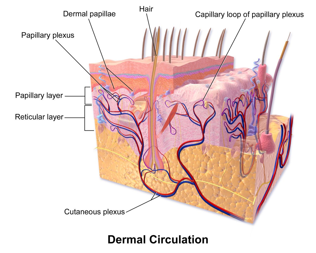
The first breath: fluid out, air in
Before birth the lung is not collapsed — it is full. Roughly 20 to 30 mL/kg of lung liquid, secreted by the pulmonary epithelium itself, keeps the airways distended and lets the lung grow. That fluid has to disappear in minutes. The old teaching that vaginal squeeze empties the chest explains only a small fraction of it. The dominant mechanism is chemical: in the days and hours before birth, the surge of fetal cortisol, thyroid hormone and catecholamines flips the alveolar epithelium from chloride secretion to sodium absorption through epithelial sodium channels (ENaC). Water follows sodium out of the airspace into the interstitium and then into the pulmonary capillaries and lymphatics. This is why a baby born by scheduled caesarean before labour begins — without the catecholamine surge that labour supplies — is at higher risk for transient tachypnoea of the newborn. Labour is not an inconvenience the fetus survives; it is preparation the fetus needs.
The first inspiratory effort is triggered by a convergence of stimuli. Cord clamping removes placental gas exchange, so carbon dioxide rises and oxygen falls; the resulting mild hypercapnia and acidosis stimulate the medullary respiratory centre. Cold air on wet skin, gravity, light, sound and tactile stimulation add to the drive. That first breath demands enormous negative pressure — opening pressures of 40 to 80 cm H₂O are typical, far above the 5 to 10 cm H₂O a settled newborn uses — because the infant must overcome the surface tension of fluid-lined alveoli and the resistance of remaining lung liquid. Surfactant, produced by type II pneumocytes from about 24 weeks and reaching mature quantities near 34 to 36 weeks, lowers that surface tension and, crucially, keeps small alveoli from emptying into large ones at end-expiration. Without adequate surfactant the lung re-collapses with every breath, and the infant works progressively harder for progressively less gas exchange — the mechanism of respiratory distress syndrome.
Normal newborn respirations are 30 to 60 breaths per minute, irregular, diaphragmatic and predominantly nasal. Newborns are obligate nose breathers for the first several months, which is why nasal obstruction is never trivial. Brief pauses of up to 15 seconds without colour or heart-rate change are periodic breathing and are normal. A pause of 20 seconds or more, or any pause accompanied by cyanosis, pallor or bradycardia, is apnoea and is not.
The circulatory switch: three shunts close
Fetal circulation is designed to bypass two organs the fetus does not need: the lungs and, largely, the liver. Oxygenated blood arrives from the placenta in the umbilical vein, and roughly half of it is shunted past the hepatic sinusoids through the ductus venosus into the inferior vena cava. In the right atrium, the foramen ovale lets the better-oxygenated stream cross directly to the left atrium and on to the coronary and cerebral circulations. Whatever blood does enter the right ventricle meets a pulmonary vascular bed held in intense hypoxic vasoconstriction, so about 90% of it is diverted through the ductus arteriosus into the descending aorta.
Birth reverses the pressure gradients that hold that pattern in place. Lung expansion, rising alveolar oxygen tension and rising pH cause pulmonary vascular resistance to fall steeply, so pulmonary blood flow increases eight- to tenfold within minutes. Simultaneously, clamping the cord removes the placenta — a very large low-resistance circuit — so systemic vascular resistance rises. Left atrial pressure now exceeds right atrial pressure and the flap of the foramen ovale is pressed shut; functional closure occurs within minutes to hours, though anatomic fusion takes months and a probe-patent foramen persists in roughly a quarter of adults. The ductus arteriosus constricts in response to two changes: rising arterial oxygen tension, which is a direct constrictor of ductal smooth muscle, and the abrupt fall in prostaglandin E₂, which the placenta had been supplying. Functional closure usually occurs by 12 to 24 hours and is essentially complete by 72 hours; anatomic obliteration to the ligamentum arteriosum takes 2 to 3 weeks. The ductus venosus closes functionally within minutes of cord clamping and becomes the ligamentum venosum; the umbilical vein becomes the ligamentum teres and the umbilical arteries become the medial umbilical ligaments.
Two clinical consequences follow directly. First, hypoxia and acidosis reopen the ductus arteriosus and re-constrict the pulmonary bed — the newborn can fall back into a fetal circulatory pattern. This is the physiology behind persistent pulmonary hypertension of the newborn, and it is why cold stress, hypoglycaemia and delayed resuscitation are dangerous well beyond their immediate effects. Second, in duct-dependent congenital heart lesions the closing ductus is what kills the baby, typically at 24 hours to 2 weeks of age when a previously well newborn suddenly becomes grey, mottled and shocked. Those infants need the ductus held open with prostaglandin E₁ (alprostadil) at 0.05 to 0.1 mcg/kg/min; apnoea is the expected adverse effect, so intubation equipment stays at the bedside.
What the birth itself hands you: obstetric emergencies and the newborn
The newborn nurse inherits whatever happened in the last ten minutes of labour. In each of the emergencies below the correct first action is often not the intuitive one, and the sequence itself is what saves the life. Learn these as sequences, not as lists.
| Emergency | Recognition | What you do FIRST | What follows — and what the newborn needs |
|---|---|---|---|
| Postpartum haemorrhage (cumulative loss ≥1000 mL, or any loss with signs of hypovolaemia, within 24 h) | Boggy uterus displaced above and to the right of the umbilicus; saturating a pad in under 15 minutes; rising pulse before falling pressure | Massage the fundus. Uterine atony causes 70 to 80% of postpartum haemorrhage, and a firm fundus stops the bleeding faster than any drug you can draw up | Empty the bladder, call for help, large-bore IV, oxytocin 10 to 40 units in 500 to 1000 mL infusion; methylergonovine 0.2 mg IM (avoid if hypertensive); carboprost 250 mcg IM (avoid in asthma); misoprostol 600 to 1000 mcg; tranexamic acid 1 g IV within 3 hours. The newborn may need a second nurse assigned so care is not interrupted |
| Magnesium sulfate toxicity in the birthing parent | Therapeutic range for seizure prophylaxis is 4 to 8 mg/dL. Above it, deep tendon reflexes disappear before respiratory depression: loss of patellar reflex at roughly 9 to 12 mg/dL, respiratory depression 12 to 18, cardiac arrest above 25 to 30. A lost reflex is your warning that the next thing to go is the breathing | Stop the magnesium infusion. Then give the antidote — calcium gluconate 1 g (10 mL of a 10% solution) IV over about 3 minutes | Monitor respiratory rate (hold for fewer than 12/min), urine output (hold below 30 mL/h), reflexes and level of consciousness. Anticipate a floppy, sleepy, hypotensive newborn with respiratory depression — have full resuscitation ready at the birth |
| Umbilical cord prolapse | Sudden prolonged deceleration or bradycardia, often just after membranes rupture; a pulsating cord palpated or seen at the introitus | Relieve the pressure. With a gloved hand, lift the presenting part off the cord and keep it there; place in knee-chest or Trendelenburg with hips elevated. Never push the cord back in | Call for immediate help, oxygen 10 L/min by non-rebreather, stop oxytocin, prepare for emergency caesarean; cover an exposed cord with saline-moistened sterile gauze. Your hand stays in place through transport and prep. Expect a newborn who may be acidotic and need resuscitation |
| Shoulder dystocia | The turtle sign — the head delivers and retracts against the perineum; restitution fails; gentle traction achieves nothing | Call for help and note the time, then McRoberts (hyperflex the parent’s hips onto the abdomen) plus suprapubic pressure. Never fundal pressure — it drives the shoulder harder into the symphysis and risks uterine rupture | Then Gaskin all-fours, Rubin, Woods screw, delivery of the posterior arm. Assess the newborn for brachial plexus injury (Erb palsy, C5 to C6 — arm adducted, internally rotated, “waiter’s tip” posture, absent Moro on that side), clavicle fracture (crepitus, asymmetric Moro) and hypoxic injury |
| Uterine rupture | Most common sign is an abnormal fetal heart pattern — prolonged deceleration or bradycardia. Sudden tearing abdominal pain, contractions stop, presenting part rises and station is lost, fetal parts palpable abdominally, shock | Stop oxytocin and mobilise for immediate surgical delivery. There is no medical management — this is an operating-room diagnosis | Two large-bore IVs, oxygen, type and crossmatch, notify the neonatal team. Expect a severely asphyxiated newborn: full resuscitation team, cord gases, and screening for therapeutic hypothermia if criteria are met |
Resuscitation in the golden minute
At every birth, ask three questions: is this a term gestation, does the infant have good tone, and is the infant breathing or crying? Three yeses buy skin-to-skin care with the birthing parent, with drying, warming and ongoing observation. Any no sends the infant to the radiant warmer for the initial steps — warm, dry, position the airway, stimulate — and if the infant remains apnoeic or gasping, or the heart rate is below 100 beats per minute, positive-pressure ventilation must be established within the first 60 seconds of life. Ventilation is the single most effective intervention in neonatal resuscitation, and inflating the lungs is what raises the heart rate.
If the heart rate does not rise, the fault is almost always in the ventilation, not the baby. Run the MRSOPA corrective sequence: Mask adjustment, Reposition the airway, Suction mouth then nose, Open the mouth, Pressure increase, Airway alternative (endotracheal tube or laryngeal mask). If the heart rate remains below 60 after 30 seconds of demonstrably effective ventilation through an advanced airway with 100% oxygen, begin chest compressions at a ratio of 3 compressions to 1 breath — 90 compressions plus 30 breaths, that is 120 events per minute. If the heart rate stays below 60 after 60 seconds of coordinated compressions, give epinephrine 0.01 to 0.03 mg/kg IV or intraosseous (0.1 to 0.3 mL/kg of the 0.1 mg/mL concentration, formerly written 1:10,000), preferably by umbilical venous catheter. For hypovolaemia give normal saline or emergency-release O-negative blood at 10 mL/kg over 5 to 10 minutes. Start resuscitation of term infants in 21% oxygen (21 to 30% for infants under 35 weeks) and titrate to targeted preductal saturations: about 60 to 65% at 1 minute, 70 to 75% at 3 minutes, 80 to 85% at 5 minutes and 85 to 95% at 10 minutes. A pink newborn at one minute of life is the exception, not the goal.
Aeration of the lung is the engine of the entire transition — it drops pulmonary vascular resistance, closes the fetal shunts and raises the heart rate — which is why effective ventilation within the first 60 seconds outranks every other resuscitation manoeuvre.
Section 2APGAR, Gestational Age and the Newborn Examination
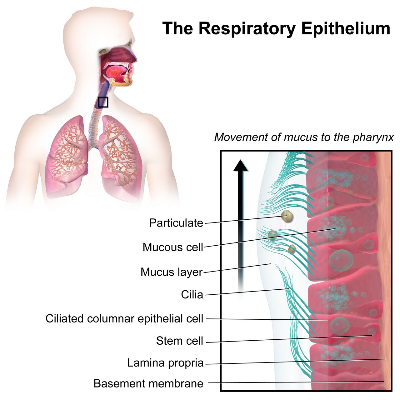
APGAR, component by component
Virginia Apgar built the score in 1952 to standardise what people meant when they said a baby “looked bad.” It has five components, each scored 0, 1 or 2, for a maximum of 10. Assign it at 1 minute and 5 minutes of life. If the 5-minute score is below 7, repeat it every 5 minutes up to 20 minutes of age.
| Component | 0 | 1 | 2 | What it is telling you |
|---|---|---|---|---|
| A — Appearance (colour) | Blue or pale all over | Acrocyanosis — pink body, blue hands and feet | Completely pink | The least specific component. Acrocyanosis is normal for 24 to 48 hours and reflects peripheral vasomotor instability. Central cyanosis of lips, tongue and trunk is never normal |
| P — Pulse (heart rate) | Absent | Below 100/min | 100/min or above | The most important single component and the one that drives resuscitation decisions. Auscultate at the apex for a full 60 seconds or use ECG leads |
| G — Grimace (reflex irritability) | No response to stimulation | Grimace or feeble cry | Cry, cough, sneeze, active withdrawal | Tests brainstem and cranial-nerve integrity. Elicited by suctioning the nares or flicking the sole |
| A — Activity (muscle tone) | Limp, flaccid | Some flexion of the extremities | Active motion, well flexed | Tone falls early with hypoxia and with maternal magnesium or anaesthesia. A term newborn at rest should be in flexion |
| R — Respiration (effort) | Absent | Slow, irregular, weak cry | Good, vigorous cry | Effort, not rate. Gasping scores the same as apnoea for management purposes — both mean start ventilating |
Interpretation is banded. A total of 7 to 10 is reassuring and calls for routine care — drying, warmth, skin-to-skin and observation. A total of 4 to 6 indicates moderate difficulty: stimulate, clear the airway if needed, provide oxygen or positive-pressure ventilation and reassess. A total of 0 to 3 indicates severe depression and calls for full resuscitation, including ventilation and, by heart-rate criteria, compressions and medication. A 5-minute score of 0 to 3 is associated with increased risk of neurologic injury at a population level, but the score is a poor predictor for any individual baby and must never be used to counsel a family about long-term outcome.
Measurements, gestational age and size classification
Normal term newborn measurements: weight 2500 to 4000 g (average about 3400 g), length 45 to 55 cm (average 50 cm), head circumference 32 to 38 cm (average 34 to 35 cm) and chest circumference 30 to 36 cm, typically 2 to 3 cm less than the head. Measure head circumference at the widest point, just above the eyebrows and over the occipital prominence, and remeasure after moulding resolves. A physiologic weight loss of up to 7 to 10% of birth weight over the first 3 to 5 days is expected, with birth weight regained by 10 to 14 days. A loss exceeding 7% is a signal to evaluate feeding — a latch assessment, a weighed feed, a look at output — not a signal to label the infant or the parent.
Gestational age is scored with the New Ballard Score, which combines six neuromuscular criteria (posture, square window at the wrist, arm recoil, popliteal angle, scarf sign, heel-to-ear) with six physical criteria (skin, lanugo, plantar surface, breast bud, eye and ear, genitals). It is accurate to roughly plus or minus 2 weeks, is most reliable when performed between 12 and 24 hours of age, and spans 20 to 44 weeks; each 5-point increment corresponds to about 2 weeks of gestation. Classify gestation as preterm below 37 0/7 weeks (with late preterm 34 0/7 to 36 6/7), early term 37 0/7 to 38 6/7, full term 39 0/7 to 40 6/7, late term 41 0/7 to 41 6/7 and post-term at 42 0/7 weeks or beyond. Then plot weight for gestational age: small for gestational age (SGA) below the 10th percentile, appropriate (AGA) 10th to 90th, large (LGA) above the 90th. Both SGA and LGA infants are hypoglycaemia risks, for opposite reasons — the SGA infant has minimal glycogen stores, the LGA infant of a parent with diabetes has hyperinsulinaemia that outlasts the maternal glucose supply.
Newborn vital signs
A newborn awake heart rate runs 100 to 180 beats per minute (typically 110 to 160), respirations 30 to 60 breaths per minute, and systolic blood pressure 60 to 90 mmHg; a systolic below 60 mmHg in a term newborn is hypotensive. Normal newborn axillary temperature is 36.5 to 37.5 °C (97.7 to 99.5 °F). Count a newborn heart rate and respiratory rate for a full 60 seconds; both are irregular and a 15-second count multiplied by four is unreliable.
Head to toe: what is normal, what is not
Begin with the head, because three swellings look similar and mean very different things. Caput succedaneum is oedema of the scalp above the periosteum: it is present at birth, feels soft and pits, crosses suture lines, and resolves in 3 to 5 days. Cephalhaematoma is bleeding beneath the periosteum: it appears hours after birth, feels firm and fluctuant, does not cross suture lines because the periosteum is bound at the sutures, resolves over 2 to 8 weeks, and adds a bilirubin load as the blood breaks down. Subgaleal haemorrhage is an emergency: bleeding into the loose potential space beneath the scalp aponeurosis, which is large enough to hold most of a newborn’s blood volume. It is associated with vacuum-assisted birth, presents as a boggy, shifting, fluctuant swelling that crosses sutures and spreads toward the neck and behind the ears, and it causes hypovolaemic shock. Serial head circumference measurements every 15 to 30 minutes, with heart rate and perfusion checks, are the nursing surveillance; escalate immediately for an increase of more than 1 cm.
The anterior fontanel is diamond-shaped, 4 to 5 cm across, and closes at 12 to 18 months; the posterior fontanel is triangular, 0.5 to 1 cm, and closes at 2 to 3 months. Both should be soft and flat, and may bulge transiently with crying. A persistently bulging, tense fontanel suggests raised intracranial pressure; a sunken one suggests dehydration. Eyes may show subconjunctival haemorrhages and transient strabismus; the red reflex must be present and symmetric in both eyes — absence suggests congenital cataract or retinoblastoma and is urgent. Count the umbilical cord vessels: two arteries and one vein. A two-vessel cord warrants evaluation for renal and other anomalies. Auscultate the chest with the infant quiet; murmurs in the first 24 hours are frequently the closing ductus, but a murmur with cyanosis, poor perfusion, tachypnoea or weak femoral pulses is not. Palpate femoral pulses in every newborn — diminished or delayed femoral pulses suggest coarctation of the aorta.
Skin findings that are normal and need explanation, not treatment: acrocyanosis, erythema toxicum neonatorum (blotchy erythema with tiny yellow-white papules, day 1 to 3), milia, vernix caseosa, lanugo, cutis marmorata (mottling with cool), nevus simplex (“stork bite”) and congenital dermal melanocytosis — slate-grey macules over the sacrum and buttocks, most common in infants with darker skin. Document melanocytosis carefully with location and size at the first assessment; it is a normal variant and misreading it as bruising has real consequences for families. Never normal: central cyanosis, pallor, jaundice in the first 24 hours, and petechiae outside the presenting part.
That documentation cuts both ways, and the safeguarding rule is the mirror image of it. A true bruise on an infant who is not yet mobile is a red flag: “those who don’t cruise rarely bruise.” The test you apply to any injury is whether it is plausible for the child’s developmental stage and consistent with the history given — a skull fracture attributed to a roll off a sofa by a two-week-old who cannot roll, an injury whose explanation changes between tellings, or a significant delay in seeking care all fail that test. Other patterns that warrant concern include bruising of the torso, ears or neck in a child under four, and any burn with sharply demarcated margins. Report it. In every jurisdiction the nurse is a mandatory reporter, the duty is triggered by reasonable suspicion and not by certainty, you do not need to prove anything or identify who caused it, and you do not need the permission of a manager or a physician to make the report. Investigating is someone else’s job; reporting is yours. Document objectively — measure and describe what you see, quote the caregiver’s explanation verbatim, and avoid conclusions or accusations in the record.
Primitive reflexes give you a rapid neurologic screen. Moro (startle, symmetric abduction then embrace) fades by 4 to 6 months; asymmetry suggests clavicle fracture or brachial plexus injury. Rooting and sucking fade by 3 to 4 months awake. Palmar grasp fades by 3 to 4 months, plantar grasp by 8 to 12 months. The tonic neck (fencing) reflex fades by 3 to 4 months, stepping by 4 to 8 weeks, and the truncal incurvation (Galant) reflex by about 3 months. An upgoing plantar response (Babinski) is normal until 12 to 24 months. Reflexes that are absent, asymmetric or persist beyond their window are referrals.
The APGAR describes the transition and the physical examination explains it — and the three scalp swellings are the classic worked example: caput crosses sutures and resolves in days, cephalhaematoma respects sutures and adds bilirubin, and subgaleal haemorrhage crosses sutures, expands, and bleeds a newborn into shock.
Section 3Thermoregulation, Cold Stress and Glucose
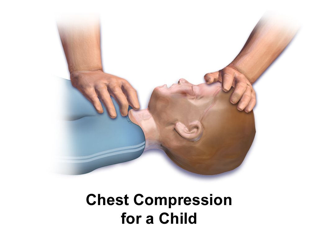
Why newborns lose heat and how they make it
A newborn is a small, wet body with a large surface area relative to mass, thin subcutaneous fat, limited ability to vasoconstrict, and no capacity to put on a sweater. Heat leaves by four routes, and each has a specific countermeasure. Evaporation — the dominant loss in the first minutes — is heat carried away as amniotic fluid vaporises; the countermeasure is immediate, thorough drying and removal of the wet linen. Conduction is loss to a cold surface in direct contact; pre-warm the scale, the mattress, the stethoscope and your hands. Convection is loss to moving air; close the door, move the cot away from vents, and use the sides of the radiant warmer. Radiation is loss to nearby cold surfaces without contact; move the cot away from exterior windows and walls.
Newborns cannot shiver effectively. They make heat by non-shivering thermogenesis in brown adipose tissue, which is deposited from about 26 to 30 weeks of gestation and sits in the interscapular region, around the neck, in the axillae, in the mediastinum and around the kidneys and adrenals. Cold sensed at the skin triggers a noradrenergic signal; norepinephrine drives lipolysis in brown fat, and the mitochondrial uncoupling protein 1 (thermogenin) dissipates the proton gradient as heat instead of capturing it as ATP. It is metabolically expensive and it is a finite resource — which is exactly why preterm and growth-restricted infants, who laid down little brown fat, decompensate first.
The cold-stress cascade
Cold stress is not simply being chilly. Follow the chain. Cold triggers norepinephrine release, which does two things at once: it burns brown fat, and it constricts pulmonary vessels. Burning brown fat raises oxygen consumption and glucose consumption, which produces hypoglycaemia. Pulmonary vasoconstriction plus rising oxygen demand produces hypoxia, which forces anaerobic metabolism, which generates lactic acid and metabolic acidosis. Acidosis and hypoxia together do two more things: they impair surfactant production and function, worsening respiratory distress, and they reopen the ductus arteriosus and re-constrict the pulmonary bed, pushing the infant back toward fetal circulation. And the free fatty acids released by brown-fat lipolysis compete with bilirubin for albumin binding sites, raising the free bilirubin available to cross into the brain — acidosis compounds this by further weakening bilirubin-albumin binding. A single unaddressed temperature of 36.0 °C can therefore end as respiratory distress, hypoglycaemia, acidosis and worsening jaundice.
Classify hypothermia by axillary or rectal temperature: cold stress or mild hypothermia 36.0 to 36.4 °C, moderate 32.0 to 35.9 °C, severe below 32.0 °C. The nursing goal is a neutral thermal environment — the narrow ambient range in which the infant maintains a normal core temperature with minimal oxygen consumption and minimal metabolic work. Rewarm a cold newborn gradually, aiming to raise the temperature by no more than about 0.5 °C per hour; rapid rewarming causes apnoea and hypotension. When using a radiant warmer, place the servo-control skin probe on the abdomen over the liver, shiny side up, and never over brown fat, a bony prominence or the area under the infant — a probe reading a warm surface will command the warmer off and let the infant cool. Delay the first bath until the temperature has been stable at 36.5 °C or above for at least 2 to 4 hours (many units now wait 12 to 24 hours), keep the room warm during the bath, and dry and dress immediately afterward.
Neonatal hypoglycaemia: who, when and how much
The fetus receives a continuous glucose infusion across the placenta. Cord clamping ends it, glucose falls to a physiologic nadir at about 1 to 2 hours of age, and the newborn then mobilises glycogen, launches gluconeogenesis and begins to use ketones and lactate as alternative brain fuels. Infants who cannot complete that switch become hypoglycaemic. The at-risk groups you screen are: infants of parents with diabetes (any type), LGA infants, SGA or growth-restricted infants, preterm and late preterm infants (34 0/7 to 36 6/7 weeks), infants with perinatal stress or asphyxia, cold-stressed infants, those with polycythaemia or suspected sepsis, infants exposed to maternal beta-blockers or terbutaline, and infants with syndromes such as Beckwith-Wiedemann.
Signs are non-specific and often absent: jitteriness or tremors, a high-pitched or weak cry, poor feeding, lethargy, hypotonia, temperature instability, tachypnoea, apnoea, cyanosis, diaphoresis and, late, seizures. Jitteriness stops when you hold the limb; a seizure does not. Because many hypoglycaemic newborns are asymptomatic, the rule is to screen the at-risk groups on schedule rather than wait for signs. A common protocol screens at 30 minutes to 1 hour after the first feed, then before feeds every 2 to 3 hours for the first 12 to 24 hours (longer for infants of parents with diabetes). Always confirm a low bedside glucometer value with a laboratory plasma glucose — but treat first and send the confirmatory sample second; do not wait on the laboratory to feed a symptomatic baby.
| Hour of life | Action threshold (plasma glucose) | Target | Intervention |
|---|---|---|---|
| First 4 hours | Below 25 mg/dL after a feed, or any symptomatic infant | Maintain 45 mg/dL or above | Intravenous dextrose. Between 25 and 40 mg/dL, feed and recheck in 1 hour |
| 4 to 24 hours | Below 35 mg/dL after a feed, or any symptomatic infant | Maintain 45 mg/dL or above | Intravenous dextrose. Between 35 and 45 mg/dL, refeed and recheck in 1 hour |
| Beyond 48 hours | Persistently below 50 to 60 mg/dL | 60 mg/dL or above | Investigate for hyperinsulinism or a metabolic or endocrine cause |
| Any age, symptomatic | Below 40 mg/dL | Treat immediately | Intravenous dextrose — do not rely on oral feeding alone in a symptomatic infant |
Thresholds vary between guidelines and units; follow the protocol where you work, but know that the operational floor most units use in the first 24 to 48 hours is a plasma glucose of 45 mg/dL (2.5 mmol/L). Management is tiered. An asymptomatic infant with a mildly low value is fed — breast or chestfeed, or supplement with expressed milk or formula — and rechecked in 30 to 60 minutes. 40% oral dextrose gel, massaged into the buccal mucosa immediately before a feed, is an evidence-based first-line adjunct that reduces the need for intravenous therapy and separation from the parent; the dose is 0.5 mL/kg (equal to 200 mg/kg of dextrose). A symptomatic infant, or one below 25 mg/dL, needs intravenous dextrose 10% in water (D10W) 2 mL/kg (also 200 mg/kg) given as a slow push over about 5 minutes, followed by a continuous infusion at a glucose infusion rate of 5 to 8 mg/kg/min so the glucose does not rebound downward.
Dextrose gel 40%: 0.5 mL/kg × 3.2 kg = 1.6 mL buccally, then feed.
D10W bolus: 2 mL/kg × 3.2 kg = 6.4 mL IV over 5 minutes.
Maintenance infusion, checking the glucose infusion rate: GIR in mg/kg/min = (dextrose percent × rate in mL/h) ÷ (6 × weight in kg). D10W at 12 mL/h gives (10 × 12) ÷ (6 × 3.2) = 120 ÷ 19.2 = 6.25 mg/kg/min — inside the 5 to 8 mg/kg/min target. Recheck the glucose 30 minutes after any intervention. Never give undiluted 25% or 50% dextrose to a newborn: the osmotic load causes rebound hypoglycaemia and, through a peripheral vein, tissue necrosis.
Cold stress and hypoglycaemia are the same emergency arriving from two directions — the cold newborn burns brown fat and glucose to make heat, and the hypoglycaemic newborn cannot make heat — so drying, warming and early feeding are simultaneously the thermal and the metabolic intervention.
Section 4Bilirubin: Physiologic versus Pathologic Jaundice
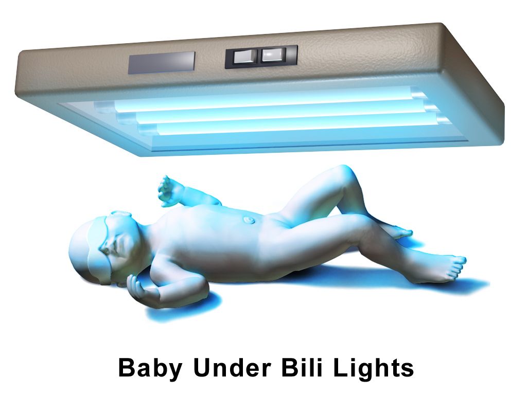
Where the bilirubin comes from
Every newborn has a bilirubin problem for four structural reasons. Newborns are polycythaemic, with haematocrits of 45 to 65%, so there is more haemoglobin to break down. Fetal red cells live only 70 to 90 days rather than 120, so they break down faster. The hepatic enzyme that conjugates bilirubin, uridine diphosphate glucuronosyltransferase (UGT1A1), is immature for the first days of life, so conjugation lags. And the newborn gut is initially sterile and slow, while it contains beta-glucuronidase that deconjugates bilirubin and returns it to the circulation — the enterohepatic circulation. Meconium is loaded with bilirubin; every stool passed is bilirubin leaving the body. This is why feeding frequency, not water, is the intervention for early jaundice.
Unconjugated (indirect) bilirubin is lipid-soluble and travels bound to albumin. Free unconjugated bilirubin — the fraction not bound — can cross the blood-brain barrier and deposit in the basal ganglia and brainstem nuclei. Acidosis, hypoxia, hypothermia, hypoalbuminaemia, sepsis and certain drugs that compete for albumin binding all increase the free fraction, which is why a sick newborn is treated at a lower bilirubin threshold than a well one. Jaundice becomes visible when the total serum bilirubin reaches roughly 5 to 7 mg/dL, and it progresses cephalocaudally — face first, then trunk, then extremities, palms and soles last. Visual estimation is unreliable, especially in infants with darker skin: blanch the skin over the sternum or check the sclerae and gums, and then get a number. Do not manage jaundice by eye.
The 24-hour rule and the other pathologic markers
Jaundice appearing in the first 24 hours of life is pathologic until proven otherwise. There is no exception to this rule and it is the single most important sentence in newborn jaundice care. Early jaundice means haemolysis is running: Rh isoimmunisation, ABO incompatibility (a parent with type O blood and an infant with type A or B, which does not require prior sensitisation and so can affect a first pregnancy), G6PD deficiency, hereditary spherocytosis, or sepsis. Get a total and direct bilirubin, blood type and Rh, direct antiglobulin (Coombs) test, haematocrit and reticulocyte count, and escalate.
| Feature | Physiologic jaundice | Pathologic jaundice |
|---|---|---|
| Onset | After 24 hours of age | Within the first 24 hours — always investigate |
| Peak | Day 3 to 5 in term infants; day 5 to 7 in preterm | Any time; often early and rising fast |
| Rate of rise | Less than 5 mg/dL per day (less than 0.2 mg/dL per hour) | More than 5 mg/dL per day, or more than 0.2 mg/dL per hour |
| Peak level | Usually below 12 to 13 mg/dL (may reach about 15 in exclusively breastfed infants) | Above the hour-specific treatment threshold on the nomogram for that gestational age and risk category |
| Type of bilirubin | Unconjugated (indirect) | A direct (conjugated) bilirubin above 1 mg/dL, or above 20% of the total, is never physiologic — think biliary atresia, neonatal cholestasis or sepsis |
| Duration | Resolves by 1 week (term), 2 weeks (preterm) | Persisting beyond 2 weeks in a term infant requires evaluation, including a direct bilirubin and thyroid screen |
| Other findings | Well infant, feeding, normal output, normal haematocrit | Pallor, hepatosplenomegaly, poor feeding, lethargy, dark urine, pale or clay-coloured stools, positive Coombs test |
Two feeding-related patterns are worth separating because they are managed oppositely. Suboptimal-intake jaundice (historically “breastfeeding jaundice”) appears on days 2 to 5 and is a volume problem: intake is not yet established, stooling is infrequent, and bilirubin is reabsorbed through the enterohepatic circulation. The management is more effective feeding — 8 to 12 feeds in 24 hours, latch assessment, lactation support, expressed milk or supplementation if indicated. Water and dextrose water are contraindicated: they displace milk, do not increase stooling, and risk hyponatraemia. Breast-milk jaundice is different: it begins around day 5 to 7, peaks in the second week, can persist for up to 12 weeks, and occurs in a thriving, well-gaining infant. Factors in the milk increase enterohepatic reabsorption and slow conjugation. Feeding continues; interruption is rarely needed and is not the routine answer.
Escalation, phototherapy and the injury you are preventing
All newborns should have a transcutaneous or serum bilirubin measured before discharge, plotted against the hour-specific nomogram together with gestational age and neurotoxicity risk factors, and paired with a follow-up appointment timed to the risk zone. A transcutaneous reading is a screen, not a diagnosis: confirm with serum when the value approaches the treatment threshold, and note that transcutaneous readings are unreliable during and shortly after phototherapy.
Phototherapy works by photoisomerisation. Blue light in the 460 to 490 nm range converts unconjugated bilirubin in the skin into water-soluble isomers, chiefly lumirubin, which are excreted in bile and urine without needing conjugation. Nursing care is specific: expose the maximum skin surface with only a nappy; apply opaque eye shields and check the eyes at least every shift for discharge or corneal irritation; remove the shields during feeding and parent holding so the infant sees faces; keep the prescribed distance between light and infant; monitor temperature frequently, since both hyperthermia and hypothermia occur; and increase feeding frequency because insensible water losses rise. Expect loose, green stools — that is bilirubin leaving and is a sign of effectiveness, but watch skin integrity and hydration. Do not apply lotions, oils or powders, which can cause burns. Bronzing of the skin (bronze baby syndrome) occurs when phototherapy is used with cholestasis and is a reason to reassess. For isoimmune haemolytic disease with bilirubin approaching exchange level, intravenous immunoglobulin at 0.5 to 1 g/kg may be given; exchange transfusion is reserved for extreme hyperbilirubinaemia or any sign of acute bilirubin encephalopathy.
The injury being prevented is acute bilirubin encephalopathy, which progresses through three phases: early — lethargy, hypotonia, poor suck; intermediate — irritability, a shrill high-pitched cry, fever, and retrocollis and opisthotonos (arching of the neck and trunk); advanced — apnoea, seizures, coma, death. Survivors develop kernicterus: athetoid cerebral palsy, sensorineural hearing loss or auditory neuropathy, upward-gaze palsy and dental enamel dysplasia. It is permanent and it is preventable, which is why the timed bilirubin and the follow-up appointment carry the weight they do.
Jaundice in the first 24 hours of life, a rise faster than 0.2 mg/dL per hour, or a direct bilirubin above 1 mg/dL are each pathologic by definition and demand investigation — everything else is managed by hour-specific bilirubin levels, effective feeding and timed follow-up.
Section 5Daily Care, Weight-Based Dosing and Discharge
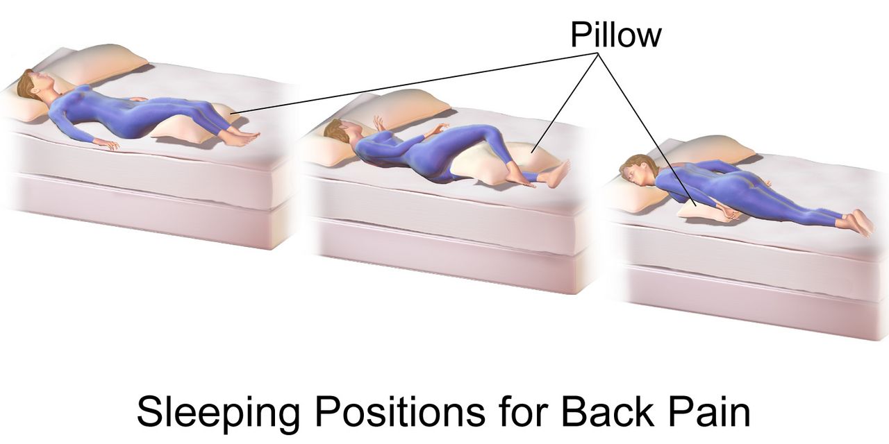
Routine prophylaxis and screening — every dose per kilogram
Three medications and three screens are standard for essentially every newborn. Vitamin K (phytonadione) 1 mg intramuscularly for infants weighing 1500 g or more, or 0.5 mg for those under 1500 g, is given within 6 hours of birth into the vastus lateralis with a 25-gauge, 5/8-inch needle at a 90-degree angle. Newborns are born with a sterile gut that cannot yet synthesise vitamin K, and placental transfer is poor; without prophylaxis, vitamin K deficiency bleeding occurs, and the late form presents at 2 to 12 weeks with intracranial haemorrhage. If a family declines, document the discussion and educate about bleeding signs — do not treat the decision as an argument to win. Erythromycin 0.5% ophthalmic ointment, a 1 cm ribbon in each lower conjunctival sac within the first hour (up to 2 hours), prevents gonococcal ophthalmia neonatorum; it does not reliably prevent chlamydial conjunctivitis, which presents at 5 to 14 days and needs systemic treatment. Hepatitis B vaccine 0.5 mL IM is given within 24 hours; if the birthing parent is HBsAg-positive, the infant receives both the vaccine and hepatitis B immune globulin 0.5 mL within 12 hours of birth, at separate sites.
Screens: the newborn metabolic screen is a heel-stick collected after at least 24 hours of feeding (with a repeat per state protocol), warming the heel first and sampling the lateral aspect to avoid the calcaneus and nerves. Hearing screening by automated auditory brainstem response or otoacoustic emissions is done before discharge. Critical congenital heart disease (CCHD) screening is a pulse-oximetry screen performed at 24 hours of age or later, with probes on the right hand (preductal) and either foot (postductal). A pass requires saturation of 95% or above in either limb and a difference of 3% or less between them. Any reading below 90% is an immediate fail requiring urgent evaluation; readings of 90 to 94%, or a difference greater than 3%, are repeated hourly for up to three screens before failing.
Weight-based dosing and maintenance fluids
Children are not small adults, and paediatric dosing is not adult dosing scaled by eye. Every paediatric dose is expressed per kilogram of actual measured weight in kilograms — never a stated weight, never pounds. Weigh the child on admission, record in kilograms only, and remove the pounds display from the scale if your unit permits. Then run three checks on every calculation: is the dose per kilogram within the reference range, is the total dose below the maximum adult dose, and does the volume make sense for the route and the child’s size. High-alert paediatric medications require an independent double-check by a second nurse.
A 22 kg child: 15 mg/kg × 22 kg = 330 mg per dose. Daily maximum 75 × 22 = 1650 mg — four 330 mg doses total 1320 mg, which is within range. Safe.
A 70 kg adolescent: 15 mg/kg × 70 kg = 1050 mg — mathematically correct, but it exceeds the maximum single adult dose of 1000 mg. Give 1000 mg. Once a child’s weight-based calculation exceeds the adult dose, the adult dose becomes the ceiling. This is the check that catches the adolescent overdose.
Maintenance intravenous fluids for infants beyond the newborn period and for children use the 4-2-1 rule: 4 mL/kg/h for the first 10 kg of body weight, plus 2 mL/kg/h for the second 10 kg, plus 1 mL/kg/h for every kilogram above 20. Worked example, a 26 kg child: (10 kg × 4) + (10 kg × 2) + (6 kg × 1) = 40 + 20 + 6 = 66 mL/h. The daily equivalent (the 100-50-20 rule) cross-checks it: (10 × 100) + (10 × 50) + (6 × 20) = 1000 + 500 + 120 = 1620 mL/day, which is 67.5 mL/h — the same answer. Use an isotonic maintenance fluid with dextrose in most children; hypotonic maintenance fluid is a recognised cause of hospital-acquired hyponatraemia. Note the exception that matters in this chapter: the 4-2-1 rule does not apply to the first days of life. Term newborns start at roughly 60 to 80 mL/kg/day on day 1 and advance by 10 to 20 mL/kg/day toward about 150 mL/kg/day by the end of the first week, because renal concentrating ability and insensible losses in the newborn follow a different curve entirely.
Feeding, elimination and the first weeks
Feed on cue, not on a clock. A breast- or chestfed newborn feeds 8 to 12 times in 24 hours; early hunger cues are rooting, hand-to-mouth movements, lip smacking and stirring, and crying is a late cue that makes latching harder. Teach latch by what you can see and hear: a wide gape, more areola visible above the upper lip than below the lower, flanged lips, the chin touching the breast, and rhythmic swallowing that is audible. Assess comfort, transfer and output rather than counting minutes. Formula-fed newborns take about 45 to 90 mL (1.5 to 3 ounces) every 3 to 4 hours in the first week, roughly 150 mL/kg/day; never prop a bottle, never heat formula in a microwave, and discard formula left after a feed. Exclusively or partially breastfed infants need vitamin D 400 IU daily starting in the first days of life.
Output is the most honest measure of intake in the first week. The first void should occur within 24 hours and the first meconium within 24 to 48 hours; failure of either is an escalation. Expect one wet nappy on day 1, increasing to six or more per day by day 4 to 5, with pale straw-coloured urine. Stools progress from meconium (sticky, black-green, first 24 to 48 hours) to transitional (loose, greenish-brown, days 3 to 4) to the yellow, seedy stools of a milk-fed infant by days 4 to 5, at least three to four times a day in a breastfed newborn. Brick-red urate crystals in the nappy in the first days are usually benign but, after day 3, prompt a feeding evaluation.
Cord care is dry cord care: keep it clean and dry, fold the nappy below it, and expect separation at 7 to 14 days. Teach parents to call for purulent drainage, foul odour, periumbilical redness spreading onto the abdomen, or active bleeding. After circumcision, apply petroleum jelly to the glans with each nappy change (except with a Plastibell, where it is not used), expect a yellowish exudate at 24 to 48 hours as normal granulation — not infection — and report bleeding larger than a coin-sized spot or failure to void within 6 to 8 hours. Take temperatures axillary, and teach the family the same.
Safe sleep, safe travel, and when to call
Safe sleep is taught at every contact and modelled in the hospital, because parents copy what they saw the nurse do. Every sleep, the infant goes down supine, on a firm, flat, non-inclined surface with a fitted sheet and nothing else — no bumpers, pillows, blankets, positioners, wedges or soft toys. Room-share without bed-sharing for at least the first 6 months. Avoid overheating and overbundling; use a wearable blanket rather than loose bedding. Offer a pacifier at sleep once feeding is established. Keep the environment smoke-free. Supervised awake tummy time from the first days prevents positional plagiocephaly and builds neck strength. Car-seat teaching is equally concrete: rear-facing in the back seat, harness straps at or below the shoulders in a rear-facing seat, chest clip at armpit level, snug enough that you cannot pinch a horizontal fold of webbing at the shoulder, and no bulky coats or aftermarket inserts between the child and the harness. Keep the child rear-facing for as long as possible — until they reach the highest weight or height allowed by the seat’s manufacturer — rather than turning them forward-facing at a set age.
| Newborn red flag | Why it matters | SBAR escalation — what you say |
|---|---|---|
| Rectal temperature 38.0 °C (100.4 °F) or above, or below 36.0 °C, in an infant under 3 months | Either direction signals sepsis in a newborn; hypothermia is at least as ominous as fever | S: “Baby Ortiz, 26 hours old, rectal temp 38.2.” B: “Term, unremarkable birth, group B strep status unknown.” A: “This meets criteria for sepsis evaluation.” R: “I need you at the bedside now for blood culture, CBC, lumbar puncture and empiric antibiotics.” |
| Central cyanosis, or a failed CCHD screen | Duct-dependent congenital heart disease presents as the ductus closes at 24 hours to 2 weeks | R: “Preductal 88%, postductal 82%. I need urgent evaluation and an echocardiogram, and I want prostaglandin availability confirmed.” |
| Jaundice in the first 24 hours | Haemolysis until proven otherwise | R: “Requesting total and direct bilirubin, type and Rh, direct Coombs, haematocrit and reticulocyte count now.” |
| Respiratory rate above 60 sustained, grunting, nasal flaring, retractions | Respiratory distress; grunting is auto-generated positive end-expiratory pressure and is a late, serious sign | R: “Sustained rate 78 with grunting and subcostal retractions. I need respiratory evaluation and a saturation target order now.” |
| Glucose below 40 mg/dL with symptoms, or below 25 mg/dL at any time | Neuroglycopenia; brain injury is silent until it is not | R: “Glucose 22 at 3 hours, infant jittery. I am feeding and requesting an order for D10W 2 mL/kg IV now.” |
| No void by 24 hours or no stool by 48 hours; weight loss above 7% | Intake problem, obstruction, or renal anomaly | R: “No void at 26 hours. Requesting evaluation and a feeding plan; I have lactation consulting now.” |
| Boggy, expanding scalp swelling that crosses sutures, especially after vacuum-assisted birth | Subgaleal haemorrhage — can exsanguinate a newborn | R: “Head circumference up 1.5 cm in 30 minutes, heart rate 190, pale. I need you at the bedside immediately and blood available.” |
Routine newborn care is a set of exact numbers — vitamin K 1 mg IM within 6 hours, hepatitis B immune globulin within 12 hours for an HBsAg-positive parent, CCHD screening at 24 hours with a 95% and 3% rule, and every paediatric dose calculated per kilogram and then capped at the adult maximum.
The chapter in one breath
Birth is a coordinated physiological handover. The first breath aerates a fluid-filled lung that has already begun absorbing its own liquid through sodium channels primed by labour, and that aeration drops pulmonary vascular resistance so the foramen ovale presses shut within hours and the ductus arteriosus constricts by 12 to 24 hours as oxygen rises and placental prostaglandin disappears. Everything that follows is surveillance of a transition that can reverse: hypoxia, acidosis and cold reopen the shunts. The APGAR at 1 and 5 minutes describes the response but never delays resuscitation, where effective ventilation in the first 60 seconds outranks every other action, compressions run 3:1 for a heart rate below 60, and epinephrine is 0.01 to 0.03 mg/kg. The newborn then faces three metabolic tests. Cold stress burns brown fat through non-shivering thermogenesis and cascades into hypoglycaemia, acidosis and reopened shunts. Hypoglycaemia is screened by hour of life, treated with feeding, 40% dextrose gel at 0.5 mL/kg or D10W at 2 mL/kg. Jaundice is physiologic after 24 hours and pathologic before it, or when the rise exceeds 0.2 mg/dL per hour or the direct fraction exceeds 1 mg/dL. In every obstetric emergency the sequence is the treatment: massage the fundus first, stop the magnesium and give calcium gluconate, lift the presenting part off a prolapsed cord, McRoberts and suprapubic pressure but never fundal pressure.
Words worth knowing
| Term | What it means |
|---|---|
| Acrocyanosis | Blue hands and feet with a pink trunk; a normal vasomotor finding for the first 24 to 48 hours, unlike central cyanosis, which is never normal. |
| APGAR score | A 0 to 10 score of Appearance, Pulse, Grimace, Activity and Respiration assigned at 1 and 5 minutes; describes the transition and never delays resuscitation. |
| Ballard score | Assessment of gestational age using six neuromuscular and six physical maturity criteria; accurate to about plus or minus 2 weeks, best performed at 12 to 24 hours. |
| Brown adipose tissue | Specialised fat laid down from 26 to 30 weeks that generates heat through uncoupling protein 1 rather than storing energy; the substrate of non-shivering thermogenesis. |
| Caput succedaneum | Soft, pitting scalp oedema present at birth that crosses suture lines and resolves in 3 to 5 days. |
| Cephalhaematoma | Subperiosteal bleeding appearing hours after birth that does not cross suture lines, resolves over weeks, and increases the bilirubin load. |
| Cold stress | The metabolic cascade triggered by heat loss: brown-fat burning, rising oxygen and glucose consumption, hypoglycaemia, acidosis and re-opening of fetal shunts. |
| Ductus arteriosus | Fetal vessel from the pulmonary artery to the aorta; closes functionally in 12 to 24 hours as oxygen rises and prostaglandin E₂ falls, becoming the ligamentum arteriosum. |
| Enterohepatic circulation | Reabsorption of deconjugated bilirubin from the newborn gut back into the bloodstream; reduced by frequent feeding and stooling, not by water. |
| Erythema toxicum neonatorum | Blotchy erythema with small yellow-white papules appearing on days 1 to 3; benign, self-limiting and requiring only explanation. |
| Foramen ovale | Fetal opening between the atria; closes functionally within minutes to hours when left atrial pressure exceeds right atrial pressure after birth. |
| Kernicterus | Permanent bilirubin-induced neurological injury — athetoid cerebral palsy, sensorineural hearing loss, upward-gaze palsy and enamel dysplasia. |
| McRoberts manoeuvre | Hyperflexion of the birthing parent’s hips onto the abdomen to flatten the sacral promontory in shoulder dystocia; paired with suprapubic — never fundal — pressure. |
| Neutral thermal environment | The ambient temperature range in which a newborn maintains a normal core temperature with minimal oxygen consumption and metabolic work. |
| Non-shivering thermogenesis | Heat production by noradrenaline-driven lipolysis in brown fat; the newborn’s main heat source because effective shivering is not available. |
| Periodic breathing | Pauses of up to 15 seconds without colour or heart-rate change; normal, and distinct from apnoea, which lasts 20 seconds or more or is accompanied by bradycardia or cyanosis. |
| Physiologic jaundice | Unconjugated hyperbilirubinaemia appearing after 24 hours, peaking on days 3 to 5, rising less than 5 mg/dL per day and resolving within a week at term. |
| Subgaleal haemorrhage | Bleeding into the space beneath the scalp aponeurosis that crosses sutures and expands; an emergency because it can hold most of a newborn’s blood volume. |
| Surfactant | Phospholipid produced by type II pneumocytes from about 24 weeks, mature near 34 to 36 weeks, that lowers alveolar surface tension and prevents end-expiratory collapse. |
| Vitamin K deficiency bleeding | Haemorrhage from absent vitamin K synthesis in the sterile newborn gut; prevented by phytonadione 1 mg intramuscularly within 6 hours of birth. |
Check yourself
The High-Risk Newborn
Most newborns make the transition from placenta to air on their own. A minority cannot, and the nursing work of the first minutes, hours and days decides how much of that minority survives intact. A high-risk newborn is one whose gestational age, growth, birth events, physiology or environment puts them at greater-than-usual risk of illness, injury or death — the 34-week late preterm baby who looks deceptively term, the macrosomic infant of a parent with diabetes whose glucose falls within ninety minutes, the meconium-stained post-term newborn whose pulmonary vasculature will not relax, the baby whose bilirubin is climbing faster than the nomogram allows. This chapter moves the way the bedside moves: mechanism first, then what you assess, then what you do, then what goes wrong if you are slow. Every number here is per kilogram, per hour or per week of gestation, because in this population a decimal point is a diagnosis.
By the end of this chapter you can
- Classify a newborn by gestational age and by birth weight, and distinguish small for gestational age, appropriate for gestational age and large for gestational age from intrauterine growth restriction.
- Sequence the Neonatal Resuscitation Program algorithm through the Golden Minute, including the corrective ventilation steps (MR SOPA), compression ratio and the weight-based dose of epinephrine.
- Differentiate respiratory distress syndrome, transient tachypnoea of the newborn, meconium aspiration syndrome and persistent pulmonary hypertension of the newborn by history, examination and radiograph.
- Calculate weight-based volumes and doses, including the 4–2–1 maintenance-fluid rule, the glucose infusion rate and a neonatal antibiotic dose, and check every result against the maximum adult dose.
- Screen, interpret and treat neonatal hypoglycaemia and hyperbilirubinaemia using hour-specific thresholds, and describe the progression to acute bilirubin encephalopathy and kernicterus.
- Recognise early-onset and late-onset neonatal sepsis and necrotising enterocolitis from their earliest, least dramatic signs.
- State the single correct first action for congenital diaphragmatic hernia, oesophageal atresia with tracheo-oesophageal fistula, myelomeningocele, gastroschisis and a ductal-dependent cardiac lesion.
- Apply non-pharmacologic Eat, Sleep, Console care to the newborn experiencing withdrawal, and describe therapeutic hypothermia for hypoxic-ischaemic encephalopathy.
- Sequence the obstetric emergency responses — postpartum haemorrhage, magnesium sulfate toxicity, cord prolapse, shoulder dystocia and uterine rupture — that decide which newborn you receive.
- Escalate deterioration using SBAR, act on a safeguarding concern at the threshold of reasonable suspicion, and delegate within scope, retaining assessment, teaching, evaluation and the unstable patient for the registered nurse.
Section 1Who Counts as High Risk: Age, Growth and the Birth Itself
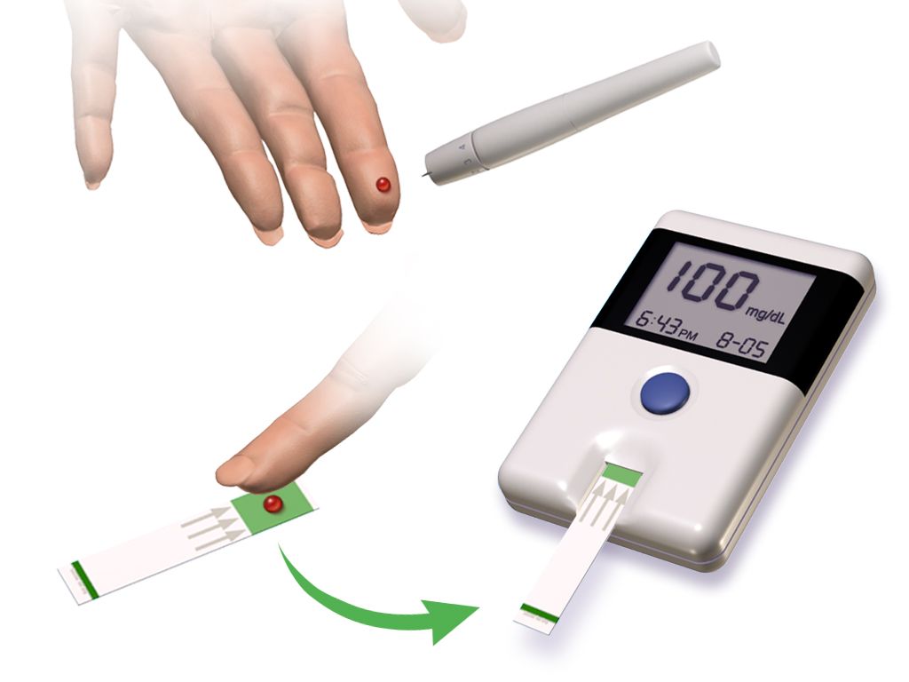
Gestational age and birth weight are two different questions
Ask them separately, because the answers do not travel together. Gestational age is counted in completed weeks from the first day of the last menstrual period, and it predicts organ maturity: lung surfactant, gut motility, hepatic conjugation of bilirubin, thermal control, the integrity of the germinal matrix. Birth weight predicts a different set of problems — substrate reserve, surface-area-to-mass ratio, and the mechanical difficulty of the birth itself. A 1,900 g newborn may be a well-grown 33-week infant or a growth-restricted 39-week infant, and those two babies need almost opposite anticipatory care.
Preterm means birth before 37 completed weeks. Within that, late preterm covers 34 0/7 to 36 6/7 weeks, moderate preterm 32 0/7 to 33 6/7, very preterm below 32 weeks, and extremely preterm below 28 weeks. Early term is 37 0/7 to 38 6/7, full term 39 0/7 to 40 6/7, late term 41 0/7 to 41 6/7, and post-term 42 0/7 weeks and beyond. Low birth weight is under 2,500 g, very low birth weight under 1,500 g, and extremely low birth weight under 1,000 g. When the dating is uncertain, physical and neuromuscular maturity are estimated with the New Ballard Score, which should be performed within 12 hours of birth for infants under 26 weeks and remains valid up to 96 hours in more mature infants, and which can assess infants as immature as 20 weeks.
The late preterm newborn is the trap of this section. At 35 weeks a baby often weighs 2,400 g, feeds a little, and looks like a small term infant to a tired family and a busy unit. Physiologically the baby is not term: brain mass is roughly two-thirds of term brain mass, hepatic uridine diphosphate glucuronosyltransferase activity is low, brown fat stores are thin, and the suck-swallow-breathe sequence is immature. Late preterm infants are several times more likely than term infants to develop hypoglycaemia, hyperbilirubinaemia requiring phototherapy, temperature instability, apnoea, feeding difficulty and readmission after discharge. Treat every 34-to-36-week newborn as a surveillance patient, not a well baby.
| Category | Definition | What it predicts at the bedside |
|---|---|---|
| Late preterm | 34 0/7 – 36 6/7 weeks | Hypoglycaemia, jaundice, cold stress, poor feeding, apnoea, readmission |
| Very preterm | Less than 32 weeks | RDS, intraventricular haemorrhage, patent ductus arteriosus, NEC, retinopathy |
| Extremely preterm | Less than 28 weeks | All of the above plus insensible water loss, BPD, long-term neurodevelopmental risk |
| Post-term | 42 0/7 weeks or more | Meconium aspiration, placental insufficiency, hypoglycaemia, birth trauma, polycythaemia |
| Low birth weight | Less than 2,500 g | Thermal instability, hypoglycaemia, feeding immaturity |
| Very low birth weight | Less than 1,500 g | Ventilatory support, central access, parenteral nutrition, ROP screening |
| Extremely low birth weight | Less than 1,000 g | Massive transepidermal water loss, humidified isolette, extreme fragility |
Growth: SGA, LGA and the two shapes of restriction
Plot the weight on a gestational-age growth curve. Below the 10th percentile is small for gestational age, between the 10th and 90th is appropriate for gestational age, and above the 90th is large for gestational age. Small for gestational age is a statistical statement about size; intrauterine growth restriction is a pathological statement about a fetus that did not achieve its own growth potential. Most growth-restricted fetuses are small for gestational age, but a constitutionally small baby with healthy parents may be perfectly well, and a genetically large fetus can be growth restricted while still plotting at the 25th percentile.
Symmetric restriction — head, length and weight all reduced proportionally — usually begins early and reflects an intrinsic problem: chromosomal abnormality, congenital infection from cytomegalovirus, toxoplasmosis, rubella or syphilis, or early exposure to alcohol. Asymmetric restriction spares the head while the abdomen and weight fall away, and it points to late uteroplacental insufficiency from chronic hypertension, pre-eclampsia, pre-gestational vascular disease, smoking or high-altitude hypoxia. The asymmetric baby has burned the hepatic glycogen and the subcutaneous fat, which is exactly why the loose-skinned, wide-eyed, scaphoid-abdomened newborn becomes hypoglycaemic and hypothermic within the first two hours.
Large for gestational age is most often the newborn of a birthing parent with diabetes. Fetal hyperinsulinaemia in response to maternal hyperglycaemia produces macrosomia, organomegaly and, occasionally, an interventricular septum thick enough to obstruct outflow. At birth the placental glucose supply stops but the pancreatic insulin does not, so glucose can fall steeply in the first 30 to 90 minutes. These newborns also carry risks of birth injury, hypocalcaemia, polycythaemia, hyperbilirubinaemia and respiratory distress syndrome even at term, because insulin antagonises cortisol-driven surfactant maturation.
The birth events that hand you a compromised newborn
The high-risk newborn is often created in the ten minutes before delivery, so the newborn nurse must understand the obstetric emergency sequences. In every one of them the correct first action is the counterintuitive one, and getting it wrong changes the baby you receive. Postpartum haemorrhage: massage the fundus first, before the oxytocin, before the bladder, before the laboratory tube — uterine atony is the cause in most cases and a contracted uterus is the treatment. Magnesium sulfate toxicity in a patient being treated for pre-eclampsia: deep tendon reflexes disappear before respiratory depression, so absent reflexes are the early warning, not a late one; stop the infusion, and the antidote is calcium gluconate 1 g intravenously over 3 minutes. Magnesium crosses the placenta, so the newborn may arrive hypotonic, hyporeflexic and hypoventilating, and needs ventilatory support rather than naloxone.
Umbilical cord prolapse: relieve the pressure of the presenting part on the cord with a gloved hand and place the birthing parent in knee-chest or steep Trendelenburg position while preparing for immediate caesarean birth. Never attempt to replace the cord into the uterus, and never clamp it; keep any exposed loop warm and moist. Shoulder dystocia: McRoberts hyperflexion of the maternal hips and suprapubic pressure, in that order, and never fundal pressure, which drives the anterior shoulder harder into the symphysis and risks uterine rupture. The newborn from a shoulder dystocia gets a careful examination for clavicular fracture, brachial plexus injury and asphyxia. Uterine rupture announces itself as sudden severe abdominal pain, loss of the fetal station, an abnormal fetal heart rate pattern with prolonged deceleration or bradycardia, and maternal signs of hypovolaemia; the response is immediate surgical birth, and the newborn arrives acidotic.
The newborn of a pregnancy complicated by pre-eclampsia is commonly growth restricted, preterm, and transiently neutropenic and thrombocytopenic.
| Obstetric emergency | First action — and the tempting wrong one | What arrives at the warmer |
|---|---|---|
| Postpartum haemorrhage | Massage the fundus FIRST; not the oxytocin bolus first | Usually a well newborn; the emergency is the parent, so protect bonding and feeding once stable |
| Magnesium sulfate toxicity | Stop infusion; calcium gluconate 1 g IV; NOT naloxone | Hypotonic, hyporeflexic, hypoventilating newborn needing PPV, not an antidote |
| Cord prolapse | Lift the presenting part off the cord, knee-chest or Trendelenburg; do NOT reduce the cord | Acidotic, possibly bradycardic newborn; full resuscitation team |
| Shoulder dystocia | McRoberts plus suprapubic pressure; NEVER fundal pressure | Clavicle fracture, Erb palsy, asphyxia; check for asymmetric Moro |
| Uterine rupture | Immediate surgical birth and volume resuscitation | Severe acidosis, candidate for therapeutic hypothermia |
Gestational age, birth weight and the events of the birth are three independent risk axes, and the newborn who looks least sick — the late preterm infant — is the one most often discharged too soon.
Section 2The Golden Minute: Resuscitation and Respiratory Distress
The Golden Minute and the numbers that drive it
Roughly one newborn in ten needs some assistance to begin breathing and about one in a hundred needs extensive resuscitation. Preparation is therefore an assessment skill: before every birth, ask the four antenatal questions — what is the gestational age, is the amniotic fluid clear, how many babies are expected, and are there additional risk factors — and agree the umbilical cord management plan. Preheat the room to 23–25 °C, turn on the radiant warmer, and check the equipment. For a vigorous newborn, delay cord clamping for 30 to 60 seconds; do not delay if immediate resuscitation is required or the placental circulation is disrupted by abruption, praevia or cord avulsion.
Three rapid questions at birth decide everything: term gestation, good tone, breathing or crying. Three yes answers mean the baby stays skin-to-skin with routine care. Any no answer moves the baby to the warmer for the initial steps — warm, dry, stimulate, position the airway in the neutral sniffing position, and clear secretions only if they obstruct. Then assess respirations and heart rate. If the newborn is apnoeic or gasping, or the heart rate is under 100 beats per minute, begin positive-pressure ventilation within the first 60 seconds of life. This is the Golden Minute, and it is a deadline, not an aspiration.
Ventilate at 40 to 60 breaths per minute. The first and most reliable sign of effective ventilation is a rising heart rate; chest rise is the second. If the heart rate does not rise, run the corrective steps in order — MR SOPA: Mask readjust, Reposition the head, Suction mouth then nose, Open the mouth, Pressure increase in increments, and Alternative airway by endotracheal tube or laryngeal mask. A laryngeal mask is an appropriate rescue airway from about 34 weeks and roughly 1,500 to 2,000 g. If the heart rate remains under 60 after 30 seconds of ventilation that visibly moves the chest, preferably through an advanced airway, start chest compressions with two thumbs on the lower third of the sternum, depressing one-third of the anterior-posterior chest diameter, in a 3:1 ratio — 90 compressions and 30 breaths, 120 events, every minute — and increase the oxygen to 100 per cent.
Reassess after 60 seconds of coordinated compressions and ventilation, using cardiac monitor leads for the most accurate heart rate. If the heart rate is still under 60, give epinephrine. Volume expansion with 10 mL/kg of normal saline or O-negative blood over 5 to 10 minutes is reserved for suspected blood loss with poor response, and is given more slowly in preterm infants because rapid volume shifts contribute to intraventricular haemorrhage. Naloxone is not recommended in the delivery room, and neither is routine sodium bicarbonate; both are classic distractors from the actual intervention, which is ventilation. If there is no detectable heart rate after 20 minutes of adequate resuscitation, the team discusses discontinuation with the family.
Oxygen is a drug: saturation targets by minute of life
A healthy newborn does not saturate 95 per cent at birth and should not be pushed there. Fetal preductal saturation is around 60 per cent and climbs over ten minutes as the lungs fill and pulmonary vascular resistance falls. Place the pulse oximeter probe preductally on the right hand or wrist. Begin resuscitation of newborns 35 weeks or more in 21 per cent oxygen, and those under 35 weeks in 21 to 30 per cent, titrating to the target range. Hyperoxia in the preterm infant fuels retinopathy of prematurity and bronchopulmonary dysplasia; hypoxia sustains pulmonary vasoconstriction. The target is a corridor, not a ceiling.
| Minutes after birth | Target preductal SpO2 |
|---|---|
| 1 minute | 60–65 per cent |
| 2 minutes | 65–70 per cent |
| 3 minutes | 70–75 per cent |
| 4 minutes | 75–80 per cent |
| 5 minutes | 80–85 per cent |
| 10 minutes | 85–95 per cent |
The Apgar score is assigned at 1 and 5 minutes and, if the 5-minute score is under 7, every 5 minutes to 20 minutes. It scores heart rate, respiratory effort, muscle tone, reflex irritability and colour from 0 to 2 each. It describes the transition and it belongs in the record, but it never dictates resuscitation — by the time the 1-minute score is calculated, ventilation should already be underway. For meconium-stained amniotic fluid, routine intubation and tracheal suctioning is no longer recommended, even for a non-vigorous newborn; perform the initial steps and, if the baby is not breathing effectively, begin positive-pressure ventilation.
Telling the respiratory syndromes apart
Respiratory distress in the newborn shows itself as tachypnoea above 60 breaths per minute, nasal flaring, intercostal and subcostal retractions, expiratory grunting, cyanosis and, in the deteriorating infant, apnoea. Grunting is the sound of a baby closing the glottis in expiration to hold open alveoli that want to collapse; it is a compensation, and when it stops without improvement elsewhere, the baby is tiring. Silverman-Andersen or a similar respiratory distress score converts these observations into a trackable number.
Respiratory distress syndrome is surfactant deficiency. It belongs to the preterm infant, is worsened by maternal diabetes, caesarean birth without labour, asphyxia and cold stress, and is prevented by antenatal betamethasone 12 mg intramuscularly given twice, 24 hours apart, between 24 and 34 weeks, and in selected patients up to 36 6/7 weeks. The radiograph shows a diffuse ground-glass reticulogranular pattern with air bronchograms and low lung volumes. Treatment is early continuous positive airway pressure at 5 to 6 cm H2O and, when required, exogenous surfactant — poractant alfa 200 mg/kg (2.5 mL/kg) as an initial dose with 100 mg/kg repeats, or beractant 100 mg/kg (4 mL/kg) up to four doses. Improvement can be abrupt, so lower the ventilator pressures and the oxygen promptly to avoid air leak and hyperoxia.
Transient tachypnoea of the newborn is retained fetal lung fluid, most often in a term or late preterm baby born by caesarean without labour. Respiratory rate can reach 100 to 120, but oxygen requirement is modest and the baby usually needs nothing more than time, warmth and, in a very tachypnoeic newborn, a pause in oral feeding to protect the airway. It resolves within 24 to 72 hours. Meconium aspiration syndrome affects post-term and growth-restricted infants; meconium obstructs airways in a ball-valve fashion, inactivates surfactant and causes chemical pneumonitis, producing coarse patchy infiltrates with hyperinflation, a high risk of pneumothorax and a high risk of persistent pulmonary hypertension.
Persistent pulmonary hypertension of the newborn is failure of the pulmonary vasculature to relax, so blood shunts right to left across the ductus arteriosus and foramen ovale. The signature finding is labile, profound hypoxaemia with a preductal-to-postductal saturation difference greater than 10 per cent between the right hand and either foot. Management is meticulous oxygenation and ventilation, correction of acidosis, minimal handling because agitation worsens shunting, inhaled nitric oxide typically at 20 parts per million, and extracorporeal membrane oxygenation when maximal therapy fails. A sudden deterioration with asymmetric chest movement and a shifted point of maximal impulse is a pneumothorax until proven otherwise, and needs immediate needle decompression.
| Condition | Typical newborn | Radiograph | Core management |
|---|---|---|---|
| Respiratory distress syndrome | Preterm; worse with maternal diabetes or asphyxia | Ground-glass, air bronchograms, low volumes | CPAP 5–6 cm H2O, surfactant, wean oxygen briskly |
| Transient tachypnoea | Term or late preterm, caesarean without labour | Fluid in fissures, perihilar streaking | Warmth, oxygen if needed, time; resolves 24–72 h |
| Meconium aspiration | Post-term or growth restricted, stained fluid | Patchy infiltrates plus hyperinflation | Ventilation, watch for air leak and PPHN |
| PPHN | Term, after asphyxia, MAS or sepsis | Often clearer than the hypoxaemia suggests | Oxygen, gentle ventilation, correct acidosis (no deliberate hyperventilation or alkali therapy), minimal handling, iNO, ECMO |
Apnoea of prematurity is a pause of more than 20 seconds, or a shorter pause with bradycardia under 100 or desaturation, in an infant under 37 weeks. It reflects an immature respiratory centre, but the first response is never to assume immaturity — new apnoea in a previously stable preterm infant is sepsis, necrotising enterocolitis, intraventricular haemorrhage, hypoglycaemia, seizure, anaemia or reflux until those are excluded. Treatment is gentle tactile stimulation, positioning, and caffeine citrate: a loading dose of 20 mg/kg intravenously over 30 minutes, then 5 to 10 mg/kg once daily. Bronchopulmonary dysplasia is defined by a continuing need for supplemental oxygen at 36 weeks postmenstrual age and is the price of oxygen, pressure and time; it is minimised by gentle ventilation, vitamin A, caffeine and avoidance of fluid overload.
Ventilation of the lungs is the intervention that resuscitates a newborn; if the heart rate is not rising, fix the ventilation with MR SOPA before reaching for compressions or drugs.
Section 3Warmth, Sugar and Fluid: The Metabolic Newborn
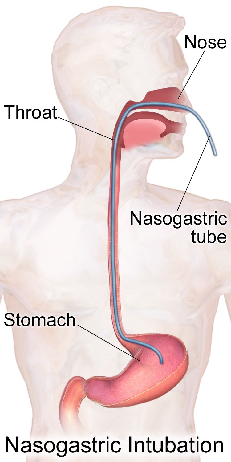
Cold stress is a metabolic emergency, not a comfort issue
Newborns lose heat by four routes: conduction to cold surfaces, convection to moving air, evaporation from wet skin and mucosa, and radiation to nearby cold objects such as a window or an incubator wall. They lose it fast because their surface-area-to-mass ratio is large, their subcutaneous fat is thin and their vasomotor control is immature. They cannot shiver. Instead they perform non-shivering thermogenesis by metabolising brown adipose tissue at the nape of the neck, between the scapulae, in the axillae and around the kidneys and adrenals — stores that the preterm and growth-restricted newborn largely do not have.
The consequences cascade in a predictable order. Heat production consumes oxygen and glucose, so cold stress produces hypoxia and hypoglycaemia. Anaerobic metabolism generates lactic acid, and metabolic acidosis causes pulmonary vasoconstriction, which reduces pulmonary perfusion and inhibits surfactant production, which worsens the hypoxia. A cold baby is also a jaundiced baby, because free fatty acids released from brown fat compete with bilirubin for albumin binding sites. Target axillary temperature is 36.5 to 37.5 °C. Below 36.5 °C is cold stress; below 36.0 °C is moderate hypothermia and warrants escalation.
Prevention is nursing work: dry the newborn immediately and remove the wet linen, put a hat on, use skin-to-skin contact with the birthing parent as the default warming device for stable infants, and for infants under 32 weeks place the baby without drying into a polyethylene wrap or bag with a thermal mattress and humidified warmed gases. Preheat surfaces, keep the isolette away from external walls, and cluster cares to limit the time the portholes are open. Rewarm a hypothermic newborn slowly, about 0.5 °C per hour, because rapid rewarming provokes apnoea and vasodilatory hypotension.
Hypoglycaemia: who, when and how much
Glucose falls physiologically in the first one to two hours of life in every newborn and then recovers as counter-regulation and feeding begin. Screening is targeted, not universal: the newborn of a parent with any form of diabetes, the large-for-gestational-age newborn, the small-for-gestational-age or growth-restricted newborn, the late preterm and preterm newborn, and any newborn who is cold, septic, asphyxiated, polycythaemic or symptomatic. Screen at approximately one hour of age, roughly 30 minutes after the first feed, then before feeds every three hours through the first 12 to 24 hours.
Signs are non-specific and easy to attribute to something else: jitteriness, high-pitched or weak cry, poor feeding, hypotonia, lethargy, temperature instability, tachypnoea, apnoea, cyanosis, diaphoresis and, late, seizure. Any point-of-care result must be confirmed on a laboratory plasma sample, but treatment starts on the bedside value — do not wait for the laboratory to treat a symptomatic baby.
Common operational thresholds in the first 24 hours: from birth to 4 hours of age, a value under 25 mg/dL (1.4 mmol/L) after a feed, or any symptomatic value, calls for intravenous dextrose; 25 to 40 mg/dL calls for a feed and a recheck in one hour. From 4 to 24 hours, treat under 35 mg/dL (1.9 mmol/L) intravenously and feed-and-recheck between 35 and 45 mg/dL. The general goal is a plasma glucose of at least 45 mg/dL (2.5 mmol/L) before routine feeds, rising toward 60 mg/dL after 48 hours. Buccal 40 per cent dextrose gel at 0.5 mL/kg, given with a feed, treats many asymptomatic episodes and keeps parent and baby together.
When intravenous treatment is needed, give a minibolus of 2 mL/kg of 10 per cent dextrose — that is 200 mg/kg of glucose — and follow immediately with a continuous infusion, because a bolus alone provokes rebound insulin secretion. Start the infusion at a glucose infusion rate of 5 to 8 mg/kg/min. The formula is worth memorising: glucose infusion rate in mg/kg/min equals the dextrose percentage multiplied by the rate in mL/hr, divided by the weight in kilograms multiplied by 6. A 3 kg newborn on 10 per cent dextrose at 12 mL/hr receives (10 × 12) ÷ (3 × 6) = 120 ÷ 18 = 6.7 mg/kg/min. Persistent hypoglycaemia requiring more than 8 to 10 mg/kg/min beyond 48 hours suggests hyperinsulinism and needs endocrine evaluation.
Weight-based dosing: the arithmetic and the ceiling
Children are not small adults, and the only safe way to express a paediatric dose is per kilogram of actual measured weight in kilograms, taken on admission, not reported by a caregiver in pounds. Write the calculation out, have a second nurse verify it independently for high-alert drugs, and then perform the step that is skipped most often: compare the calculated dose against the maximum adult dose. A weight-based formula that is correct for a 12 kg toddler becomes an overdose in a 70 kg adolescent because the milligrams-per-kilogram relationship stops scaling.
Worked example. Acetaminophen is dosed at 15 mg/kg per dose. For a 14 kg child: 14 × 15 = 210 mg. For a 62 kg adolescent: 62 × 15 = 930 mg, which is under the 1,000 mg adult single-dose maximum, so it stands. For an 84 kg adolescent: 84 × 15 = 1,260 mg, which exceeds the maximum — the dose is capped at 1,000 mg, and the daily total must stay under 75 mg/kg/day and under the 4,000 mg adult daily maximum. The same discipline applies to ibuprofen at 10 mg/kg per dose with a 400 mg per dose ceiling, and to amoxicillin at 45 to 90 mg/kg/day divided, capped at the adult daily maximum. If the calculated dose exceeds the adult dose, the calculation is not wrong — the ceiling is right.
Neonatal antibiotic arithmetic follows the same pattern. Ampicillin for suspected early-onset sepsis is 50 mg/kg per dose (100 mg/kg per dose when meningitis is suspected). A 3.2 kg newborn: 3.2 × 50 = 160 mg per dose. Gentamicin is 4 to 5 mg/kg per dose with the interval set by postmenstrual age — commonly every 24 hours at 35 weeks or more, and every 36 to 48 hours below that, because renal clearance is slower. For the same newborn at 4 mg/kg: 3.2 × 4 = 12.8 mg. Draw a trough before the next dose when therapy extends beyond 48 hours, and monitor for nephrotoxicity and ototoxicity.
Volumes follow the same per-kilogram discipline but not the same numbers at every age, and this is the pair most often confused. In the delivery room and in the neonatal unit, volume expansion is 10 mL/kg of isotonic crystalloid or O-negative blood over 5 to 10 minutes, given more slowly still in the preterm infant because rapid volume shifts contribute to intraventricular haemorrhage. Beyond the newborn period, the paediatric fluid resuscitation bolus for shock is 20 mL/kg of isotonic crystalloid — normal saline or lactated Ringer’s — given rapidly, with reassessment of heart rate, perfusion, mental state and liver edge after each bolus before the next is given. Reduce it to 10 mL/kg and give it more cautiously in diabetic ketoacidosis, in suspected cardiogenic shock and in severe acute malnutrition. Reassessment between boluses is part of the dose, not an optional extra.
Feeding the preterm gut, and necrotising enterocolitis
Coordinated sucking, swallowing and breathing matures at about 32 to 34 weeks, so infants below that are fed by orogastric or nasogastric tube while non-nutritive sucking on a pacifier during gavage builds the reflex. Feeding readiness is assessed by cues — rooting, hand-to-mouth activity, stable respiratory rate, sustained alert state — not by the clock and never by labelling a baby a poor feeder. Describe what you observe: this infant sustains four to five sucks before pausing to breathe and desaturates to 88 per cent during bursts. That sentence generates a plan; a label does not.
Human milk is the preferred enteral nutrition and is protective against necrotising enterocolitis; when the birthing parent’s own milk is unavailable, pasteurised donor human milk is preferred for very low birth weight infants. Preterm infants need fortification for protein, calcium and phosphorus to support bone mineralisation. Trophic or gut-priming feeds of 10 to 20 mL/kg/day stimulate mucosal maturation and hormone release without providing meaningful nutrition, and are advanced steadily rather than in leaps.
Necrotising enterocolitis is inflammatory and ischaemic injury to the bowel, usually presenting in the second or third week of life in a preterm infant whose feeds are advancing. The early signs are quiet: increased gastric residuals, feeding intolerance, abdominal distension with visible loops, a shiny or discoloured abdominal wall, bilious emesis, blood in the stool, and a generalised deterioration with apnoea, temperature instability and lethargy. The abdominal radiograph shows pneumatosis intestinalis, and free air means perforation. Management is immediate nil by mouth, orogastric tube to low intermittent suction for decompression, intravenous fluids and parenteral nutrition, broad-spectrum antibiotics, blood cultures, serial abdominal radiographs and girths, and surgical consultation. Do not palpate the abdomen repeatedly and never take a rectal temperature in a newborn with suspected necrotising enterocolitis.
Warmth, glucose and fluid are one physiological system: a cold newborn burns glucose and oxygen to make heat, and every dose or volume given must be per kilogram, written out, and checked against the maximum adult dose.
Section 4Bilirubin, Blood and Infection
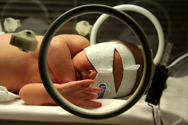
Why every newborn turns a little yellow, and when it stops being normal
Newborns have a high haematocrit, a red cell lifespan of only 70 to 90 days, and an immature hepatic conjugating enzyme, uridine diphosphate glucuronosyltransferase. Haem breakdown yields unconjugated, fat-soluble, albumin-bound bilirubin that the liver must conjugate before it can be excreted in bile. What reaches the gut is partly deconjugated by beta-glucuronidase and reabsorbed through the enterohepatic circulation, which is why delayed or inadequate feeding raises bilirubin and why stooling lowers it. Physiologic jaundice therefore appears after 24 hours of age, peaks on day three to five in the term infant and day five to seven in the preterm infant, and resolves without treatment.
Jaundice is pathologic when it appears in the first 24 hours of life, when total serum bilirubin rises faster than 0.2 mg/dL per hour or more than 5 mg/dL per day, when the direct (conjugated) fraction exceeds 1 mg/dL or 20 per cent of the total, or when visible jaundice persists beyond two weeks in a term infant. Jaundice within 24 hours is haemolysis, sepsis or both until proven otherwise, and it never waits for morning rounds. A raised direct fraction is never phototherapy-responsive and points toward biliary atresia, which is time-critical surgery, or toward sepsis, metabolic disease or parenteral-nutrition cholestasis.
Distinguish two feeding-related patterns without blaming anyone. Suboptimal-intake jaundice appears on day two to four when a chestfeeding dyad has not yet established volume; the treatment is more effective and more frequent feeding, 8 to 12 times in 24 hours, with lactation support — not the substitution of water or dextrose, which worsens it. Breast-milk jaundice appears later, around day five to ten, persists for weeks, and occurs in a thriving, well-gaining baby; feeding continues.
| Feature | Physiologic | Pathologic |
|---|---|---|
| Onset | After 24 hours of age | Within the first 24 hours |
| Rate of rise | Under 0.2 mg/dL per hour | Over 0.2 mg/dL per hour or over 5 mg/dL per day |
| Direct fraction | Under 1 mg/dL | Over 1 mg/dL or over 20 per cent of total |
| Duration | Resolves by day 7–10 (term) | Persists beyond 2 weeks (term) or 3 weeks (preterm) |
| Baby | Feeding, stooling, gaining | Lethargic, poor feeding, pale stools or dark urine, unwell |
Phototherapy, exchange transfusion and the brain
Every newborn is screened before discharge with a transcutaneous bilirubin or serum bilirubin, and the result is plotted against the exact age in hours on a curve chosen for gestational age and neurotoxicity risk factors — not compared to a single remembered number. Neurotoxicity risk factors include gestational age under 38 weeks, isoimmune haemolytic disease, glucose-6-phosphate dehydrogenase deficiency, sepsis, albumin under 3.0 g/dL and significant clinical instability in the preceding 24 hours; any of these lowers the treatment threshold. Once phototherapy has begun, transcutaneous readings are unreliable and serum values are used.
Phototherapy uses narrow-band blue light near 460 to 490 nm to photoisomerise bilirubin in the skin into water-soluble forms excreted in bile and urine without conjugation. Nursing care is specific: expose the maximum skin surface with only a nappy, apply opaque eye shields and check the eyes each shift for discharge or corneal abrasion, keep the prescribed light-to-skin distance, monitor temperature because both hyperthermia and cold stress occur, and increase attention to hydration — insensible losses rise and stools become loose and green. Continue feeding; do not interrupt phototherapy for prolonged periods, and never substitute water. Expect a transient bronze discolouration if the direct fraction is raised.
Exchange transfusion, usually double-volume at about 160 mL/kg through an umbilical venous catheter, is reserved for bilirubin at or above the exchange threshold or for any sign of acute bilirubin encephalopathy. In isoimmune haemolytic disease, intravenous immunoglobulin 0.5 to 1 g/kg may reduce the need for exchange. Acute bilirubin encephalopathy progresses through a recognisable sequence: lethargy, hypotonia and poor feeding first; then a high-pitched cry, irritability, retrocollis and opisthotonos, and fever; then apnoea, seizures, coma and death. The chronic sequel is kernicterus — athetoid cerebral palsy, sensorineural hearing loss, upward gaze palsy and dental enamel dysplasia — and it is permanent, which is why the bilirubin curve is treated as a time-critical value.
Haemolysis, polycythaemia and anaemia
Rh incompatibility arises when an Rh-negative birthing parent carries an Rh-positive fetus and becomes sensitised, usually at a previous birth or bleeding event; maternal IgG anti-D then crosses the placenta and destroys fetal red cells, causing anaemia, hepatosplenomegaly, hydrops in severe cases, and rapid postnatal jaundice. Prevention is Rho(D) immune globulin 300 micrograms intramuscularly at 28 weeks and again within 72 hours of birth of an Rh-positive newborn, with additional dosing guided by a Kleihauer-Betke test after a large fetomaternal bleed. ABO incompatibility — a group O parent with a group A or B newborn — is more common, generally milder, and can occur in a first pregnancy. A positive direct antiglobulin (Coombs) test on the newborn confirms antibody-coated red cells.
Polycythaemia is a central venous haematocrit above 65 per cent, seen after delayed cord clamping, in growth restriction, in infants of parents with diabetes, at altitude and in twin-to-twin transfusion. Hyperviscosity produces a plethoric, ruddy baby with sluggish capillary refill, respiratory distress, hypoglycaemia, hyperbilirubinaemia, poor feeding and, at worst, thrombosis. Treatment of a symptomatic infant is a partial exchange transfusion replacing blood with normal saline. At the other end, anaemia of prematurity reflects low erythropoietin, short red cell survival and phlebotomy losses, and is managed by minimising blood draws, iron supplementation and restrictive transfusion thresholds.
Sepsis: early, late and almost silent
Early-onset sepsis presents in the first 72 hours and is acquired vertically, most often from group B Streptococcus, Escherichia coli or Listeria. Risk factors are maternal group B streptococcal colonisation without adequate intrapartum antibiotic prophylaxis, rupture of membranes for 18 hours or more, intraamniotic infection with maternal fever of 38 °C or higher, and preterm labour. Adequate prophylaxis means penicillin G, or ampicillin, given at least four hours before birth. Late-onset sepsis appears after 72 hours and is usually healthcare-associated or environmental — coagulase-negative staphylococci, Staphylococcus aureus, Gram-negative organisms and Candida — and central lines are the dominant risk.
The newborn does not localise infection and rarely mounts a convincing fever; the classic presentation is that the baby simply is not right. Temperature instability, and more often hypothermia than hyperthermia, poor feeding, lethargy, hypotonia, tachypnoea or apnoea, tachycardia or bradycardia, mottling, delayed capillary refill, hypoglycaemia or hyperglycaemia, jaundice within 24 hours and irritability are the vocabulary. Neonatal meningitis does not produce nuchal rigidity; look for a bulging fontanelle, a high-pitched shrill cry, seizures and opisthotonos. Investigation is blood culture before antibiotics, complete blood count with differential including the immature-to-total neutrophil ratio (0.2 or higher is concerning), C-reactive protein trends and lumbar puncture when meningitis is suspected. Antibiotics are started empirically within the first hour of suspicion and narrowed when cultures return.
Jaundice in the first 24 hours and a newborn who is subtly not right are both emergencies: plot bilirubin against the hour-specific curve, and draw the blood culture and start antibiotics before the baby looks septic.
Section 5Injury, Anomaly, Withdrawal and the Family
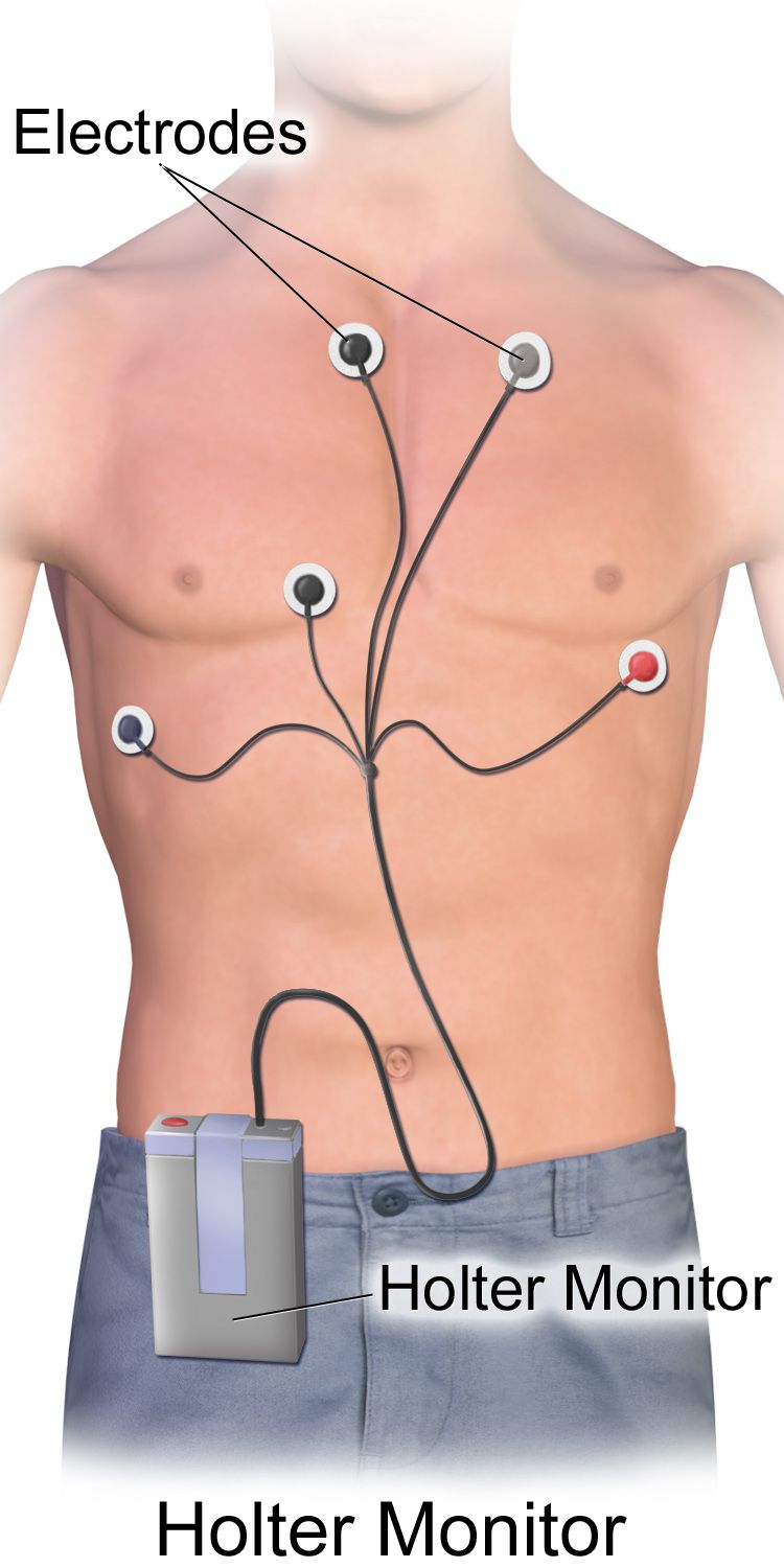
Birth injury and hypoxic-ischaemic encephalopathy
Three scalp swellings must be told apart, because one of them is a resuscitation. Caput succedaneum is oedema of the presenting scalp, present at birth, soft, pitting, and it crosses suture lines; it resolves within days. Cephalohaematoma is subperiosteal bleeding that appears hours after birth, is firm, and does not cross suture lines; it resolves over weeks and raises the bilirubin load as it breaks down. Subgaleal haemorrhage is bleeding into the potential space between the aponeurosis and the periosteum, it crosses suture lines and shifts with position, it feels boggy and fluctuant, and it can hold 20 to 40 per cent of the newborn’s blood volume. It is associated with vacuum-assisted birth and it is a haemorrhagic shock emergency: measure occipitofrontal circumference hourly, watch for tachycardia, pallor, falling haematocrit and hypotension, and escalate at once.
A clavicular fracture after shoulder dystocia presents with crepitus, a lump, crying on movement of the arm and an asymmetric Moro reflex; treatment is gentle handling and pinning the sleeve to the shirt. Erb-Duchenne palsy from C5 to C6 stretch produces an arm held adducted and internally rotated with the forearm pronated — the waiter’s tip position — an absent Moro on that side, and an intact grasp reflex, which is the finding that distinguishes it from a lower plexus injury. Facial nerve palsy after forceps shows as asymmetric crying with an eye that will not close on the affected side.
Safeguarding belongs in this section, because birth injury is where nurses learn to read an injury against a mechanism. Every injury must be consistent with both the history given and the child’s developmental stage. A cephalohaematoma after a vacuum-assisted birth is consistent; a bruise, a torn frenulum, a rib or long-bone fracture, a retinal haemorrhage or a burn in an infant who is not yet mobile is not. Use the milestones as the yardstick: an infant rolls at about 4 to 6 months, sits unsupported at about 6 months, crawls at about 8 to 9 months, pulls to stand and cruises at about 9 to 12 months, and walks alone at about 12 to 15 months — so bruising in a pre-mobile infant is a red flag until explained. A history that changes between tellings, an unexplained delay in seeking care, or an injury that outruns what the child can actually do raises the same duty. Mandatory reporting is a legal obligation, not a clinical judgement call. The threshold is reasonable suspicion, not proof, not certainty and not a completed diagnosis; you report to the statutory child-protection authority yourself rather than passing the concern to someone else to file, you inform the treating clinician, you keep the infant safe in the meantime, and you document objectively — what you saw, what you measured, and what you were told, in quotation marks where it was said. Investigating, interrogating the family or deciding whether the concern is founded is not the nurse’s role.
Hypoxic-ischaemic encephalopathy follows a sentinel event such as cord prolapse, abruption or uterine rupture. Candidacy for therapeutic hypothermia typically requires 36 weeks or more of gestation and about 1,800 g or more, plus evidence of perinatal acidosis such as a cord pH of 7.0 or less or a base deficit of 12 mmol/L or more, or an Apgar of 5 or less at 10 minutes or continued resuscitation at 10 minutes, together with moderate or severe encephalopathy on a structured neurological examination. Cooling to a core temperature of 33.5 °C must begin within 6 hours of birth and continues for 72 hours, followed by rewarming at no more than 0.5 °C per hour. Nursing during cooling means expecting sinus bradycardia in the 80 to 100 range as normal, monitoring amplitude-integrated EEG for subclinical seizures, watching for coagulopathy, thrombocytopenia, hypotension and subcutaneous fat necrosis, and above all preventing hyperthermia, which worsens injury.
Congenital anomalies: the first action is the whole answer
Congenital diaphragmatic hernia is the emergency that punishes reflexes. The abdominal contents herniate into the chest, so the baby has a scaphoid abdomen, a barrel chest, absent breath sounds and audible bowel sounds on the affected side, and often mediastinal shift. Do not give bag-mask positive-pressure ventilation: it fills the herniated stomach and bowel with gas and compresses the lung further. Intubate immediately, place a large-bore orogastric tube to low suction, ventilate gently, position with the affected side down, keep the baby nil by mouth, and transfer to a surgical and extracorporeal membrane oxygenation centre.
Oesophageal atresia with tracheo-oesophageal fistula presents with copious frothy oral secretions, choking, coughing and cyanosis with the first feed, and an orogastric tube that will not pass. Keep the baby nil by mouth, place a sump catheter in the upper pouch on continuous low suction, elevate the head of the bed 30 to 45 degrees to reduce reflux of gastric acid through the fistula into the trachea, and arrange surgery. Myelomeningocele demands prone or side-lying positioning to keep pressure off the sac, a sterile non-adherent dressing moistened with warm sterile normal saline, a strictly latex-free environment because of the high rate of latex sensitisation, daily occipitofrontal circumference measurement for hydrocephalus, and surgical closure typically within 24 to 72 hours.
Gastroschisis and omphalocele both need the exposed viscera covered immediately with a sterile saline-moistened dressing and a bowel bag, a nil-by-mouth order, orogastric decompression, side-lying positioning to protect the mesenteric blood supply, and a very high fluid requirement — often two to three times maintenance for gastroschisis because evaporative and third-space losses are extreme. Choanal atresia produces a newborn who is pink while crying and cyanotic at rest, relieved by an oral airway. A ductal-dependent congenital heart lesion deteriorates as the ductus closes over the first hours to days; the treatment is prostaglandin E1 at 0.01 to 0.05 micrograms/kg/min, and the nurse must anticipate its predictable side effects of apnoea, hypotension and fever with intubation equipment at the bedside. Critical congenital heart disease screening by pulse oximetry is performed at 24 hours or later in the right hand and either foot, and a pass requires 95 per cent or higher in either limb with a difference of 3 per cent or less.
The newborn experiencing withdrawal
Neonatal opioid withdrawal, still often called neonatal abstinence syndrome, is a physiological consequence of in-utero exposure, not a moral event, and the family deserves language that says so: a parent with a substance use disorder in treatment, a newborn experiencing withdrawal. Timing depends on the drug half-life — short-acting opioids typically 24 to 48 hours, methadone and buprenorphine 48 to 72 hours and sometimes as late as day five to seven, selective serotonin reuptake inhibitors within one to two days. Signs cluster in four systems: central nervous (high-pitched persistent cry, tremors, hypertonia, irritability, poor sleep, exaggerated Moro, seizures), autonomic (sweating, mottling, nasal stuffiness, frequent sneezing and yawning, low-grade fever), gastrointestinal (uncoordinated suck, regurgitation, loose stools, excoriated buttocks, poor weight gain) and respiratory (tachypnoea).
Care is non-pharmacologic first, and it is nursing care rather than pharmacy care. Room the baby in with the parent, maximise skin-to-skin contact, dim the lights and cluster the cares, swaddle in flexion, offer a pacifier, feed small volumes frequently with a high-calorie formula or human milk, and support chestfeeding when the parent is engaged in treatment and there is no contraindication such as untreated HIV or ongoing illicit polysubstance use. Protect the skin of the buttocks with a barrier ointment and change nappies promptly. The Eat, Sleep, Console approach evaluates function rather than tallying symptoms: can the newborn eat at least an ounce or chestfeed well, sleep undisturbed for at least an hour, and be consoled within ten minutes? If not, optimise the non-pharmacologic care first, then consider medication. Where a scored approach is used, the Finnegan tool commonly triggers treatment at three consecutive scores of 8 or more, or two scores of 12 or more. When medication is needed, oral morphine at approximately 0.04 to 0.05 mg/kg per dose every three to four hours is typical, weaned by protocol, with phenobarbital or clonidine as adjuncts.
Priorities, delegation and going home
A newborn compensates with tachycardia and vasoconstriction and keeps the blood pressure normal until very late, so hypotension is a pre-arrest finding, not an early one, and the deterioration you are actually meant to catch is the compensated one — rising heart rate, rising respiratory rate, a narrowing pulse pressure, capillary refill over 2 seconds, cool mottled peripheries and any change in level of consciousness. Read the newborn’s own ranges rather than an adult range: a term newborn breathes 30 to 60 times a minute, runs a heart rate of roughly 100 to 205 awake, and is hypotensive below a systolic of 60 mmHg. Priority then follows airway, breathing and circulation, and the acute problem outranks the chronic one: the preterm infant who has just become apnoeic is assessed before the stable infant whose phototherapy is overdue, and the newborn with grunting and retractions is assessed before the newborn with a rising but sub-threshold bilirubin.
Delegation in the neonatal unit follows the same rule as everywhere else, and the high-risk newborn stretches it hard. The registered nurse retains assessment, teaching, evaluation and any unstable patient. That means the admission and shift assessments, interpretation of a bilirubin or glucose result, the decision to begin or stop a feed, phototherapy assessment, discharge teaching about safe sleep and car seats, the first feed of an at-risk newborn, and every aspect of the care of an infant on respiratory support, receiving therapeutic hypothermia or being resuscitated. Assistive personnel may weigh and measure, bathe a stable newborn, change nappies and record output, stock the bed space, transport a stable infant with a nurse, and hold or bottle-feed a stable term newborn after the nurse has assessed and specified the plan. Licensed practical nurse scope varies by jurisdiction and by employer, and unit policy governs.
Discharge from a neonatal unit is a teaching event, not a paperwork event. Confirm the newborn metabolic screen was collected at 24 to 48 hours and repeated when required, the hearing screen was performed, critical congenital heart disease screening passed, and immunisations are given by chronological age — a preterm infant is vaccinated on the calendar, not on corrected age — while developmental milestones are judged by corrected age until about two years. The routine schedule is hepatitis B at birth, then diphtheria-tetanus-acellular pertussis, inactivated polio, Haemophilus influenzae type b, pneumococcal conjugate and rotavirus vaccines beginning at 2 months and repeated at 4 and 6 months according to the product used, with measles-mumps-rubella and varicella at 12 to 15 months. Doses are never reduced or split for a small infant. The single accepted exception to the calendar is the birth dose of hepatitis B vaccine in an infant under 2,000 g whose birthing parent is hepatitis B surface antigen negative, which is deferred to 1 month of age or to discharge; if that status is positive or unknown, both the vaccine and hepatitis B immune globulin are given within 12 hours of birth whatever the weight. Preterm infants under 37 weeks need a car seat tolerance screen of 90 to 120 minutes before discharge. Teach safe sleep without exception: supine, alone, on a firm flat surface, no soft bedding, room-sharing without bed-sharing, and no smoking. Arrange follow-up for bilirubin and weight within 24 to 48 hours, and ensure the family knows exactly who to call, for what, and at what number.
Finally, the family is the patient too. Support parental presence and unrestricted access, teach kangaroo care and let parents provide it as care rather than as a reward, involve them in cares and in decision-making, and protect lactation with early and frequent expression when the baby cannot yet feed. Name the baby, not the diagnosis or the bed number. When an infant is dying or has a life-limiting condition, palliative care is offered alongside, not instead of, active treatment: symptom control, memory-making, sibling inclusion, cultural and spiritual practices honoured, and bereavement follow-up planned before the family leaves.
For every high-risk newborn there is one correct first action, and it is usually the one that overrides instinct — intubate rather than bag, cool rather than warm, console rather than medicate, and always assess the family alongside the infant.
The chapter in one breath
A newborn becomes high risk through gestational age, growth, the events of the birth, or the physiology of the first hours — and the late preterm infant, who looks well, is the one most often underestimated. Resuscitation is a timed sequence: initial steps, then positive-pressure ventilation within the Golden Minute if the newborn is apnoeic or the heart rate is under 100, MR SOPA when the heart rate will not rise, 3:1 compressions when it stays under 60 despite chest-moving ventilation, and epinephrine 0.02 mg/kg intravenously only after that. Saturation targets climb minute by minute from 60–65 per cent at one minute to 85–95 per cent at ten. Warmth protects glucose, glucose protects the brain, and every volume and dose is per kilogram, written out and checked against the adult maximum — with 4–2–1 for maintenance beyond the newborn period and mL/kg/day within it. Jaundice before 24 hours, a bilirubin climbing faster than the hour-specific curve, and a baby who is simply not right are all emergencies. Congenital anomalies each have one counterintuitive first action. Hypotension in a newborn is a pre-arrest sign rather than an early one, and an injury that does not fit the history or the child’s developmental stage is reported on reasonable suspicion, not on proof. Throughout, the language is family-centred, the escalation is SBAR, and assessment, teaching, evaluation and the unstable patient stay with the registered nurse.
Words worth knowing
| Term | What it means |
|---|---|
| Late preterm | Birth at 34 0/7 to 36 6/7 weeks; physiologically immature despite a term-like appearance. |
| Small for gestational age | Birth weight below the 10th percentile for gestational age; a size statement, not a diagnosis. |
| Intrauterine growth restriction | Failure of a fetus to achieve its own growth potential; symmetric (early, intrinsic) or asymmetric (late, placental). |
| Golden Minute | The first 60 seconds of life, within which initial steps are completed and positive-pressure ventilation begun if needed. |
| MR SOPA | Corrective ventilation steps: Mask, Reposition, Suction, Open mouth, Pressure increase, Alternative airway. |
| Surfactant | Phospholipid-protein complex that lowers alveolar surface tension; deficient in respiratory distress syndrome. |
| Persistent pulmonary hypertension | Failure of pulmonary vascular resistance to fall, causing right-to-left shunting and a preductal-postductal saturation gap over 10 per cent. |
| Bronchopulmonary dysplasia | Chronic lung disease defined by continued oxygen need at 36 weeks postmenstrual age. |
| Non-shivering thermogenesis | Heat production by metabolism of brown adipose tissue; the newborn’s only means of generating heat. |
| Neutral thermal environment | The ambient conditions in which oxygen consumption and metabolic demand are minimal, keeping axillary temperature 36.5–37.5 °C. |
| Glucose infusion rate | Milligrams of glucose per kilogram per minute; (dextrose per cent × mL/hr) ÷ (kg × 6). |
| 4–2–1 rule | Hourly maintenance fluid: 4 mL/kg for the first 10 kg, 2 mL/kg for the next 10 kg, 1 mL/kg thereafter; not for the first days of life. |
| Necrotising enterocolitis | Ischaemic-inflammatory bowel injury of the preterm infant; pneumatosis intestinalis on radiograph, treated with nil by mouth, decompression and antibiotics. |
| Kernicterus | Permanent bilirubin-induced neurological damage: athetoid cerebral palsy, sensorineural hearing loss, upward gaze palsy, enamel dysplasia. |
| Direct antiglobulin test | Coombs test detecting antibody bound to newborn red cells; positive in Rh and ABO isoimmune haemolysis. |
| Subgaleal haemorrhage | Bleeding beneath the scalp aponeurosis that crosses sutures and can hold 20–40 per cent of blood volume; a shock emergency. |
| Therapeutic hypothermia | Cooling to 33.5 °C within 6 hours of birth for 72 hours in moderate to severe hypoxic-ischaemic encephalopathy at 36 weeks or more. |
| Eat, Sleep, Console | Function-based assessment of the newborn experiencing withdrawal; non-pharmacologic care is optimised before medication is considered. |
| Corrected age | Chronological age minus the weeks of prematurity; used to judge development to about two years, but never to delay immunisations. |
Check yourself
Paediatric Growth, Development and Health Promotion
A child is not a small adult, and the difference is not sentimental — it is physiological, pharmacological and numerical. The airway is narrower, the metabolic rate faster, the body water fraction larger, the compensatory reserve deeper and then, without warning, gone. Every dose is calculated per kilogram; every vital sign is read against the child’s age band, not an adult range; every conversation is pitched to the child’s stage of cognitive development and routed through the family. This chapter builds the scaffolding on which the rest of paediatric nursing hangs: how children grow and how you measure it, what development looks like month by month and which absences demand referral, what health promotion actually consists of at each age, and — the safety spine of paediatric practice — how to calculate a weight-based dose and a maintenance fluid rate without harming the child you are trying to help. Recognition of the deteriorating child closes the chapter, because in paediatrics the earliest sign is almost never the dramatic one.
By the end of this chapter you can
- Describe the cephalocaudal and proximodistal patterns of growth and explain why development is sequential but individually paced.
- Plot and interpret weight-for-age, length/height-for-age, head circumference and BMI-for-age percentiles, and identify growth faltering by percentile crossing.
- Match Erikson’s psychosocial stages, Piaget’s cognitive stages and stage-typical play and fears to nursing interventions for each age band.
- State expected developmental milestones from 2 months to 5 years and name the developmental red flags that require prompt referral, including loss of previously acquired skills.
- Deliver age-appropriate anticipatory guidance on feeding, safe sleep, oral health, immunisation, screen use and injury prevention, using teach-back.
- Calculate a weight-based dose in mg/kg, show the arithmetic, and verify it against the maximum adult dose.
- Apply the 4-2-1 rule to compute an hourly maintenance fluid rate and a 20 mL/kg resuscitation bolus, with a worked example.
- Interpret vital signs by age band, recognise compensated shock, and escalate using the Paediatric Assessment Triangle, a PEWS score and structured SBAR communication.
- Distinguish nursing actions that may be delegated to assistive personnel from those that remain with the registered nurse.
Section 1How Children Grow: Principles, Patterns and Measurement
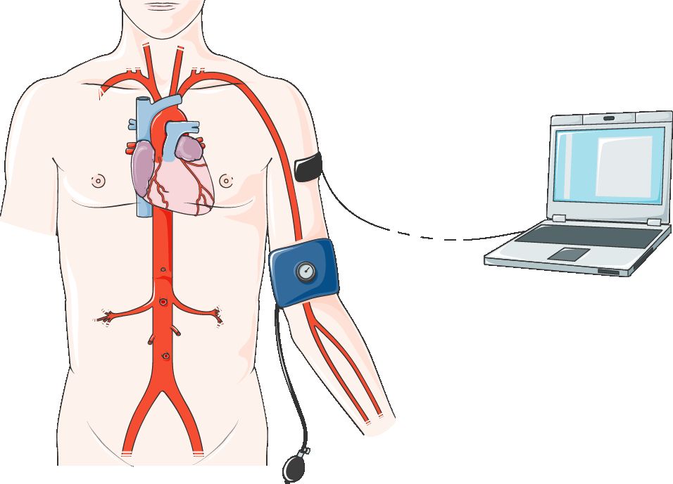
The rules that growth follows
Growth is the measurable increase in size; development is the progressive acquisition of function. They run together but they are not the same thing, and a child can be entirely normal in one and delayed in the other. Both follow directional rules that hold across every healthy child. Growth is cephalocaudal — head to toe. The newborn head is roughly one quarter of body length and the brain reaches about eighty per cent of adult size by three years, which is why head control precedes trunk control, which precedes walking. Growth is also proximodistal — centre outward. Shoulder and arm control arrive before the crude palmar rake, which arrives before the neat pincer grasp at around twelve months. And development moves from general to specific: the infant who waves both arms in undifferentiated excitement at three months is the toddler who isolates one index finger to point at eighteen months.
Two more principles matter at the bedside. First, the sequence is fixed but the rate is individual. Every child sits before standing and stands before walking, but the age at which each happens spreads across a wide normal window. A parent who reports that a cousin walked at nine months and their own child is not walking at fourteen months needs reassurance grounded in the actual range, not in comparison. Second, there are critical periods — windows in which a particular system is uniquely vulnerable to insult or uniquely responsive to stimulation. Neural tube closure in the first four weeks after conception, the surge of synaptic formation in the first three years, and language exposure before school age are all such windows. This is the biological argument for early intervention: the same effort applied at two years buys more than it buys at seven.
Growth is not smooth. Infancy is the fastest period of postnatal growth, followed by a long, steady, comparatively slow stretch through the school years, followed by the adolescent growth spurt. Because the tempo differs so much, a single measurement tells you very little. What tells you something is a trajectory — the same child measured the same way over time, plotted on a chart.
Anthropometrics and the growth chart
Four measurements form the routine paediatric anthropometric set. Weight is recorded in kilograms only; the pound scale should not exist on a paediatric unit, because unit confusion is one of the classic routes to a catastrophic overdose. Infants are weighed nude on a calibrated infant scale, older children in light clothing without shoes. Length is measured recumbent with a length board until two years; height is measured standing with a stadiometer from two years, and the switch produces a small apparent drop of roughly half a centimetre that should not be misread as a loss. Head circumference (occipitofrontal circumference) is measured at the widest point, above the brows and over the occipital prominence, routinely to at least twenty-four months and often to thirty-six. BMI-for-age is calculated and plotted from two years; below two years the corresponding measure is weight-for-length.
Growth charts are population references, and which chart you use matters. The World Health Organization charts describe how healthy, adequately fed, largely breastfed children should grow and are the standard from birth to twenty-four months. Country-specific reference charts, such as the CDC charts in the United States, are typically used from two to twenty years. Preterm infants are plotted using corrected age — chronological age minus the number of weeks born before forty weeks — usually until two to three years for weight and length and eighteen months for head circumference. Failing to correct will make a perfectly healthy former twenty-eight-week infant look profoundly delayed.
Interpretation rests on percentiles, not on averages. A child steadily tracking the third percentile with parents who are both short is likely constitutionally small and entirely well. A child who has tracked the seventy-fifth percentile for a year and now measures at the twenty-fifth has crossed two major percentile lines downward and needs assessment. That pattern — sustained downward crossing, or weight-for-length below the fifth percentile — is what older texts called failure to thrive. Say growth faltering instead. The older phrase reads as an accusation aimed at a parent, and the causes are overwhelmingly medical, nutritional or social rather than a matter of anyone failing. For BMI-for-age from two years, the conventional bands are underweight below the fifth percentile, healthy weight from the fifth to the eighty-fourth, overweight from the eighty-fifth to the ninety-fourth, and obesity at or above the ninety-fifth percentile.
| Age | Weight | Length / height | Head circumference |
|---|---|---|---|
| Birth (term) | Average about 3.4 kg (range 2.5–4.0 kg); loses up to 7–10 per cent in the first days and regains birth weight by 10–14 days | Average about 50 cm | Average 33–35 cm; exceeds chest circumference until about 12 months |
| 0–6 months | Gains roughly 20–30 g/day; doubles birth weight by 4–6 months | Gains about 2.5 cm/month | Gains about 2 cm/month for 3 months, then about 1 cm/month |
| 6–12 months | Gains roughly 10–15 g/day; triples birth weight by 12 months | Length increases about 50 per cent over the year (about 75 cm at 1 year) | Gains about 0.5 cm/month; about 45–47 cm at 1 year |
| Toddler (1–3 y) | Gains about 2–3 kg/year; quadruples birth weight by 24 months | Gains about 7.5 cm/year; birth length doubles by about 4 years | Gains about 2 cm/year; anterior fontanelle closes 12–18 months |
| Preschool (3–5 y) | Gains about 2–3 kg/year | Gains about 6–8 cm/year | Not routinely measured after 36 months |
| School-age (6–12 y) | Gains about 2–3 kg/year | Gains about 5 cm/year | — |
| Adolescence | Gains 7–25 kg across the spurt; body composition diverges by sex | Peak height velocity about 8–9 cm/year in girls, 9–10 cm/year in boys | — |
Teeth, senses and the maturing organ systems
Dentition follows a predictable clock that is useful both as a developmental marker and as a health-promotion trigger. The first primary tooth — usually a lower central incisor — erupts at about six to ten months. A serviceable rule of thumb up to about two years is that the number of teeth equals the age in months minus six. The full set of twenty primary teeth is in place by about thirty months, and the first permanent molars arrive at about six years, often mistaken by parents for primary teeth and therefore neglected. Oral health promotion begins at the first tooth, not at the first dental complaint.
Vision matures over years. The newborn sees best at about twenty to thirty centimetres — roughly the distance to a feeding caregiver’s face — with acuity in the region of 20/400. Binocular fixation and following past the midline consolidate by three to four months, and adult acuity of about 20/20 is reached at around six years. Persistent misalignment beyond four months warrants ophthalmology referral, because untreated strabismus causes amblyopia and the treatment window closes. Hearing, by contrast, is essentially mature at birth, which is why universal newborn hearing screening is done before discharge and why any failed screen needs diagnostic follow-up rather than a wait-and-see approach.
Organ maturation explains most of what makes paediatric physiology distinctive. Renal concentrating ability is limited in the first year, so infants dehydrate quickly and excrete drugs more slowly. Hepatic enzyme systems are immature at birth, mature through infancy, and in early childhood may actually clear some drugs faster per kilogram than in adults. Total body water is about seventy-five per cent of body weight in the newborn compared with about sixty per cent in the adult, and a much greater proportion of it is extracellular — which is precisely why a diarrhoeal illness that inconveniences an adult can kill an infant. Immunoglobulin G transferred across the placenta wanes over the first six months, opening the well-known window of increased infection risk that the primary immunisation series is timed to close.
Puberty and Tanner staging
Puberty is staged by Tanner (sexual maturity rating) stages one to five, based on breast development and pubic hair in girls and genital development and pubic hair in boys. The first physical sign in girls is breast budding (thelarche, Tanner stage 2), typically between eight and thirteen years. The first sign in boys is testicular enlargement, typically between nine and fourteen years. Menarche arrives late in the sequence — usually about two to two and a half years after thelarche, most often at Tanner stage 4, at an average of around twelve to twelve and a half years. Critically, peak height velocity precedes menarche in girls; after menarche most girls grow only a further five to seven centimetres. In boys, peak height velocity comes later in the sequence, around Tanner stage 4, which is why boys who are shorter than their female classmates at thirteen commonly overtake them by sixteen.
Two thresholds trigger referral. Sexual development before eight years in girls or before nine years in boys is precocious puberty. Absence of breast development by thirteen years in girls, absence of menarche by fifteen years, or absence of testicular enlargement by fourteen years in boys is delayed puberty. Both deserve evaluation, and both carry a psychosocial load that the nurse should name openly with the adolescent rather than leave unspoken.
A single measurement is a data point; a plotted series is a diagnosis, and sustained downward crossing of two major percentile lines is the signal that something has changed and must be explained.
Section 2Developmental Theory and the Child at Each Age
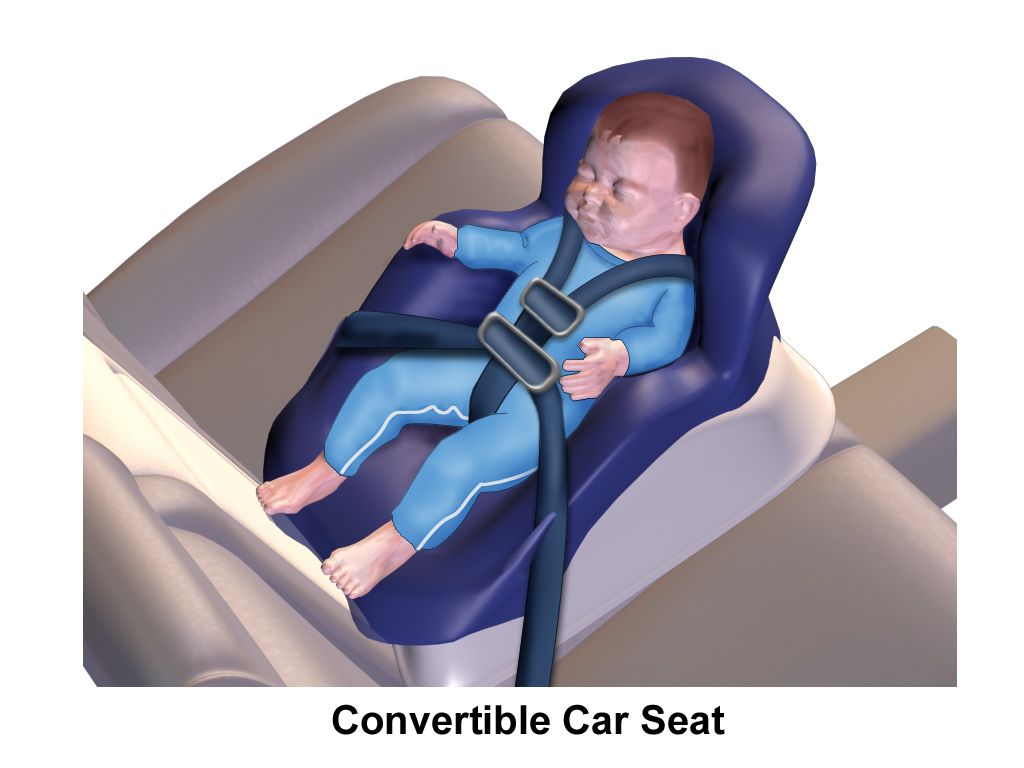
Frameworks that change what you actually do
Developmental theory earns its place in nursing only if it changes an intervention. It does. Erikson frames each stage as a psychosocial task. The infant is resolving trust versus mistrust, so consistent caregivers, prompt response to crying and a parent kept at the bedside are clinical interventions, not courtesies. The toddler is resolving autonomy versus shame and doubt, so you offer two acceptable choices — “this arm or that arm” — and never a false choice about whether the injection happens. The preschooler is in initiative versus guilt and is prone to believing that illness is punishment for wrongdoing, so you say explicitly that nothing the child did caused this. The school-age child is in industry versus inferiority and needs jobs: hold the tape, count the breaths, choose the finger. The adolescent is in identity versus role confusion and needs privacy, confidential interview time without a parent, and involvement in decisions about their own body.
Piaget tells you how the child will interpret what you say. In the sensorimotor stage (birth to two years) the infant learns through the senses and acquires object permanence at around eight to nine months — the understanding that a person or object still exists when it is out of sight. That is exactly why separation anxiety appears then: the infant now knows the parent continues to exist somewhere else and protests the absence. Before object permanence, a parent who left the room genuinely ceased to exist for the infant. In the preoperational stage (two to seven years) thinking is egocentric, magical and literal. Tell a preschooler you are going to “take” their blood pressure and they may believe you are taking something away; say the medicine will “kill” the germs and expect a nightmare. In concrete operational thinking (seven to eleven years) the child grasps cause and effect, conservation and reversibility, and responds well to diagrams and to handling the equipment. In formal operational thinking (eleven years and up) abstraction is available, so long-term consequences and hypothetical reasoning become usable teaching tools — though the adolescent brain’s reward and risk systems mature on different timetables, which is why knowing the risk does not reliably prevent the behaviour.
Play progresses in parallel and is a direct assessment window. Infants engage in solitary play; toddlers in parallel play alongside but not with peers; preschoolers in associative play, sharing materials without shared goals; school-age children in cooperative play with rules and roles. So do fears. Stranger anxiety emerges at six to eight months. Separation anxiety peaks between about ten and eighteen months and progresses through protest, despair and detachment when separation is prolonged — detachment can look like settled behaviour and is frequently misread as improvement. Preschoolers fear bodily mutilation, the dark and loss of control, which is why a plaster over a puncture site genuinely matters to them. School-age children fear loss of control, death and exclusion. Adolescents fear body-image change, loss of independence and peer rejection more than they fear the diagnosis.
Infancy: birth to twelve months
Infancy compresses more change into twelve months than any subsequent year of life. Motor development runs cephalocaudal: lifting the head briefly when prone at about two months, steady head control and lifting head and chest prone by three to four months, rolling front to back at about four months and both directions by six, sitting in a tripod at six months and sitting unsupported by eight to nine, crawling around eight to nine months, pulling to stand and cruising by ten to twelve, and independent walking anywhere from nine to fifteen months. Fine motor runs proximodistal: an ulnar palmar rake at six months, transferring objects hand to hand at six to seven months, a crude pincer at nine months, and a neat pincer grasp using thumb and index finger by twelve months — which is simultaneously a developmental triumph and the reason the choking risk climbs sharply in the second half of the first year.
Language moves from cooing at two months to babbling at four to six months, to non-specific “mama” and “dada” at eight to nine months, to specific use of one to three meaningful words plus gestures such as waving and pointing at twelve months. Social development moves from the responsive social smile at about two months — a genuine milestone with real prognostic weight — through laughing at four months, stranger awareness at six to eight months, and simple imitative games and separation protest by the end of the year.
Primitive reflexes are the other half of the infant neurological examination, and their disappearance is as informative as their presence. The Moro (startle) reflex should be gone by four to six months, the palmar grasp by five to six months, the asymmetric tonic neck reflex by four to six months, and the rooting reflex by three to four months. A primitive reflex that persists beyond its window suggests a central nervous system problem and requires referral. So does a hand preference established before twelve to eighteen months, which may indicate weakness on the non-preferred side rather than early talent.
Toddler, preschool, school-age and adolescence
The toddler years (one to three) are dominated by mobility, language explosion and the drive for autonomy. Independent walking consolidates by fifteen months, running by eighteen, stair climbing with a rail and kicking a ball by two years. Vocabulary goes from roughly ten to twenty-five words at eighteen months to fifty or more words and two-word phrases at twenty-four months, with speech about fifty per cent intelligible to a stranger at two years and seventy-five per cent at three. Toilet training generally becomes physiologically possible between eighteen and twenty-four months once the child can walk to the toilet, stay dry for two hours, and communicate the need — readiness is the child’s, not the calendar’s. Negativism, ritualism and the tantrum are developmentally normal expressions of autonomy, and the nursing guidance for parents is consistency and ignoring the behaviour where safe, not escalation.
Preschoolers (three to five) refine everything. The three-year-old pedals a tricycle, copies a circle and speaks in three-word sentences with about seventy-five per cent intelligibility. The four-year-old hops on one foot, copies a cross, draws a person with two to four parts and should be essentially one hundred per cent intelligible to strangers. The five-year-old skips, copies a triangle or square, draws a person with six parts and prints some letters. Imaginative play peaks, and with it magical thinking and imaginary companions, both normal. School-age children (six to twelve) gain competence, rule-based reasoning and peer orientation; chronic illness at this age is felt largely as difference from peers, and school reintegration is a nursing concern in its own right. Adolescence (twelve to eighteen) brings the growth spurt, sexual maturation, abstract reasoning and a shift of primary attachment toward peers. Confidentiality, a structured psychosocial interview and non-judgemental discussion of sexual health, substance use, mood and online life belong in every adolescent encounter.
| Age | Gross motor | Fine motor | Language / social |
|---|---|---|---|
| 2 months | Lifts head briefly when prone | Hands often fisted; briefly follows to midline | Coos; responsive social smile |
| 4 months | Steady head control; rolls front to back | Brings hands together; grasps a rattle | Laughs aloud; babbles; follows past midline |
| 6 months | Sits with support (tripod); rolls both ways | Transfers hand to hand; ulnar rake | Babbles consonants; stranger awareness begins |
| 9 months | Sits unsupported; crawls; pulls to stand | Crude pincer; bangs two objects together | “Mama/dada” non-specific; object permanence; separation protest |
| 12 months | Cruises; may walk alone | Neat pincer grasp; releases object into a cup | 1–3 meaningful words; waves; follows a one-step request with gesture |
| 18 months | Walks well; runs stiffly; climbs stairs with help | Scribbles; stacks 3–4 cubes; uses a spoon | 10–25 words; points to body parts; parallel play |
| 24 months | Kicks a ball; walks up stairs holding a rail; begins to jump | Stacks 6 cubes; turns single pages | 50-plus words; 2-word phrases; about 50 per cent intelligible |
| 3 years | Pedals a tricycle; alternates feet upstairs | Copies a circle; stacks 9–10 cubes | 3-word sentences; about 75 per cent intelligible; takes turns |
| 4 years | Hops on one foot; catches a bounced ball | Copies a cross; draws a person with 2–4 parts; uses scissors | Fully intelligible; tells a simple story; associative play |
| 5 years | Skips; balances on one foot for about 10 seconds | Copies a triangle or square; prints some letters; ties a knot | Draws a person with 6 parts; cooperative play with rules |
| Red flag | Why it matters | Action |
|---|---|---|
| Loss of any previously acquired skill, at any age | Regression suggests a neurological, metabolic or neurodevelopmental process, not a plateau | Urgent referral — never “watch and wait” |
| No responsive social smile by 3 months | Earliest reliable social milestone | Developmental evaluation |
| Poor head control by 4 months | Suggests hypotonia or central injury | Neurology / early intervention referral |
| Not sitting unsupported by 9 months | Gross motor delay | Early intervention referral |
| No babbling, pointing or gesture by 12 months | Strong early marker of communication disorder | Hearing test plus developmental evaluation |
| No single words by 16 months; no 2-word phrases by 24 months | Expressive language delay | Hearing test plus speech and language referral |
| Not walking independently by 18 months | Beyond the normal window | Evaluation, including creatine kinase in boys |
| Hand preference before 12–18 months | May reflect weakness of the opposite side | Neurology referral |
| Persistent Moro or asymmetric tonic neck reflex beyond 6 months | Suggests central nervous system abnormality | Neurology referral |
Developmental stage dictates the intervention — two choices for the toddler, honesty about blame for the preschooler, a job for the school-age child and confidentiality for the adolescent — and loss of an acquired skill at any age is an urgent finding.
Section 3Nutrition, Feeding, Sleep and Immunisation

Feeding through the first year
Human milk is the reference standard for infant nutrition. Exclusive breastfeeding or chestfeeding is recommended for about the first six months, with continued human milk alongside complementary foods through the first year and beyond for as long as the parent and child wish. Iron-fortified formula is a complete and appropriate alternative, and the nurse’s role when a family formula-feeds is to teach safe preparation, correct dilution and paced bottle feeding — not to relitigate the decision. Language matters here: a newborn who latches poorly is not “a difficult feeder”; there is a feeding problem to solve, and it usually has a mechanical or positional cause.
Two supplements are routine. All breastfed and partially breastfed infants need vitamin D 400 international units daily beginning in the first days of life, continued until they are taking about one litre daily of vitamin D fortified formula or milk. Term breastfed infants also need supplemental iron at about 1 mg/kg/day from four months until iron-rich complementary foods are established; preterm infants generally need about 2 mg/kg/day from one month. For a 6 kg four-month-old, 1 mg/kg/day is 6 mg of elemental iron per day — the arithmetic is trivial, but writing it out is the habit that protects the child when the numbers are larger.
Volume and frequency follow appetite, not a schedule. Newborns feed eight to twelve times in twenty-four hours; formula-fed infants typically take about 150 to 200 mL/kg/day in the early months, divided across feeds. Adequacy is judged by output and growth, not by the bottle: six or more wet nappies daily after the first week, appropriate stooling, and return to birth weight by ten to fourteen days. Teach parents the hunger cues — rooting, hand-to-mouth, stirring — and that crying is a late cue. Never prop a bottle and never put a bottle in the cot: it causes early childhood caries, raises the risk of otitis media, and removes the human being from the feed.
Complementary foods, allergy and the toddler plate
Complementary foods begin at around six months and not before four, when the infant can sit with support, has good head control, has lost the extrusion (tongue-thrust) reflex and shows interest in food. Start with iron-rich foods — iron-fortified cereal, pureed meat, lentils — because infant iron stores are running out at exactly this point. Introduce one new single-ingredient food at a time and wait three to five days before the next, so a reaction can be attributed. Early introduction of common allergens, including peanut in an age-appropriate form at around four to six months, reduces the risk of peanut allergy in high-risk infants and should be discussed with the family rather than avoided.
Several absolute rules belong in every anticipatory guidance conversation. No honey before twelve months, because Clostridium botulinum spores cause infant botulism. No whole cow’s milk before twelve months, because of occult gastrointestinal blood loss, high renal solute load and insufficient iron. No fruit juice before twelve months, and thereafter limited to about 120 mL daily for ages one to three. After twelve months, whole milk is appropriate and should be capped at roughly 480 to 720 mL daily, because excess milk displaces iron-rich food and is a leading cause of toddler iron deficiency anaemia. Choking hazards must be named specifically for children under four: whole grapes, round slices of sausage, nuts, popcorn, hard sweets, raw carrot, marshmallows and thick spoonfuls of nut butter. Grapes are quartered lengthwise; sausage is cut lengthwise, never into coins.
Toddler appetite falls sharply as growth slows — physiological anorexia — and parents commonly interpret it as illness. The guidance is a division of responsibility: the adult decides what is offered and when; the child decides whether and how much. Offer roughly one tablespoon of each food per year of age, serve at a table without screens, and expect food jags and neophobia. A new food may need ten to fifteen neutral exposures before acceptance. Food should never be used as reward or punishment.
Sleep, oral health and screens
Sleep requirement falls with age: roughly fourteen to seventeen hours in twenty-four for the newborn, twelve to sixteen hours including naps for the infant, eleven to fourteen for the toddler, ten to thirteen for the preschooler, nine to twelve for the school-age child and eight to ten for the adolescent. Adolescent circadian phase genuinely shifts later at puberty, so “go to bed earlier” is poor advice; consistent wake times, morning light and screens out of the bedroom work better. For infants, safe sleep is a survival intervention: supine for every sleep, on a firm flat separate surface, room-sharing without bed-sharing for at least the first six months, no soft bedding, pillows, bumpers or positioners, avoidance of overheating, a smoke-free environment, and a pacifier offered at sleep once feeding is established.
Oral care starts before teeth — wipe the gums — and becomes brushing with fluoride toothpaste at the first tooth, using a rice-grain smear under three years and a pea-sized amount from three years. Fluoride varnish is applied from tooth eruption at well-child visits, and the first dental visit should occur by twelve months or within six months of the first tooth. Screen guidance is simple to state and hard to live: avoid screen media other than video calls before eighteen to twenty-four months, limit to about one hour daily of high-quality co-viewed content from two to five years, and thereafter agree consistent family limits that protect sleep, physical activity and meals.
Immunisation: principles, timing and contraindications
Immunisation schedules are set nationally and differ between countries, so always administer according to the current national schedule rather than from memory. The underlying principles are constant. The primary series is timed to close the gap left by waning maternal immunoglobulin G, which is why most schedules cluster doses at approximately two, four and six months, with boosters in the second year and again before school entry, and adolescent doses for tetanus-diphtheria-pertussis, meningococcal and human papillomavirus protection. Live vaccines — measles-mumps-rubella, varicella, rotavirus and live attenuated influenza — are generally deferred until twelve months for the first two and are contraindicated in severe immunosuppression and in pregnancy. If two live parenteral vaccines are not given on the same day, they must be separated by at least four weeks.
Most of what families and even staff believe to be contraindications are not. Mild illness with or without low-grade fever, current antibiotic therapy, prematurity (immunise by chronological age, at full dose, not corrected age), breastfeeding, a family history of adverse events and a mild local reaction to a previous dose are not reasons to defer. True contraindications are few: anaphylaxis to a previous dose or to a vaccine component, encephalopathy within seven days of a pertussis-containing vaccine, severe immunosuppression for live vaccines, and a history of intussusception or severe combined immunodeficiency for rotavirus vaccine. Technique matters too: infants under twelve months receive intramuscular injections in the vastus lateralis of the anterolateral thigh, not the deltoid, which lacks sufficient muscle mass until the toddler years.
Vaccine conversations should be respectful and specific. A parent asking questions is not “non-compliant”; they are a parent asking questions. Presumptive framing (“Today Ella is due for three vaccines”), an honest description of common minor reactions, a clear statement of your own recommendation, and a willingness to answer the actual concern raised achieve far more than a leaflet.
Health promotion in childhood is a short list of high-yield, age-specific instructions — supine sleep, vitamin D and iron, no honey or cow’s milk before one year, fluoride from the first tooth, and on-time immunisation — delivered without blame and confirmed with teach-back.
Section 4Anticipatory Guidance, Injury Prevention and Screening
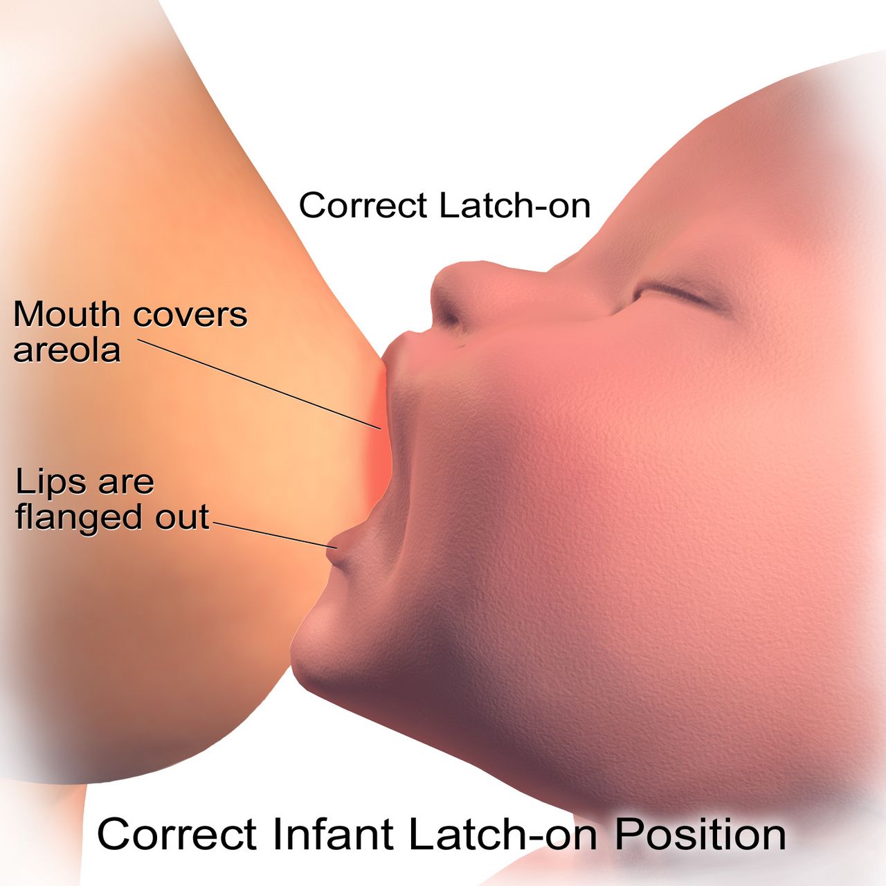
Anticipatory guidance and the well-child visit
Anticipatory guidance means teaching families about what is coming before it arrives, and it is the single most cost-effective thing done in paediatrics. Its structure follows development. You discuss rolling and falls from surfaces at the two-month visit, because rolling starts at four months and a parent who has already heard it will not leave the infant on the changing table. You discuss choking, poisoning and stair gates at the six-month visit, because crawling and the pincer grasp are about to arrive together. You discuss firearm storage, water safety and traffic before the summer, and you discuss consent, contraception and online life with adolescents before they need it rather than after.
Every well-child visit combines measurement and plotting, developmental surveillance, a physical examination, immunisations due, screening appropriate to age, and guidance. Developmental surveillance — asking about concerns and observing — happens at every visit; standardised developmental screening with a validated tool is generally performed at nine, eighteen and thirty months, with autism-specific screening at eighteen and twenty-four months. Any parental concern about development is itself a screening result and should trigger a formal tool.
Injury prevention matched to development
Injury is the leading cause of death after the first year of life in most countries, and every mechanism maps onto a developmental capability the child has just acquired. The infant who has learned to roll falls; the toddler who has learned to walk drowns and is poisoned; the school-age child who has learned to ride is struck by a vehicle; the adolescent who has learned to drive crashes. Prevention advice that is not matched to the child’s current stage is wasted breath.
Passenger safety follows a fixed progression. Rear-facing in the back seat for as long as possible — until the child outgrows the height or weight limit the seat’s manufacturer sets for rear-facing use, rather than turning at any particular age — then forward-facing with a five-point harness, then a belt-positioning booster until the adult belt fits correctly (typically at about 145 cm, often between eight and twelve years), then the adult belt, with the back seat used until about thirteen years. Harness straps sit at or below the shoulders when rear-facing and at or above the shoulders when forward-facing, with the chest clip at armpit level. Water safety means constant touch supervision for young children, four-sided isolation pool fencing with self-closing self-latching gates, emptying buckets and paddling pools, and swim lessons as a supplement to — never a substitute for — supervision. Poison prevention means locked storage above eye level, original containers, and the poison centre number in the phone. Where firearms are present in the home, storage advice is delivered without judgement: unloaded, locked, ammunition locked separately, and the question asked of any home where the child plays.
| Age band | Dominant risk mechanism | Priority guidance |
|---|---|---|
| Under 1 year | Sleep-related death, suffocation, falls, choking, scalds, motor-vehicle | Supine on a firm separate surface; never leave unattended on a raised surface; set water heater to about 49 °C; rear-facing car seat; small objects out of reach |
| 1–4 years | Drowning (leading unintentional cause in this band), poisoning, falls, burns, choking, driveway run-overs | Touch supervision near any water; four-sided pool fence; locked cupboards; turn pot handles inward; cut food safely; stair gates |
| 5–9 years | Pedestrian and cycle injury, motor-vehicle passenger, playground falls, burns | Correctly fitted helmet; road-crossing skills; booster seat until the belt fits; supervised play equipment; fire escape plan |
| 10–14 years | Sport injury, cycling, drowning, early substance exposure, self-harm | Protective equipment; concussion recognition and return-to-play rules; swimming competence; mood screening; secure medication and firearm storage |
| 15–18 years | Motor-vehicle crashes, substance use, self-harm and suicide, interpersonal violence | Graduated licensing, no phone and no passengers early on; seatbelt every trip; confidential screening for mood, substances and safety; means restriction counselling |
The same developmental logic runs in reverse, and it is the safeguarding question every paediatric nurse holds open: does this injury fit the child’s developmental stage and the history given? Bruising in a baby who is not yet cruising is a sentinel injury — pre-mobile infants do not bruise themselves. A spiral long-bone fracture in a child who cannot yet walk, a scald with a sharp stocking or glove line and no splash marks, bruising of the torso, ears or neck in a young child, a torn frenulum in an infant, unexplained delayed presentation, or a history that changes between tellings are all findings that do not match the mechanism claimed. Document exactly what you observed and what was said in the caregiver’s own words, body-map or photograph according to local policy, and do not interrogate the family or try to prove anything. Referral to the statutory child protection service is mandatory, is a personal duty that cannot be handed upward or talked out of, and requires only a reasonable suspicion — not certainty, not proof and not a diagnosis. Investigating is somebody else’s job; noticing and reporting is yours.
Screening across childhood
Screening is what converts a routine visit into detection of disease that has not yet declared itself. The newborn period carries the densest schedule: the blood-spot screen for metabolic, endocrine and haemoglobin disorders, universal hearing screening, and pulse oximetry screening for critical congenital heart disease at or after twenty-four hours of age. Beyond the newborn period, haemoglobin is checked at around twelve months for iron deficiency anaemia, lead screening is performed at twelve and twenty-four months where risk warrants, blood pressure is measured at least annually from three years and interpreted against age, sex and height percentiles rather than a single adult cut-off, and vision screening begins at about three years with instrument-based screening available earlier.
Adolescent screening is largely psychosocial and requires confidential time alone with the young person. A structured interview covering home, education and employment, eating, activities, drugs, sexuality, suicide and mood, and safety gives a reproducible framework. Annual screening for depression from about twelve years using a validated tool, screening for anxiety, screening for substance use, and sexual health screening according to risk are standard. Explain the limits of confidentiality up front — that you keep what is said private unless there is risk of serious harm to the young person or to someone else — because an adolescent who discovers the limits afterwards will never disclose again.
Partnering with families
Family-centred care is not a slogan; it is a set of behaviours. Parents are present for procedures if they wish to be and are given a role (holding a hand, comforting) rather than a task that makes them the person who restrains their child. Information is shared in the family’s preferred language through a professional interpreter, never through a sibling. Care plans are negotiated around what the family can actually do, and when a plan is not being followed the first question is what makes it hard — cost, work shifts, transport, literacy, competing caregiving — rather than a label. Health literacy is addressed by plain language, one to three key points per encounter, written material at a low reading level, demonstration of any device, and teach-back: “So I can be sure I explained it clearly, tell me how you will give this medicine at home.”
Injury prevention advice works only when it is delivered before the child acquires the capability that creates the risk, and it is delivered to a family whose actual constraints you have asked about.
Section 5Medication Safety, Fluids and Recognising the Deteriorating Child
Weight-based dosing: the arithmetic and the guard-rails
Paediatric doses are calculated from body weight in kilograms, and there is no such thing as a paediatric dose that can be estimated by eye. The sequence is always the same: obtain an accurate current weight in kilograms on a calibrated scale; identify the safe dose range in mg/kg/dose or mg/kg/day from an authoritative reference; multiply; divide by the number of doses per day if the reference is a daily dose; compare your answer with the maximum adult dose; and then convert the milligram dose into a volume using the concentration on the bottle in front of you.
Work an example. A child weighs 12 kg and paracetamol (acetaminophen) is ordered at 15 mg/kg per dose. Dose equals 15 mg/kg multiplied by 12 kg, which is 180 mg. The suspension is 160 mg per 5 mL, so the volume is 180 divided by 160, multiplied by 5 mL, which is 5.6 mL. Check the daily ceiling: the maximum is five doses in twenty-four hours and no more than 75 mg/kg/day, so 75 multiplied by 12 is 900 mg per day, and five doses of 180 mg is exactly 900 mg — at the ceiling, with no room for a sixth dose or for any paracetamol hidden in a combination cough preparation. Now scale the same order to a 75 kg sixteen-year-old: 15 mg/kg multiplied by 75 kg equals 1125 mg, which exceeds the usual adult single dose of 1000 mg. The dose given is 1000 mg. This is the guard-rail that must never be skipped: a paediatric dose is the smaller of the weight-based calculation and the maximum adult dose. Adolescents routinely reach adult weight, and a mg/kg formula applied without a ceiling is a straightforward route to hepatotoxicity.
Two more worked examples fix the pattern. Ibuprofen at 10 mg/kg per dose for the same 12 kg child is 120 mg; with a suspension of 100 mg per 5 mL, that is 6 mL, given no more often than every six to eight hours, with a daily cap of 40 mg/kg and an over-the-counter adult ceiling of 1200 mg per day. Ibuprofen is avoided under six months of age and in dehydration, because renal perfusion is already compromised. High-dose amoxicillin for acute otitis media at 90 mg/kg/day divided every twelve hours in an 18 kg child is 90 multiplied by 18, which equals 1620 mg per day, divided into two doses of 810 mg — checked against the usual adult daily maximum before it is given. And for anaphylaxis, intramuscular adrenaline (epinephrine) is 0.01 mg/kg of the 1 mg/mL concentration: for a 22 kg child that is 0.22 mg, which is 0.22 mL, injected into the vastus lateralis, with a maximum single paediatric dose of 0.3 mg. Auto-injectors are dispensed in fixed strengths — approximately 0.15 mg for children of roughly 7.5 to 25 kg and 0.3 mg at or above about 25 to 30 kg — so the device strength is chosen by weight band.
The systems guard-rails are as important as the arithmetic. Weigh in kilograms only and remove pounds from the record. Use a leading zero and never a trailing zero: 0.5 mg, never .5 mg; 5 mg, never 5.0 mg — a missed decimal point is the classic tenfold error. Write “unit” in full rather than “U”. Express liquid volumes in millilitres, never in cubic centimetres or household spoons. Use oral syringes for all oral liquids, and never a parenteral syringe for an oral dose. Obtain an independent double check by a second nurse — both calculating separately, not one reading aloud to the other — for high-alert medications including insulin, opioids, anticoagulants, chemotherapy, concentrated electrolytes and any infusion in a child. In an emergency, use a length-based resuscitation tape for weight estimation; if none is available, a serviceable estimate for children one to ten years is weight in kilograms equals age in years multiplied by two, plus eight. A stated or remembered weight is not acceptable for a calculated dose once the emergency is over.
Maintenance and replacement fluids: the 4-2-1 rule worked
Maintenance fluid replaces ongoing insensible and urinary losses in a child who cannot drink. The 4-2-1 rule gives an hourly rate directly: 4 mL/kg/h for the first 10 kg, plus 2 mL/kg/h for the next 10 kg, plus 1 mL/kg/h for every kilogram above 20 kg. Work a 26 kg child. First 10 kg at 4 mL/kg/h equals 40 mL/h. Next 10 kg at 2 mL/kg/h equals 20 mL/h. Remaining 6 kg at 1 mL/kg/h equals 6 mL/h. Total equals 40 plus 20 plus 6, which is 66 mL/h. The equivalent daily formula gives 100 mL/kg for the first 10 kg, 50 mL/kg for the next 10 kg and 20 mL/kg thereafter: 1000 plus 500 plus 120 equals 1620 mL/day, which divided by twenty-four is about 67.5 mL/h — the small difference between the two methods is expected and clinically unimportant. For an 8 kg infant, the whole calculation is 4 multiplied by 8, which is 32 mL/h — about 768 mL over twenty-four hours, or 800 mL/day by the daily formula (100 mL/kg multiplied by 8 kg).
Maintenance fluid in children is now given as an isotonic solution with appropriate dextrose and potassium once urine output is established, because hypotonic maintenance fluid caused hospital-acquired hyponatraemia and cerebral oedema. Resuscitation is a separate calculation: a fluid bolus for shock is 20 mL/kg of isotonic crystalloid given rapidly over five to twenty minutes and reassessed after each bolus — for a 14 kg child that is 280 mL. Smaller boluses of 10 mL/kg are used in neonates and in specific situations such as diabetic ketoacidosis, severe malnutrition and known cardiac disease, where rapid volume can do harm. All paediatric infusions run on a volumetric pump with the volume to be infused limited, and burette or syringe-pump systems are used for small volumes.
Dehydration is quantified by acute weight loss, which is the most sensitive available measure: a loss of 1 kg equals a deficit of approximately 1 litre. Mild dehydration is roughly three to five per cent of body weight, moderate six to nine per cent, and severe ten per cent or more, with slightly higher thresholds conventionally applied to infants. Clinical signs progress from mildly dry mucous membranes through decreased skin turgor, sunken eyes and fontanelle, tachycardia and reduced urine output to lethargy, mottling and cool extremities. Oral rehydration solution is first-line for mild to moderate dehydration: about 50 mL/kg over four hours for mild and 100 mL/kg over four hours for moderate, plus roughly 10 mL/kg replaced for each additional loose stool, given in small frequent volumes by syringe or spoon even if the child is vomiting. Expected urine output is about 2 mL/kg/h in infants and toddlers, 1 mL/kg/h in older children and 0.5 to 1 mL/kg/h in adolescents; falling below this is an early, quantifiable warning.
Vital signs by age and the physiology of compensated shock
There is no single normal paediatric vital sign range, and reading a child’s numbers against an adult range is one of the most reliable ways to miss deterioration. Heart rate and respiratory rate fall with age; blood pressure rises. Interpret every number against the child’s age band, their own baseline, and their state — a sleeping child’s heart rate differs materially from the same child screaming.
| Age band | Heart rate (awake, beats/min) | Respiratory rate (breaths/min) | Systolic BP (mmHg, approximate) |
|---|---|---|---|
| Neonate (0–1 month) | 100–180 | 30–60 | 60–90 |
| Infant (1–12 months) | 100–160 | 30–53 | 72–104 |
| Toddler (1–2 years) | 90–150 | 22–37 | 86–106 |
| Preschool (3–5 years) | 80–140 | 20–28 | 89–112 |
| School-age (6–12 years) | 70–120 | 18–25 | 97–115 |
| Adolescent (13–18 years) | 60–100 | 12–20 | 110–131 |
| Age | Systolic BP defining hypotension |
|---|---|
| Term neonate (0–28 days) | Below 60 mmHg |
| Infant 1–12 months | Below 70 mmHg |
| Child 1–10 years | Below 70 mmHg plus twice the age in years (a 4-year-old: below 78 mmHg) |
| Over 10 years | Below 90 mmHg |
The single most important paediatric physiological fact is this: children compensate for shock by increasing heart rate and systemic vascular resistance, and they do it extremely well. Tachycardia is the earliest sign of shock. Hypotension is a late and pre-terminal sign, often appearing only after about twenty-five to thirty per cent of circulating volume has been lost. A child with a normal blood pressure, a rising heart rate, capillary refill beyond two to three seconds, cool mottled peripheries, a narrowing pulse pressure, weak peripheral pulses with strong central pulses and reduced urine output is in compensated shock and needs intervention now. Waiting for the blood pressure to fall means waiting for cardiac arrest, and paediatric arrest is predominantly respiratory or hypovolaemic in origin, with outcomes far worse once it occurs than in the preceding hour.
Recognise, escalate, delegate
Structured recognition tools exist because intuition is unevenly distributed. The Paediatric Assessment Triangle is a doorway assessment requiring no equipment and under thirty seconds: Appearance (tone, interactivity, consolability, look or gaze, speech or cry), Work of breathing (abnormal sounds, positioning, retractions, nasal flaring, head bobbing) and Circulation to skin (pallor, mottling, cyanosis). Any abnormal side means the child is sick until proven otherwise. A Paediatric Early Warning Score then aggregates behaviour, cardiovascular and respiratory parameters into a number with a defined escalation threshold, and the score must be acted on, not merely documented. Age-appropriate pain assessment runs alongside: a neonatal scale for the newborn, the FLACC behavioural scale from about two months to seven years, the faces scale from about three years, and a numeric rating scale from about seven or eight years.
Escalation is communicated with SBAR. For example: “Situation — this is the nurse on the children’s ward calling about Amir, eight months, 8 kg, admitted with gastroenteritis; I am concerned he is deteriorating. Background — admitted six hours ago, has had ongoing vomiting and eight loose stools, has taken minimal oral fluid. Assessment — heart rate has risen from 140 to 186, respiratory rate 60, blood pressure 74 systolic, capillary refill four seconds, mottled peripheries, sunken fontanelle, no wet nappy for six hours, PEWS has gone from 2 to 6; I believe he is in compensated shock. Recommendation — please come now; I have intravenous access, I would like to give a 20 mL/kg bolus, which is 160 mL of isotonic crystalloid, and I have checked a point-of-care glucose.” Name the concern explicitly, state the numbers and their trend, and make a specific request.
Delegation follows a firm boundary. The registered nurse retains assessment, teaching, evaluation of response, clinical judgement, the first dose of a medication, blood administration and the unstable child. Assistive personnel may take routine vital signs on a stable child, record intake and output, weigh, bathe, position, assist with feeding a stable child who is not at aspiration risk, and transport — and must be told exactly what to report back and how quickly. A licensed practical or vocational nurse may provide ongoing care for a stable child and, within scope, administer many medications and reinforce — but not initiate — teaching. If the child’s status is changing, the task returns to the registered nurse regardless of how routine it looks.
Finally, the “what do you do first” question. In a febrile infant twenty-eight days old or younger with a rectal temperature of 38.0 °C or above, the first action is not to give an antipyretic and observe; it is to escalate immediately for a full sepsis evaluation, because a neonate can look well and be bacteraemic, and empiric antibiotics are time-critical. In a child with respiratory distress, the first action is positioning and oxygen, not obtaining a full set of observations. In a child with suspected shock, the first action is to establish access and give a 20 mL/kg bolus, not to await a blood pressure trend. The correct first action is very often the one that treats physiology rather than the one that gathers more data.
Every paediatric dose is calculated per kilogram, written out, and capped at the maximum adult dose; every fluid rate comes from the 4-2-1 rule; and every vital sign is read against the child’s age band, because tachycardia is early and hypotension is nearly too late.
The chapter in one breath
Children grow head to toe and centre outward, in a fixed sequence at an individual pace, and the only meaningful measure of that growth is a trajectory plotted over time on the correct chart, corrected for prematurity where relevant — with sustained downward crossing of two major percentile lines defining growth faltering. Development follows recognisable milestones from the social smile at two months through the neat pincer grasp and first words at twelve months to two-word phrases at twenty-four months and copying a triangle at five years, and a small set of red flags — above all the loss of any previously acquired skill — demands prompt referral rather than reassurance. Erikson, Piaget, stage-typical play and stage-typical fears are practical instruments that dictate how you approach a toddler, a preschooler and an adolescent differently. Health promotion is a short list executed reliably: supine sleep, vitamin D and iron, complementary foods at six months, nothing containing honey or cow’s milk before twelve months, fluoride from the first tooth, immunisation on the national schedule, and injury-prevention advice matched to the capability the child is about to acquire. Safety rests on two calculations: every dose in mg/kg checked against the maximum adult dose, and every fluid rate from the 4-2-1 rule. And every vital sign is read against the child’s age band, because tachycardia is the first sign of shock and hypotension is the last.
Words worth knowing
| Term | What it means |
|---|---|
| Cephalocaudal | The head-to-toe direction of growth and motor control; head control precedes sitting, which precedes walking. |
| Proximodistal | The centre-outward direction of development; shoulder and arm control precede the fine pincer grasp. |
| Corrected age | Chronological age minus the weeks born before 40 weeks gestation; used for plotting growth and judging development in preterm infants, but never for timing immunisations. |
| Growth faltering | Sustained downward crossing of two or more major percentile lines, or weight-for-length below the fifth percentile; the non-blaming replacement for “failure to thrive”. |
| Anterior fontanelle | The diamond-shaped soft spot closing at 12–18 months; bulges with raised intracranial pressure, sinks with dehydration. |
| Object permanence | The understanding, acquired at about 8–9 months, that an object still exists when out of sight; its arrival explains separation anxiety. |
| Parallel play | The toddler pattern of playing alongside but not with other children; associative play follows in the preschool years. |
| Separation anxiety | Distress at parental absence peaking at about 10–18 months, progressing through protest, despair and detachment; detachment is often misread as settling. |
| Physiological anorexia | The normal fall in toddler appetite as growth velocity slows; managed by the division of feeding responsibility, not by pressure. |
| Thelarche | Breast budding, Tanner stage 2, and the first physical sign of puberty in girls; peak height velocity precedes menarche. |
| Tanner staging | Sexual maturity rating from stage 1 (prepubertal) to stage 5 (adult), based on breast and pubic hair development in girls and genital and pubic hair development in boys. |
| 4-2-1 rule | Hourly maintenance fluid: 4 mL/kg/h for the first 10 kg, 2 mL/kg/h for the next 10 kg, 1 mL/kg/h for each kilogram above 20 kg. |
| Fluid bolus | 20 mL/kg of isotonic crystalloid given over 5–20 minutes for shock, reassessed after each bolus; 10 mL/kg in neonates, diabetic ketoacidosis, severe malnutrition and known cardiac disease. |
| Maximum adult dose | The ceiling that a weight-based paediatric calculation must never exceed; the dose given is the smaller of the two figures. |
| Independent double check | Two clinicians calculating a high-alert dose separately and comparing answers, rather than one reading the calculation aloud to the other. |
| Compensated shock | Tachycardia, delayed capillary refill, cool mottled skin, narrowed pulse pressure and falling urine output with a still-normal blood pressure; requires immediate treatment. |
| Paediatric Assessment Triangle | A doorway assessment of appearance, work of breathing and circulation to skin, completed in under 30 seconds without equipment. |
| PEWS | Paediatric Early Warning Score; an aggregate of behavioural, cardiovascular and respiratory parameters with a defined escalation threshold. |
| FLACC scale | Behavioural pain tool scoring Face, Legs, Activity, Cry and Consolability from 0 to 10, used from about 2 months to 7 years. |
| Teach-back | Asking the family to explain or demonstrate the plan in their own words to confirm the teaching was clear, framed as a check on the teacher. |
Check yourself
PAEDIATRIC RESPIRATORY AND CARDIOVASCULAR CONDITIONS
A child in trouble looks well until the moment they do not. The paediatric airway is narrower, softer and more easily occluded than an adult’s; the paediatric heart compensates for lost stroke volume almost entirely by going faster; and the paediatric blood pressure holds a deceptively normal number until the compensation collapses. That is why the nursing skill in this chapter is not pattern-matching a diagnosis but reading a trajectory — work of breathing, perfusion, and mental status — and acting on the trend before the number moves. Everything here is age-banded and weight-based. A respiratory rate of 40 is normal in a two-month-old and alarming in a ten-year-old. A dose is never a number; it is a number per kilogram, checked against the maximum adult dose. This chapter builds the mechanism, then the assessment, then the management, then the complications, for the airway and the circulation of the growing child.
By the end of this chapter you can
- Explain how paediatric airway and myocardial anatomy produce rapid decompensation, and state normal respiratory rate, heart rate and systolic blood pressure ranges by age band.
- Calculate a weight-based drug dose and a 4-2-1 maintenance fluid rate, show the arithmetic, and verify the result against the maximum adult dose.
- Distinguish croup from epiglottitis and state the single first action for each, including what must never be done to a child with suspected epiglottitis.
- Prioritise nursing care in bronchiolitis, asthma exacerbation, community-acquired pneumonia and cystic fibrosis, including the therapies that evidence does not support.
- Classify congenital heart lesions by pulmonary blood flow and obstruction, and describe the role of prostaglandin E1 in a ductal-dependent lesion.
- Manage a hypercyanotic (tet) spell in correct sequence, beginning with the knee-chest position.
- Recognise Kawasaki disease, acute rheumatic fever and paediatric heart failure, and identify the complication each one threatens.
- Differentiate compensated from hypotensive shock, apply age-specific hypotension thresholds, and state when to begin compressions for bradycardia with poor perfusion.
- Escalate deterioration using SBAR and apply delegation limits — assessment, teaching, evaluation and the unstable child remain with the registered nurse.
Section 1The Paediatric Airway, the Paediatric Heart and What the Numbers Mean
Why children decompensate faster than adults
Resistance to airflow is inversely proportional to the fourth power of the radius, so as the radius falls the resistance climbs steeply. An infant trachea is roughly 4 mm across, a radius of 2 mm; one millimetre of circumferential oedema halves that radius, removes about three-quarters of the cross-sectional area, and multiplies resistance roughly sixteen-fold. The same millimetre in an adult trachea is barely noticeable. This single fact explains why viral swelling that gives an adult a hoarse voice gives a toddler stridor at rest, and why suctioning an obligate nose-breathing infant’s nares is a genuine respiratory intervention rather than comfort care.
Beyond the lumen, the mechanics work against the child. The infant chest wall is cartilaginous and compliant, so intercostal and subcostal retractions appear early and the chest paradoxically collapses inward as the diaphragm pulls down. The ribs sit horizontally, limiting bucket-handle expansion, so the only way to increase minute ventilation is to increase the rate. Infants have fewer type I fatigue-resistant diaphragmatic fibres, which is why a baby who has been breathing at 80 breaths per minute for three hours can stop — not because the disease worsened, but because the muscle tired. Oxygen consumption is roughly 6 to 8 mL/kg/min compared with 3 to 4 in an adult, and functional residual capacity is proportionally smaller, so desaturation after apnoea is measured in seconds.
The heart has its own constraint. The neonatal and infant myocardium contains less contractile tissue and more non-contractile mass, so stroke volume is relatively fixed. Cardiac output equals heart rate multiplied by stroke volume, and when a child cannot raise stroke volume, output is almost purely rate-dependent. Two consequences follow and both are testable at the bedside. First, tachycardia is the earliest and most faithful sign of trouble — hypovolaemia, fever, pain, hypoxia, anxiety — and it must never be dismissed as “just crying” without re-checking when the child is settled. Second, bradycardia in a child is an emergency, not a reassuring return to normal; it usually means profound hypoxia and it precedes arrest.
Vital signs by age band, and the values that define shock
There is no single paediatric normal. Comparing a child against an adult range is one of the commonest sources of missed deterioration, so the ranges below should be at hand every time you record a set of observations. Rates are for the awake, afebrile, uncrying child; sleeping rates run lower and every degree Celsius of fever adds roughly 10 beats per minute and a few breaths per minute.
| Age band | Respiratory rate (per min) | Heart rate, awake (bpm) | Systolic BP (mmHg) |
|---|---|---|---|
| Neonate (birth to 28 days) | 30–60 | 100–205 | 67–84 |
| Infant (1–12 months) | 30–53 | 100–180 | 72–104 |
| Toddler (1–2 years) | 22–37 | 98–140 | 86–106 |
| Preschool (3–5 years) | 20–28 | 80–120 | 89–112 |
| School age (6–11 years) | 18–25 | 75–118 | 97–115 |
| Adolescent (12–15 years) | 12–20 | 60–100 | 110–131 |
Blood pressure is the last thing to fail. A child in compensated shock has tachycardia, cool mottled extremities, capillary refill beyond 2 seconds, a narrowing pulse pressure, weak peripheral pulses with preserved central pulses, and falling urine output — with a blood pressure that reads normal. By the time the systolic pressure drops, roughly 30 to 40 per cent of circulating volume is already gone. Learn the hypotension thresholds as hard floors, not as goals.
| Age | Hypotension defined as systolic BP below |
|---|---|
| Term neonate, 0–28 days | 60 mmHg |
| Infant, 1–12 months | 70 mmHg |
| Child, 1–10 years | 70 mmHg plus twice the age in years (a 6-year-old: 70 + 12 = 82 mmHg) |
| Over 10 years | 90 mmHg |
Urine output is a cheap, continuous perfusion monitor: expect 1.5 to 2 mL/kg/hour in infants and toddlers, at least 1 mL/kg/hour in older children, and at least 0.5 mL/kg/hour in adolescents. Weigh nappies — 1 gram equals 1 millilitre.
Weight-based dosing and maintenance fluids: always show the arithmetic
Children are not small adults, and every paediatric medication error begins with a number that was not converted. Three habits prevent nearly all of them. Use the weight in kilograms only, measured today, never a pound figure the family recalls and never a weight from a previous admission. Express the order as milligrams per kilogram per dose or per day, and write out the multiplication so a second nurse can follow it. Then perform the step that is skipped most often: compare the calculated paediatric dose with the maximum adult dose. A heavy 14-year-old on a milligram-per-kilogram formula can generate a number no adult would ever receive; the safe dose is the lower of the two.
Worked example. A 14 kg toddler with moderate croup is ordered dexamethasone 0.6 mg/kg as a single dose. 0.6 mg × 14 kg = 8.4 mg. The usual maximum is 16 mg, so 8.4 mg is within limits and is given. Second example. An 18 kg child with community-acquired pneumonia is ordered high-dose amoxicillin 90 mg/kg/day divided every 12 hours. 90 mg × 18 kg = 1620 mg/day; divided by two doses = 810 mg every 12 hours. The daily maximum is 4 g, so this is safe. Third example. A 46 kg adolescent with severe asthma is ordered prednisolone 2 mg/kg/day. 2 mg × 46 kg = 92 mg — but the maximum is 60 mg/day, so 60 mg is given and the discrepancy is documented and communicated.
Maintenance intravenous fluid uses the 4-2-1 rule, the hourly form of the Holliday–Segar method: 4 mL/kg/hour for the first 10 kg, plus 2 mL/kg/hour for the second 10 kg, plus 1 mL/kg/hour for every kilogram above 20. Worked example for a 26 kg child: the first 10 kg gives 10 × 4 = 40 mL/h; the next 10 kg gives 10 × 2 = 20 mL/h; the remaining 6 kg gives 6 × 1 = 6 mL/h. Total = 40 + 20 + 6 = 66 mL/hour. For an 8 kg infant, all weight falls in the first band: 8 × 4 = 32 mL/hour. Maintenance fluid is an isotonic solution — typically 0.9% sodium chloride or lactated Ringer’s with dextrose and, once the child voids, added potassium — because hypotonic maintenance fluid in an acutely unwell child causes hyponatraemia. Maintenance is not resuscitation: a child in hypovolaemic or septic shock receives a bolus of 20 mL/kg of isotonic crystalloid over 5 to 20 minutes with reassessment after each bolus, reduced to 10 mL/kg for neonates and for a child with known cardiac disease, severe malnutrition or diabetic ketoacidosis.
Assessment sequence, red flags, escalation and delegation
Begin every paediatric encounter from the doorway with the three sides of the assessment triangle — appearance (tone, interactiveness, consolability, gaze, cry), work of breathing (position, retractions, nasal flaring, grunting, audible noise) and circulation to skin (pallor, mottling, cyanosis). This takes seconds, requires no equipment and identifies the sick child before any monitor does. Then move to the hands-on airway, breathing, circulation, disability and exposure survey. Grunting in an infant is expiration against a partially closed glottis to generate auto-PEEP; it is not fussing and it signals alveolar disease. A child who stops fighting the oxygen mask has usually not improved.
Red flags that require immediate escalation: stridor at rest; grunting; head bobbing; a silent chest in a child who was wheezing; a respiratory rate that falls toward normal while the work of breathing rises or mental status drops; oxygen saturation under 90 per cent on supplemental oxygen; apnoea, especially in an infant under two months; capillary refill beyond 3 seconds with cool mottled skin; heart rate under 60 with poor perfusion; a systolic pressure below the age threshold; cyanosis that does not improve with oxygen; and any parental statement that the child is not behaving like themselves. Escalate with SBAR: Situation — “Room 4, Maya, 8 months, 7.2 kg, RSV bronchiolitis, now having apnoeic pauses.” Background — “Admitted 6 hours ago, on 2 L nasal cannula, feeding poorly.” Assessment — “Two pauses over 20 seconds with desaturation to 82 per cent, heart rate dropped to 88, subcostal retractions and grunting.” Recommendation — “I need you to see her now; I am setting up high-flow and requesting PICU review.”
Delegation limits. The registered nurse retains the initial and ongoing assessment, all teaching, all evaluation of response to therapy, and care of the unstable child. Assistive personnel may take vital signs on a stable child, weigh nappies, record intake and output, obtain daily weights, position and bathe, and feed a stable infant who has an established feeding pattern — but they may not perform the first set of vitals on a newly admitted or deteriorating child, and they may not interpret what they measure. Licensed practical or vocational nurses may administer most oral, subcutaneous and intramuscular medications within their scope, perform tracheostomy and gastrostomy care per policy, and reinforce teaching the RN has already given. Titrating oxygen, giving intravenous push medications, administering blood, first-dose immunoglobulin infusions and any assessment of a child in respiratory distress or shock stay with the RN.
In children the compensating vital sign is the heart rate and the failing one is the blood pressure, so treat tachycardia with poor perfusion as shock long before the pressure falls — and treat bradycardia as pre-arrest.
Section 2Upper Airway Obstruction: Croup, Epiglottitis and Aspiration
Croup (laryngotracheobronchitis)
Croup is viral inflammation and oedema of the subglottic larynx and trachea, most often caused by parainfluenza virus type 1, and it peaks between 6 months and 3 years of age. The mechanism is the fourth-power law made visible: mucosal swelling at the cricoid ring, the narrowest fixed point in the paediatric airway, narrows the lumen so that inspiratory airflow becomes turbulent. The result is the classic triad of a barking, seal-like cough, inspiratory stridor and hoarseness, typically preceded by one to three days of coryza and worst at night and when the child is upset. Fever is usually low-grade, the child is drinking, and the cough is prominent.
Assessment is clinical and severity is graded by the presence of stridor and retractions at rest, using the Westley croup score in many units. Mild croup means an occasional barking cough with no stridor at rest and no retractions. Moderate croup means stridor at rest with mild to moderate retractions but the child remains interactive. Severe croup means stridor at rest with marked retractions, agitation or lethargy, and decreased air entry. A child who becomes quiet with diminishing stridor and rising drowsiness is not improving — reduced airflow makes less noise. Do not obtain radiographs to make the diagnosis, and do not upset the child for tests that will not change management; crying increases turbulent flow and worsens obstruction.
Management begins with keeping the child calm on a caregiver’s lap. Every severity receives corticosteroid: dexamethasone 0.6 mg/kg as a single oral, intramuscular or intravenous dose, usual maximum 16 mg, which reduces oedema over the following 2 to 4 hours and shortens the illness even in mild disease. For stridor at rest, add nebulised epinephrine — racemic epinephrine 2.25%, 0.05 mL/kg to a maximum 0.5 mL diluted in saline, or L-epinephrine 1 mg/mL at 0.5 mL/kg to a maximum 5 mL. Nebulised epinephrine works within 10 to 30 minutes by alpha-mediated mucosal vasoconstriction, but its effect wears off, so the child must be observed for 3 to 4 hours for rebound obstruction before discharge. Humidified air, once a ward ritual, has no demonstrated benefit. Complications are respiratory fatigue and, rarely, secondary bacterial tracheitis.
Epiglottitis and bacterial tracheitis
Epiglottitis is bacterial cellulitis of the supraglottic structures, historically caused by Haemophilus influenzae type b and now uncommon where Hib immunisation is routine — which means the children who develop it are disproportionately under-immunised, and the nurse who has never seen a case is the one most likely to meet one. Onset is measured in hours, not days. The child, typically 2 to 7 years old, appears toxic with a high fever, sits forward in the tripod position with the chin thrust out and the tongue protruding, and shows the four Ds: drooling, dysphagia, dysphonia (a muffled “hot potato” voice) and distressed inspiratory effort. There is characteristically no barking cough. Stridor here is a late and ominous finding.
What you do first is what you do not do. Never inspect the throat, never insert a tongue depressor or swab, never place the child supine, never start an intravenous line or draw blood, and never leave the child alone. Any of these may provoke laryngospasm and complete obstruction. Keep the child upright in the position they have chosen, on the caregiver’s lap, keep them nil by mouth, offer blow-by oxygen only if it does not distress them, and summon the most skilled airway team available — anaesthesia and otolaryngology — for controlled intubation in the operating theatre with equipment for emergency tracheostomy at hand. Cultures, intravenous access and intravenous antibiotics (a third-generation cephalosporin such as ceftriaxone) follow after the airway is secured. Bacterial tracheitis presents as croup that fails to respond to epinephrine, with high fever, a toxic appearance and thick purulent secretions; it is usually staphylococcal, requires antibiotics, and frequently requires intubation for suctioning.
| Feature | Croup | Epiglottitis |
|---|---|---|
| Onset | Gradual, 1–3 days after coryza | Abrupt, over hours |
| Typical age | 6 months–3 years | 2–7 years |
| Cough | Barking, seal-like, prominent | Absent |
| Voice | Hoarse | Muffled, minimal speech |
| Drooling | No | Yes — will not swallow |
| Fever | Low-grade | High, toxic appearance |
| Posture | Any; comfortable upright | Tripod, chin forward, refuses to lie down |
| Imaging (if ever obtained) | Steeple sign, AP neck | Thumb sign, lateral neck |
| First action | Calm the child; dexamethasone 0.6 mg/kg | Do not touch the throat; call the airway team |
Foreign body aspiration and obstructive sleep apnoea
Aspiration peaks between 1 and 3 years, when a child has molars enough to bite but not to grind and a habit of running with food in the mouth. High-risk items are round and compressible — whole grapes, hot dog rounds, nuts, popcorn, hard sweets, raw carrot coins — and small objects such as coins, button batteries and small magnets. Classic presentation is a sudden choking episode followed by coughing and unilateral wheeze or decreased breath sounds; a partially obstructing object may masquerade for weeks as “asthma that only responds a little”. If the child is coughing forcefully, speaking or crying, the obstruction is partial: encourage coughing and do not intervene. If the child cannot cough, speak or cry, act — for an infant under one year, five back blows followed by five chest thrusts, repeated; for a child over one year, abdominal thrusts. Blind finger sweeps are never performed. Definitive removal is by rigid bronchoscopy. A swallowed button battery is a separate emergency: lodged in the oesophagus it causes caustic injury within two hours and requires immediate imaging and endoscopic removal.
Obstructive sleep apnoea in children is usually caused by adenotonsillar hypertrophy rather than obesity, though obesity compounds it. Parents report snoring, witnessed pauses, mouth breathing, restless sleep and night sweats; the daytime picture in children is often hyperactivity, inattention and poor school performance rather than sleepiness. Untreated, chronic nocturnal hypoxaemia produces pulmonary vasoconstriction and, eventually, cor pulmonale. Adenotonsillectomy is the usual treatment. Post-tonsillectomy nursing focuses on the bleeding risk: watch for frequent swallowing, throat clearing, restlessness and a rising pulse — the earliest signs of haemorrhage in a child who is swallowing the blood. Position the child prone or side-lying to allow drainage, keep suction available but do not suction the oropharynx deeply, avoid straws, red or brown fluids, and coughing or throat clearing. Bleeding risk is highest in the first 24 hours and again at 7 to 10 days when the eschar separates.
Croup is treated by calming the child and giving dexamethasone 0.6 mg/kg; suspected epiglottitis is treated by keeping instruments, tongue depressors and supine positioning away from the child until a skilled airway team is present.
Section 3Lower Airway and Parenchymal Disease
Bronchiolitis and respiratory syncytial virus
Bronchiolitis is the leading cause of hospital admission in infants under one year. Respiratory syncytial virus causes most cases, with rhinovirus, human metapneumovirus and parainfluenza accounting for the rest. The virus infects and sloughs the ciliated epithelium of the terminal bronchioles; the resulting debris, oedema and mucus form plugs that obstruct airways of one to two millimetres. Because obstruction is patchy and greater on expiration, air trapping and hyperinflation dominate, producing wheeze, prolonged expiration, crackles and a barrel-shaped chest. The illness follows a predictable arc: two to three days of coryza and low-grade fever, then increasing cough and work of breathing, with days 3 to 5 the worst, and slow resolution over one to three weeks.
Assess feeding as a respiratory sign. An infant who cannot sustain a feed, who takes fewer than half of the usual volumes, or who has fewer than half the usual wet nappies, is telling you the work of breathing has exceeded what they can sustain. Note apnoea risk: infants under two months, those born preterm, and those with congenital heart or chronic lung disease may present with apnoea before significant wheeze. Nasal obstruction alone can cause meaningful distress in an obligate nose-breather.
Treatment is supportive and the evidence is unusually clear about what does not help. Nasal suctioning before feeds and sleep, small frequent feeds or nasogastric or intravenous hydration, upright positioning, and supplemental oxygen if saturations persistently fall below 90 per cent are the core. High-flow nasal cannula at 1 to 2 L/kg/min is used for escalating work of breathing. Routine bronchodilators, corticosteroids, nebulised hypertonic saline in the emergency setting, antibiotics and chest physiotherapy are not recommended, and continuous pulse oximetry in a stable improving infant leads to longer stays without better outcomes. Infection control is nursing’s largest contribution: RSV survives on surfaces for hours, so contact and droplet precautions, dedicated equipment, cohorting, and rigorous hand hygiene protect every other infant on the unit. Prevention for high-risk infants uses monoclonal antibody — palivizumab 15 mg/kg intramuscularly monthly through the season, or a single dose of nirsevimab (50 mg for infants under 5 kg, 100 mg for 5 kg and over). These are passive antibodies, not vaccines, and do not create lasting immunity.
Asthma in the child
Asthma is chronic airway inflammation with bronchial hyper-responsiveness producing reversible obstruction through three simultaneous mechanisms — bronchospasm, mucosal oedema and mucus plugging. Only the first responds to a bronchodilator within minutes; the other two are why corticosteroids are given early and why a child can look better on albuterol and still be deteriorating. Triggers include viral infection, exercise, cold air, tobacco smoke, allergens, and, in a subset of older children, aspirin and non-steroidal anti-inflammatory drugs.
Assessment combines the physical picture with objective numbers. Peak expiratory flow zones, calibrated to the child’s own personal best, run green at 80 per cent or above, yellow at 50 to 79 per cent (take the reliever, follow the action plan) and red below 50 per cent (reliever now and seek urgent care). Reading the blood gas requires care: early in an attack the tachypnoeic child blows off carbon dioxide, so a low PaCO2 with respiratory alkalosis is expected. A normalising or rising PaCO2 in a child still working hard signals impending respiratory failure, not improvement. So does a silent chest, an inability to speak in full sentences, altered mental status and a saturation under 90 per cent on oxygen.
Acute management: oxygen to keep saturations at or above 92 per cent, then short-acting beta-2 agonist — nebulised albuterol 2.5 mg for a child under 20 kg and 5 mg for 20 kg and over, or weight-based at 0.15 mg/kg/dose with a 2.5 mg minimum, given every 20 minutes for three doses or continuously in severe disease. A metered-dose inhaler with a valved spacer, four to eight puffs, is at least as effective as a nebuliser for mild-to-moderate exacerbations and delivers less systemic side-effect. Add ipratropium for the first hour in moderate-to-severe attacks, and systemic corticosteroid early — prednisolone 1 to 2 mg/kg/day, maximum 60 mg/day, for 3 to 5 days. In severe exacerbation add magnesium sulfate 25 to 50 mg/kg intravenously over 20 minutes, maximum 2 g, monitoring for hypotension. Expect tremor, tachycardia and hypokalaemia from beta agonists and reassure families that these are pharmacological, not a sign of worsening disease. Chronic control rests on inhaled corticosteroids, and teaching must cover spacer use, rinsing the mouth after inhaled steroid to prevent oral candidiasis, distinguishing controller from reliever, trigger control, and a written asthma action plan shared with the school.
Pneumonia
In children under five, most community-acquired pneumonia is viral. The commonest bacterial cause across childhood is Streptococcus pneumoniae; in children over five, Mycoplasma pneumoniae causes a more insidious “walking pneumonia” with headache, malaise and a persistent dry cough. Bacterial pneumonia typically brings abrupt high fever, a toxic appearance, focal crackles or bronchial breathing and focal consolidation; viral pneumonia brings a more gradual course with diffuse findings and often coexisting wheeze. Tachypnoea is the single most sensitive sign of pneumonia in a child — more sensitive than auscultation. Note that lower-lobe pneumonia in a child frequently presents as abdominal pain, and upper-lobe pneumonia as neck stiffness.
First-line outpatient therapy for presumed bacterial pneumonia in an immunised child is high-dose amoxicillin 90 mg/kg/day divided every 12 hours, maximum 4 g/day; a macrolide is added or substituted where atypical organisms are suspected. Nursing care centres on oxygenation, hydration, antipyretics dosed by weight (acetaminophen 10 to 15 mg/kg every 4 to 6 hours, maximum 75 mg/kg/day and never exceeding the 4 g adult ceiling; ibuprofen 10 mg/kg every 6 to 8 hours in children over 6 months), positioning with the affected side down or upright as tolerated, and encouraging deep breathing through play — bubbles, pinwheels, incentive spirometry as a game. Watch for the complications: parapneumonic effusion or empyema, presenting as persistent fever beyond 48 to 72 hours of appropriate antibiotics with worsening pleuritic pain and diminished breath sounds.
Cystic fibrosis
Cystic fibrosis is an autosomal recessive disorder of the CFTR gene on chromosome 7, most commonly the F508del variant. The defective chloride channel produces dehydrated, viscous secretions in every exocrine organ. In the lung this means impaired mucociliary clearance, chronic infection with Staphylococcus aureus and later Pseudomonas aeruginosa, bronchiectasis and progressive obstruction. In the pancreas, plugged ducts cause exocrine insufficiency with steatorrhoea, fat-soluble vitamin deficiency and poor weight gain despite a voracious appetite. Sweat glands cannot reabsorb chloride, producing the salty-tasting skin families often report first. Ten to twenty per cent of affected newborns present with meconium ileus.
Diagnosis begins with the newborn screening immunoreactive trypsinogen assay and is confirmed by the sweat chloride test: 60 mmol/L or above is diagnostic, 30 to 59 mmol/L is intermediate and requires genetic testing, and below 30 mmol/L is normal. Daily management is demanding and family-centred: airway clearance one to four times daily by high-frequency chest wall oscillation vest, positive expiratory pressure device or manual physiotherapy, performed before meals or at least an hour afterwards to avoid vomiting; inhaled dornase alfa and hypertonic saline; inhaled antibiotics for chronic Pseudomonas; and CFTR modulator therapy for eligible genotypes. Nutrition is treatment, not a side issue — a high-calorie, high-protein diet with unrestricted fat, added salt, supplemental fat-soluble vitamins A, D, E and K, and pancreatic enzyme replacement taken with every meal and every snack, never crushed or chewed, and given within 30 minutes of eating. Adolescents need explicit conversation about fertility (most males have congenital absence of the vas deferens), CF-related diabetes, transition to adult services, and the isolation rules that keep people with CF apart from one another to prevent cross-infection.
Bronchiolitis is managed by suctioning, hydration and oxygen — not bronchodilators or steroids — while asthma demands early systemic corticosteroid, because only bronchospasm reverses in minutes and oedema and mucus plugging do not.
Section 4Congenital Heart Disease: Shunts, Obstruction and Cyanosis
The circulation changes at birth, and some lesions depend on that change not finishing
Congenital heart disease affects roughly 8 to 10 of every 1000 live births. In fetal life the lungs are fluid-filled and high-resistance, so blood bypasses them through the foramen ovale and the ductus arteriosus. With the first breaths, pulmonary vascular resistance falls, systemic resistance rises when the placenta is removed, the foramen ovale functionally closes, and the ductus arteriosus constricts in response to rising oxygen and falling prostaglandin, usually functionally closing within 24 to 72 hours. This transition is why many left-to-right shunts are silent at birth and only declare themselves at four to eight weeks, once pulmonary resistance has fallen enough to let blood cross the defect. It is also why a ductal-dependent lesion produces a catastrophic collapse on day two or three of life, precisely when the family have been discharged home.
Screening reflects this timing. Pulse oximetry screening for critical congenital heart disease is performed at 24 to 48 hours of age, measuring the right hand (preductal) and either foot (postductal). The screen passes when both readings are 95 per cent or higher and the difference between them is 3 per cent or less. It fails immediately at any reading below 90 per cent. Readings of 90 to 94 per cent in both limbs, or a difference greater than 3 per cent, prompt repeat measurement in one hour, up to three times, before failing. A failed screen requires echocardiography, not reassurance. The hyperoxia test helps distinguish cause: if the PaO2 stays below about 100 mmHg after 10 minutes on 100 per cent oxygen, the cyanosis is likely cardiac rather than pulmonary, because oxygen cannot fix a right-to-left shunt.
When a lesion is ductal-dependent — that is, when either pulmonary or systemic blood flow requires the duct to stay open — the treatment is prostaglandin E1 (alprostadil) as a continuous infusion, 0.01 to 0.1 mcg/kg/min, usually started around 0.05 mcg/kg/min. The nursing implications are specific and urgent: PGE1 causes apnoea (be prepared to intubate and have the equipment at the bedside), hypotension, fever, flushing, jitteriness and bradycardia. It is given through a secure dedicated line, never interrupted, and never bolused. Oxygen in these babies is used with caution — in a duct-dependent single-ventricle circulation, high oxygen lowers pulmonary resistance and can steal flow away from the body.
Lesions with increased pulmonary blood flow
When a defect connects the higher-pressure left side to the lower-pressure right side, blood recirculates through the lungs. These children are acyanotic but volume-overloaded: they show tachypnoea, diaphoresis with feeding, poor weight gain, recurrent respiratory infections and, over years, pulmonary vascular disease. Ventricular septal defect is the commonest congenital lesion and produces a harsh holosystolic murmur at the left lower sternal border; small muscular defects frequently close spontaneously in the first year. Atrial septal defect gives a softer systolic ejection murmur at the second left intercostal space with a characteristic fixed, widely split second heart sound. Patent ductus arteriosus produces a continuous machinery murmur, bounding pulses and a widened pulse pressure; in preterm infants it is closed pharmacologically with indomethacin, ibuprofen or acetaminophen, and surgically or by catheter device if that fails. Atrioventricular septal defect is strongly associated with trisomy 21 and causes early heart failure.
The complication to understand is Eisenmenger syndrome. Years of high-flow, high-pressure pulmonary circulation remodel the pulmonary arterioles until pulmonary resistance exceeds systemic resistance and the shunt reverses. A previously pink child becomes cyanotic, and the lesion becomes inoperable. This is the reason a “simple” shunt is repaired on schedule rather than watched indefinitely.
Obstructive lesions
Coarctation of the aorta is narrowing of the aorta, typically near the ductal insertion. The examination finding that makes the diagnosis costs nothing: bounding pulses and higher blood pressure in the arms, with weak or absent femoral pulses and lower pressure in the legs. Any infant with a murmur, and any child with hypertension, needs four-extremity blood pressures and femoral pulse palpation. Older children may report leg fatigue, cool feet and headaches or epistaxis from upper-body hypertension. A severe coarctation is ductal-dependent for systemic flow and presents with shock as the duct closes. Aortic stenosis causes exertional chest pain, syncope and a systolic ejection murmur radiating to the neck, and it is one of the causes of sudden death in young athletes. Pulmonary stenosis produces a systolic ejection murmur at the upper left sternal border and, when severe, right ventricular hypertrophy and cyanosis through a stretched foramen ovale.
Cyanotic lesions and the hypercyanotic (tet) spell
Cyanotic disease means deoxygenated blood reaches the systemic circulation. The classic group is remembered as the five Ts — Tetralogy of Fallot, Transposition of the great arteries, Truncus arteriosus, Tricuspid atresia and Total anomalous pulmonary venous return — to which hypoplastic left heart syndrome is added. Central cyanosis becomes visible when roughly 5 g/dL of haemoglobin is desaturated, so a polycythaemic child looks blue earlier and an anaemic child may be dangerously hypoxaemic while still looking pink. Chronic hypoxaemia drives erythropoiesis, raising haematocrit and blood viscosity, which in turn raises the risk of thrombosis and stroke — which is why maintaining hydration in a cyanotic child is a genuine neurological safeguard. Clubbing appears after months of desaturation.
| Lesion | Physiology | Hallmark finding | Priority management |
|---|---|---|---|
| Ventricular septal defect | Left-to-right shunt | Harsh holosystolic murmur, left lower sternal border | Calorie support, diuretics; surgical patch if large |
| Atrial septal defect | Left-to-right shunt | Fixed split S2 | Device or surgical closure |
| Patent ductus arteriosus | Left-to-right shunt | Machinery murmur, bounding pulses, wide pulse pressure | Indomethacin or ibuprofen in preterm; ligation or device |
| Coarctation of the aorta | Systemic obstruction | Arm-to-leg BP gradient, weak femoral pulses | PGE1 if ductal-dependent; balloon or surgical repair |
| Tetralogy of Fallot | Right-to-left shunt | Tet spells, boot-shaped heart, single S2 | Knee-chest first in a spell; complete repair in infancy |
| Transposition of the great arteries | Parallel circulations | Profound cyanosis within hours of birth | PGE1, balloon atrial septostomy, arterial switch |
| Hypoplastic left heart | Ductal-dependent systemic flow | Grey, shocked infant as the duct closes | PGE1; staged Norwood, Glenn, Fontan |
A hypercyanotic or “tet” spell is an acute increase in right ventricular outflow obstruction plus a fall in systemic vascular resistance, so more deoxygenated blood crosses the ventricular septal defect into the aorta. It is triggered by crying, feeding, defaecation, a warm bath or waking, and it looks like sudden deepening cyanosis, hyperpnoea, irritability and, if unbroken, limpness, syncope and seizure. The sequence is fixed and the first step requires no equipment: place the infant in the knee-chest position (an older child squats spontaneously), which kinks the femoral arteries and raises systemic vascular resistance so blood is pushed forward into the pulmonary artery instead of across the defect. Then calm the child and comfort rather than stimulate — agitation feeds the spell. Then give 100 per cent oxygen by blow-by, then morphine 0.1 mg/kg intravenously, intramuscularly or subcutaneously to reduce hyperpnoea and relax the infundibulum, then an intravenous fluid bolus to increase preload, and finally phenylephrine or a beta-blocker per protocol. Oxygen alone will not break a spell, because the problem is not gas exchange but where the blood is going.
Classify every congenital lesion by whether pulmonary blood flow is increased, decreased or obstructed — and in a hypercyanotic spell, the knee-chest position comes before oxygen, because the problem is shunt direction, not gas exchange.
Section 5Acquired Cardiac Disease, Heart Failure, Shock and Arrest
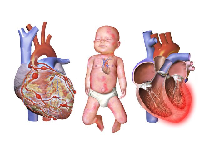
Kawasaki disease
Kawasaki disease is an acute self-limited vasculitis of medium-sized arteries, most common under five years of age, and it is the leading cause of acquired heart disease in children in high-income countries. The danger is not the rash but the coronary arteries: without treatment, 20 to 25 per cent of children develop coronary artery aneurysms; with timely treatment, that falls to about 3 to 5 per cent. Diagnosis is clinical — fever lasting 5 days or more plus at least four of five features: bilateral non-exudative conjunctival injection; oral mucosal changes (cracked red lips, strawberry tongue); polymorphous rash; extremity changes (erythema and firm oedema of the hands and feet acutely, then periungual desquamation in the subacute phase); and cervical lymphadenopathy, usually unilateral and greater than 1.5 cm. The fever is high, prolonged and characteristically unresponsive to antipyretics and antibiotics, and the child is exceptionally irritable — irritability out of proportion to everything else is the sign experienced nurses trust.
Treatment is intravenous immunoglobulin 2 g/kg as a single infusion over 10 to 12 hours, given ideally within 10 days of fever onset and preferably by day 7, together with aspirin — high dose during the acute febrile phase, then reduced to a single antiplatelet dose of 3 to 5 mg/kg/day once the child has been afebrile for 48 to 72 hours, continued 6 to 8 weeks pending echocardiography. Echocardiograms are performed at diagnosis, at one to two weeks and at four to six weeks. Nursing priorities during IVIG are cardiac monitoring, strict intake and output because the infusion is a substantial volume load, and vigilance for infusion reaction. Two teaching points are frequently missed: live vaccines (measles-mumps-rubella and varicella) must be deferred for 11 months after IVIG because passive antibody blunts the response; and while the child is on aspirin, the family should report influenza or varicella exposure and the child should receive annual influenza vaccination, given the theoretical Reye syndrome risk. Warn families that peeling of the fingers and toes and joint pain are expected in weeks two and three and are not a relapse.
Acute rheumatic fever, infective endocarditis and myocarditis
Acute rheumatic fever is an immune-mediated sequel of untreated group A streptococcal pharyngitis, appearing one to five weeks after the sore throat, most often between 5 and 15 years. Antibodies raised against streptococcal M protein cross-react with cardiac, joint, skin and neural tissue. Diagnosis uses the Jones criteria: major manifestations are carditis, migratory polyarthritis, Sydenham chorea, erythema marginatum and subcutaneous nodules; minor manifestations include fever, arthralgia, prolonged PR interval and raised inflammatory markers — with evidence of preceding streptococcal infection required. Carditis is the manifestation that matters long-term, because valvulitis (most often mitral) scars into rheumatic heart disease. Chorea may appear months later and presents as clumsiness, deteriorating handwriting and emotional lability, which is easily mistaken for behavioural change. Acute treatment eradicates the streptococcus and manages inflammation; the enduring nursing message is secondary prophylaxis — intramuscular benzathine penicillin G every four weeks (600,000 units for children under 27 kg, 1.2 million units for 27 kg and over) for years, because a second attack causes far worse valve damage. Prevention is simpler still: complete the full course of antibiotics for streptococcal pharyngitis even after the throat feels better.
Infective endocarditis should be suspected in a child with structural heart disease, a prosthetic valve or a central line who develops unexplained prolonged fever, a new or changed murmur, splenomegaly, or embolic phenomena. Blood cultures are drawn from separate sites before antibiotics. Antibiotic prophylaxis before dental procedures is now reserved for the highest-risk groups — prosthetic valves or material, previous endocarditis, unrepaired cyanotic disease, and repairs with residual defects — but meticulous daily oral hygiene is advised for every child with a cardiac lesion. Myocarditis, usually viral, presents deceptively as a “viral illness” with disproportionate tachycardia, exercise intolerance, chest pain and an enlarged heart on chest radiograph, and it can progress to dilated cardiomyopathy and arrhythmia.
Paediatric heart failure and its medications
Heart failure in children arises from volume overload (left-to-right shunts), pressure overload (obstructive lesions), or myocardial failure (myocarditis, cardiomyopathy, arrhythmia, sepsis, severe anaemia). The clinical picture divides usefully into three groups: impaired myocardial function — tachycardia, gallop rhythm, weak pulses, cool extremities, sweating, fatigue; pulmonary congestion — tachypnoea, wheeze, cough, retractions, cyanosis, exercise intolerance; and systemic venous congestion — hepatomegaly, periorbital and facial oedema (dependent ankle oedema is an older-child sign, since infants lie flat), weight gain and neck vein distension in older children.
Nursing management is a set of concrete daily actions: daily weight on the same scale, same time, minimal clothing — a gain of more than 0.5 kg in 24 hours in a child is fluid, not growth; strict intake and output; clustering care to protect rest; upright or semi-Fowler positioning; and feeding support as described earlier. Diuresis with furosemide 1 mg/kg/dose intravenously requires monitoring of potassium, since hypokalaemia potentiates digoxin toxicity. Afterload reduction with an ACE inhibitor requires blood pressure and renal function monitoring and teaching about the dry cough. Digoxin demands specific nursing discipline: maintenance is roughly 5 to 10 mcg/kg/day divided twice daily, the therapeutic serum range is 0.8 to 2 ng/mL, and the apical pulse is counted for a full minute before every dose. Hold the dose and notify the prescriber if the apical rate is below 90 to 110 in an infant, below 70 in a child, or below 60 in an adolescent. Early toxicity in children shows as vomiting, anorexia, bradycardia and arrhythmia — the visual disturbances taught for adults are unreliable in a preverbal child. Teach families to give doses one hour before or two hours after feeds, never to mix digoxin into a bottle of formula, never to repeat a dose the child vomits, to give water afterwards to protect the teeth, and to store the medication out of reach, since the paediatric elixir is potent in very small volumes.
Shock, supraventricular tachycardia and cardiac arrest
Shock is inadequate delivery of oxygen and substrate to meet tissue demand. In children the commonest cause worldwide is hypovolaemia from gastroenteritis, followed by distributive shock from sepsis or anaphylaxis, then cardiogenic and obstructive causes. Because a child compensates with tachycardia and intense vasoconstriction, the early picture is a fast heart, cool mottled peripheries, delayed capillary refill, a narrow pulse pressure and irritability progressing to lethargy — with a normal blood pressure. Warm shock in early sepsis is the exception: bounding pulses, flash capillary refill and a widened pulse pressure. Treatment is oxygen, vascular access within five minutes — intraosseous access is the correct choice when two peripheral attempts fail — isotonic fluid boluses of 20 mL/kg (10 mL/kg for neonates, cardiac disease, malnutrition or DKA) with reassessment for hepatomegaly, crackles and worsening work of breathing after each, antibiotics within one hour in septic shock, glucose checked and corrected early because children deplete glycogen fast, and vasoactive infusions if fluid alone does not restore perfusion.
Supraventricular tachycardia is the commonest symptomatic arrhythmia of childhood. Suspect it when the rate exceeds roughly 220 bpm in an infant or 180 bpm in a child, is unvarying beat-to-beat, does not change with crying or comfort, and has no visible P waves — whereas sinus tachycardia varies with activity and has an identifiable cause. Infants may present after hours of undetected SVT with pallor, poor feeding and heart failure. If the child is stable, attempt vagal manoeuvres first: for infants, a plastic bag of iced water applied to the forehead and bridge of the nose for 15 to 30 seconds; for older children, blowing through an obstructed straw or bearing down. Never apply ocular pressure and never perform carotid massage in a child. If vagal manoeuvres fail, give adenosine 0.1 mg/kg by rapid intravenous push, maximum 6 mg, immediately followed by a rapid saline flush through a proximal large-bore line using a two-syringe or stopcock technique — the half-life is under 10 seconds, so a slow push simply does not work. A second dose of 0.2 mg/kg, maximum 12 mg, may follow. Warn the child and family that a brief pause and an unpleasant flushing sensation are expected, and run a continuous rhythm strip. If the child is unstable — hypotensive, altered, poorly perfused — the answer is synchronised cardioversion at 0.5 to 1 J/kg, escalating to 2 J/kg, with sedation if it does not delay treatment.
Paediatric cardiac arrest is usually the end of a respiratory or circulatory failure, not a sudden electrical event, so the rhythm you will most often meet is bradycardia progressing to asystole or pulseless electrical activity rather than ventricular fibrillation. This changes the priority: oxygenate and ventilate first. If the heart rate is below 60 with signs of poor perfusion despite adequate oxygenation and ventilation, begin chest compressions. Compress at 100 to 120 per minute to a depth of one third of the anteroposterior chest diameter — about 4 cm in an infant, 5 cm in a child — with full recoil, at 30 compressions to 2 breaths for a single rescuer and 15 to 2 for two rescuers. With an advanced airway in place, give continuous compressions with one breath every 2 to 3 seconds. Epinephrine is 0.01 mg/kg (0.1 mL/kg of the 0.1 mg/mL concentration) intravenously or intraosseously every 3 to 5 minutes, maximum single dose 1 mg. Defibrillation for a shockable rhythm is 2 J/kg first, then 4 J/kg, then up to 10 J/kg or the adult dose. Use a length-based resuscitation tape for every weight-based decision when the weight is unknown, and support the family’s presence at the resuscitation with a dedicated staff member assigned to them — family presence is associated with better bereavement outcomes and does not impede the team.
Paediatric arrest is usually respiratory in origin, so the resuscitation priority is oxygenation and ventilation — and compressions begin at a heart rate under 60 with poor perfusion, not at zero.
The chapter in one breath
Children compensate brilliantly and then fail suddenly, and almost everything in this chapter follows from that. A narrow airway makes small amounts of oedema catastrophic; a compliant chest wall and a fatigue-prone diaphragm mean sustained tachypnoea ends in exhaustion; a relatively fixed stroke volume means the heart rate carries the whole burden of compensation, so tachycardia is the earliest alarm and bradycardia is pre-arrest. Assess from the doorway — appearance, work of breathing, circulation to skin — then confirm with age-banded numbers rather than adult ranges, and remember that blood pressure is the last value to fall. Every dose is per kilogram, written out, and checked against the maximum adult dose; maintenance fluid follows 4-2-1, resuscitation follows 20 mL/kg (10 mL/kg in neonates, cardiac disease, malnutrition and DKA). Learn the sequences that save lives: dexamethasone and calm in croup, hands off the throat in epiglottitis, suction and hydration rather than bronchodilators in bronchiolitis, early steroid in asthma, knee-chest before oxygen in a tet spell, immunoglobulin within ten days in Kawasaki disease, vagal manoeuvre before adenosine, and compressions at a heart rate under 60 with poor perfusion. Escalate with SBAR, keep assessment, teaching, evaluation and the unstable child with the registered nurse, and keep the family beside the bed.
Words worth knowing
| Term | What it means |
|---|---|
| Compensated shock | Inadequate tissue perfusion with a still-normal blood pressure, revealed by tachycardia, delayed capillary refill, narrow pulse pressure and falling urine output. |
| 4-2-1 rule | Hourly maintenance fluid: 4 mL/kg/h for the first 10 kg, 2 mL/kg/h for the next 10 kg, 1 mL/kg/h for each kilogram beyond 20. |
| Stridor | Harsh, usually inspiratory noise from upper airway narrowing; at rest it signifies significant obstruction. |
| Grunting | Expiration against a partly closed glottis to generate end-expiratory pressure — an infant sign of alveolar disease and impending failure. |
| Westley croup score | Severity scale for croup combining stridor, retractions, air entry, cyanosis and level of consciousness. |
| Steeple sign / thumb sign | Radiographic subglottic tapering of croup; radiographic swollen epiglottis of epiglottitis. |
| Tripod position | Sitting forward on the hands with chin thrust out to maximise airway diameter; classic in epiglottitis. |
| Bronchiolitis | Viral obstruction of terminal bronchioles in infants, most often RSV, treated with suction, hydration and oxygen. |
| Silent chest | Absence of wheeze in a severe asthma exacerbation, indicating airflow too poor to generate sound — an emergency. |
| Peak flow zones | Green 80 per cent or more, yellow 50–79 per cent, red under 50 per cent of the child’s personal best. |
| Sweat chloride test | Diagnostic test for cystic fibrosis; 60 mmol/L or above is diagnostic, 30–59 intermediate, under 30 normal. |
| Ductal-dependent lesion | A congenital defect in which pulmonary or systemic blood flow requires a patent ductus arteriosus, maintained with prostaglandin E1. |
| Prostaglandin E1 (alprostadil) | Infusion at 0.01–0.1 mcg/kg/min that keeps the ductus open; causes apnoea, hypotension and fever — keep intubation equipment ready. |
| Critical CHD pulse oximetry screen | Right hand and foot saturations at 24–48 hours; passes at 95 per cent or above in both with a difference of 3 per cent or less. |
| Hypercyanotic (tet) spell | Acute right-to-left shunting in tetralogy of Fallot; treat with knee-chest position first, then calm, oxygen, morphine and fluid. |
| Eisenmenger syndrome | Reversal of a left-to-right shunt once pulmonary vascular resistance exceeds systemic resistance, rendering the lesion inoperable. |
| Kawasaki disease | Medium-vessel vasculitis with fever of five days or more; treated with IVIG 2 g/kg within 10 days to protect the coronary arteries. |
| Jones criteria | Diagnostic framework for acute rheumatic fever, requiring evidence of preceding group A streptococcal infection. |
| Digoxin hold parameters | Count the apical pulse for a full minute; hold and notify if under 90–110 in infants, under 70 in children, under 60 in adolescents. |
| Intraosseous access | Rapid vascular route into the marrow, indicated when peripheral access fails within about five minutes in a shocked child. |
Check yourself
Paediatric Systemic and Chronic Conditions
A child is not a small adult, and nowhere is that truer than in the systemic and chronic conditions of childhood. A toddler carries a larger proportion of body water in the extracellular compartment, turns that water over faster, has immature renal concentrating ability, and can therefore lose a tenth of their body weight to a two-day gastroenteritis while an adult with the same illness merely feels unwell. A child compensates for shock with tachycardia and vasoconstriction and holds blood pressure nearly to the point of arrest, so hypotension in a child is a late and ominous finding. Every drug is weighed against kilograms, every fluid rate is calculated rather than ordered by habit, and every dose must still be checked against the maximum an adult would ever receive. This chapter moves system by system — gut, kidney and endocrine gland, blood and marrow, brain, bone and skin — and then returns to the two things that hold all of it together: the arithmetic, and the family.
By the end of this chapter you can
- State normal paediatric vital sign ranges by age band and calculate the age-specific threshold for hypotension, explaining why hypotension is a late sign of shock in children.
- Grade dehydration by percentage weight loss, select oral versus intravenous rehydration, and calculate an isotonic fluid bolus at 20 mL/kg.
- Calculate maintenance fluids using the 4-2-1 rule and verify the result against the Holliday-Segar daily method.
- Perform a weight-based safe-dose-range check, show the arithmetic, and compare the result with the maximum adult dose before administering.
- Prioritise nursing actions in diabetic ketoacidosis, vaso-occlusive crisis, acute chest syndrome, febrile neutropenia, status epilepticus and rising intracranial pressure, naming the first action in each.
- Differentiate nephrotic syndrome from acute glomerulonephritis and describe the monitoring that detects deterioration first.
- Recognise compartment syndrome, splenic sequestration and Hirschsprung enterocolitis as time-critical events and escalate using SBAR.
- Apply delegation boundaries in paediatrics, retaining assessment, teaching, evaluation and the unstable child within registered nurse practice.
- Partner with families in chronic illness using non-blaming, family-centred language and developmentally matched teaching.
Section 1Fluid Balance, Dehydration and the Gastrointestinal Tract
Why children destabilise faster
Water is distributed differently in a child. A newborn is roughly 75 to 80 percent water with about half of that in the extracellular compartment, compared with an adult who is around 60 percent water with only a third extracellular. Extracellular fluid is the fluid that is lost first in vomiting, diarrhoea, fever and burns, so the child has proportionally more of the vulnerable compartment and less of the protected intracellular reserve. Add a metabolic rate two to three times that of an adult, a body surface area to mass ratio that magnifies insensible loss, and renal tubules that cannot concentrate urine efficiently until around two years of age, and the picture is of an organism with a very small margin.
The compensation is cardiovascular and it is quiet. A child in early shock increases heart rate and clamps down peripherally. Stroke volume in an infant is relatively fixed, so cardiac output is almost entirely rate-dependent — which is why bradycardia in a sick infant is a pre-arrest rhythm, not a reassuring one. Blood pressure holds until roughly 25 to 30 percent of circulating volume is gone. The findings that appear before hypotension are the ones worth chasing: tachycardia at rest, capillary refill longer than two seconds, cool mottled extremities, weak peripheral pulses with a stronger central pulse, and a falling level of alertness or an unusual disinterest in the parent.
Urine output is the cheapest and most honest monitor available. Expect at least 2 mL/kg/h in an infant, 1 mL/kg/h in a child, and 0.5 to 1 mL/kg/h in an adolescent. Weigh nappies to measure rather than estimate: 1 gram of gained nappy weight equals 1 mL of urine. Weight itself is the single most sensitive index of acute fluid change, because 1 kg of acute weight loss equals 1 litre of fluid deficit. Weigh on the same scale, at the same time, in the same clothing or none, once daily — and in acute illness, twice.
| Age band | Heart rate (awake, per min) | Respiratory rate (per min) | Systolic BP (mmHg) | Hypotension if systolic below |
|---|---|---|---|---|
| Newborn (0–28 days) | 100–180 | 30–60 | 60–90 | 60 |
| Infant (1–12 months) | 100–160 | 30–53 | 70–100 | 70 |
| Toddler (1–2 years) | 90–150 | 22–37 | 80–110 | 70 + (2 × age in years) |
| Preschool (3–5 years) | 80–140 | 20–28 | 80–110 | 70 + (2 × age in years) |
| School age (6–11 years) | 70–120 | 18–25 | 90–120 | 70 + (2 × age in years) through age 10; below 90 from age 10 onward |
| Adolescent (12–18 years) | 60–100 | 12–20 | 100–130 | 90 |
Grading dehydration and rehydrating
Grade dehydration by percentage of body weight lost, using a recent pre-illness weight when one exists: percent deficit equals pre-illness weight minus current weight, divided by pre-illness weight, times 100. When no recent weight exists, grade clinically. Mild dehydration is under 5 percent, moderate is 5 to 10 percent, and severe is over 10 percent. In a 10 kg toddler, 10 percent is 1 kg — one litre — and that child looks profoundly unwell.
| Finding | Mild (under 5%) | Moderate (5–10%) | Severe (over 10%) |
|---|---|---|---|
| Mental state | Alert, thirsty | Irritable or listless | Lethargic, obtunded |
| Heart rate | Normal | Tachycardic | Marked tachycardia, weak pulses |
| Capillary refill | Under 2 s | 2–3 s | Over 3 s, mottled |
| Mucous membranes | Slightly dry | Dry | Parched, no tears |
| Anterior fontanelle (infant) | Flat | Slightly sunken | Markedly sunken |
| Urine output | Slightly reduced | Under 1 mL/kg/h | Minimal or anuric |
| First-line therapy | ORS 50 mL/kg over 4 h | ORS 100 mL/kg over 4 h | IV isotonic bolus 20 mL/kg |
Oral rehydration solution is the treatment for mild and moderate dehydration in a child who is alert and able to drink, including a child who is vomiting. The technique matters more than the volume ordered: 5 mL by syringe or spoon every one to two minutes, escalating as tolerated, gives roughly 150 to 300 mL in an hour. Replace ongoing losses with an extra 10 mL/kg for each diarrhoeal stool and about 2 mL/kg for each vomit. Continue breastfeeding or chestfeeding throughout, and reintroduce the usual diet within four to six hours — there is no evidence for prolonged clear-liquid diets, and early refeeding shortens illness. Plain water, fruit juice, sports drinks and carbonated drinks are the wrong osmolality and can worsen diarrhoea and drop sodium.
Severe dehydration is intravenous. The first action is to establish vascular access — peripheral intravenous, and if two attempts or 90 seconds fail in a decompensated child, intraosseous — and give an isotonic bolus of 0.9 percent sodium chloride or lactated Ringer solution at 20 mL/kg over 5 to 20 minutes. Reassess after every bolus: heart rate, perfusion, mental state, urine output. Repeat to a total of 40 to 60 mL/kg, and if perfusion has not improved by then, the problem is not simple hypovolaemia. Bolus volumes are reduced to 10 mL/kg in neonates, in diabetic ketoacidosis, in severe malnutrition and in known cardiac disease. Dextrose-containing and hypotonic fluids are never used for a resuscitation bolus.
Obstruction and the acute abdomen
Hypertrophic pyloric stenosis presents between two and eight weeks of age, most often in a firstborn male infant, with forceful non-bilious vomiting that becomes projectile, an infant who is ravenously hungry immediately afterwards, visible left-to-right peristaltic waves, and sometimes a firm olive-shaped mass in the right upper quadrant. Persistent loss of gastric hydrochloric acid produces the classic hypochloraemic, hypokalaemic metabolic alkalosis. The single most important teaching point is one of sequence: this is a medical emergency before it is a surgical one. The infant is kept nil by mouth with a nasogastric tube to low intermittent suction, and fluid and electrolyte derangement is corrected before pyloromyotomy, because operating on an alkalotic hypokalaemic infant invites post-operative apnoea. Feeds resume in small volumes four to six hours after surgery and some vomiting for the first day or two is expected.
Intussusception occurs when one segment of bowel telescopes into another, most often at the ileocaecal valve, in children between three months and three years with a peak around five to ten months. The child screams and draws the knees to the chest in sudden colicky episodes lasting a few minutes, then between episodes may appear entirely well or unusually lethargic. A sausage-shaped mass may be palpable in the right upper quadrant. Stool mixed with blood and mucus — the “currant jelly” stool — is a late finding and its absence proves nothing. Air or hydrostatic contrast enema under fluoroscopy is both diagnostic and therapeutic in most cases. After reduction, the passage of a normal brown stool suggests the bowel has reduced and must be reported to the provider immediately, because it changes the plan.
Appendicitis in a child begins as periumbilical pain that migrates to McBurney point in the right lower quadrant, with anorexia, low-grade fever, guarding and rebound tenderness. A sudden and complete relief of pain is not improvement — it is perforation, and it will be followed within hours by rigidity, rising fever and shock. Never apply heat to the abdomen and never give an enema or laxative for undiagnosed abdominal pain in a child. Hirschsprung disease presents with failure to pass meconium within 48 hours of birth, abdominal distension and ribbon-like foul-smelling stools; its emergency is enterocolitis — fever, explosive foul diarrhoea and a tense distended abdomen — which is life-threatening and demands immediate escalation.
Chronic conditions of the gut
Gastro-oesophageal reflux is physiological in most infants and resolves as the lower oesophageal sphincter matures by around 12 months. Management is conservative: smaller more frequent feeds, thorough burping, holding the infant upright for 20 to 30 minutes after feeds, and avoiding seat positions that flex the abdomen. Infants still sleep supine — reflux does not override safe-sleep guidance. Escalate the infant who is not gaining weight, who has haematemesis, apnoea, arching with feeds or recurrent respiratory illness, because that pattern is disease rather than physiology.
Cleft lip and cleft palate make feeding, not appearance, the first nursing priority. Use a wide-based or one-way-valve cleft bottle, hold the infant upright at 45 to 90 degrees, direct the nipple toward the intact side of the palate, and burp frequently because these infants swallow a great deal of air. Lip repair is usually undertaken around two to three months and palate repair by six to twelve months so that speech develops over a closed palate. After repair, protect the suture line absolutely: no straws, no pacifiers, no spoons or hard-tipped toys in the mouth, elbow immobilisers with scheduled removal one arm at a time for range of motion, and positioning supine or side-lying rather than prone after a lip repair.
Coeliac disease requires strict lifelong exclusion of gluten from wheat, barley and rye; rice, corn, quinoa and potato are safe, and oats are permitted provided they are certified gluten-free, since the problem with oats is contamination during growing and milling rather than gluten itself — a small minority who remain symptomatic on oats are reviewed individually. Untreated, it produces steatorrhoea, abdominal distension, wasted buttocks and growth faltering. The family needs label-reading skills, a plan for school meals and birthday parties, and the reassurance that a single exposure is a setback rather than a failure.
Grade a child’s dehydration by percentage weight lost, treat mild and moderate loss orally with 50 to 100 mL/kg of oral rehydration solution over four hours, and reserve the 20 mL/kg isotonic bolus for the child who is severely dehydrated or poorly perfused — remembering that hypotension in a child is a late sign, never an early one.
Section 2Renal, Genitourinary and Endocrine Conditions
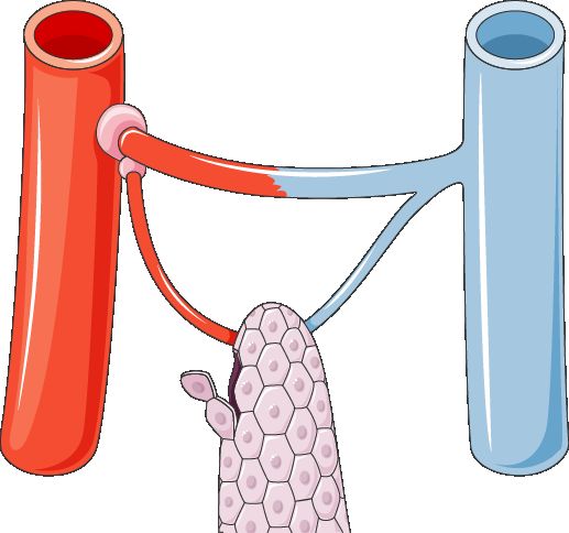
Urinary tract infection and vesicoureteral reflux
An infant with a urinary tract infection rarely complains of dysuria. The presentation is systemic and vague: fever without an obvious source, irritability, poor feeding, vomiting, foul-smelling urine, poor weight gain, and in the neonate, jaundice or hypothermia. Any infant under three months with a fever of 38.0 °C or higher needs a full evaluation that includes urine. In toilet-trained children the picture becomes recognisable — urgency, frequency, dysuria, new daytime wetting, suprapubic or flank pain.
Specimen technique determines whether the result means anything. In a child who is not toilet trained, a culture must come from a catheterised or suprapubic specimen; bag specimens are contaminated so frequently that a positive culture cannot be interpreted, though a negative bag specimen is useful for ruling out. Treat with the prescribed antibiotic for the full course, push fluids, and teach front-to-back wiping, cotton underwear, avoidance of bubble baths, complete and unhurried voiding roughly every two to three hours, and treatment of constipation, which is one of the most under-recognised drivers of recurrent infection.
Vesicoureteral reflux, graded I through V, allows infected bladder urine to reach the kidney and scar it. Lower grades often resolve with growth on surveillance with or without prophylactic antibiotics; higher grades may need surgical reimplantation. The nursing message to families is that fever in a child with reflux is not a wait-and-see event.
Nephrotic syndrome and acute glomerulonephritis
These two look superficially similar — a swollen child with abnormal urine — and they are managed almost oppositely, so the distinction is worth holding clearly.
| Nephrotic syndrome (minimal change) | Acute post-streptococcal glomerulonephritis | |
|---|---|---|
| Typical age | 2–6 years | 5–12 years |
| Trigger | Often none identified; may follow viral illness | Group A streptococcal pharyngitis 1–2 weeks prior, or impetigo 3–6 weeks prior |
| Proteinuria | Massive (over 50 mg/kg/day; urine protein-to-creatinine ratio above 2) | Mild to moderate |
| Haematuria | Usually absent | Gross — tea- or cola-coloured urine |
| Blood pressure | Normal or low | Elevated — hypertension is the hallmark |
| Serum albumin | Low (under 2.5 g/dL) | Normal or mildly reduced |
| Oedema | Periorbital on waking, becoming generalised with ascites | Periorbital and mild peripheral |
| Key labs | Hyperlipidaemia, hypoalbuminaemia | Elevated antistreptolysin-O titre, low serum C3, raised urea and creatinine |
| Mainstay of therapy | Corticosteroids: prednisone 2 mg/kg/day, maximum 60 mg/day, 4–6 weeks then taper | Supportive: blood pressure control, sodium and fluid restriction; antibiotics if infection persists |
| Watch for | Infection, especially peritonitis; thrombosis; hypovolaemia during diuresis | Hypertensive encephalopathy, acute kidney injury, circulatory congestion |
For both, the daily weight taken at the same time on the same scale is the most informative single measurement on the chart — more sensitive than abdominal girth, more reliable than a visual impression of puffiness, and quicker to change than serum albumin. Strict intake and output, daily abdominal girth in nephrotic syndrome, urine dipstick for protein each morning, and blood pressure four-hourly in glomerulonephritis complete the picture. The corticosteroid-treated child is immunosuppressed: screen visitors, avoid crowds, delay live vaccines, and treat fever as urgent. Teach families that steroids cause hunger, mood swings, a rounded face and weight gain, that these are expected and reversible, and that the drug is never stopped abruptly.
Type 1 diabetes and diabetic ketoacidosis
Type 1 diabetes is autoimmune destruction of pancreatic beta cells, and in children it frequently announces itself as ketoacidosis. Diabetic ketoacidosis is defined by a blood glucose above 200 mg/dL (11.1 mmol/L), venous pH below 7.3 or serum bicarbonate below 15 mmol/L, and ketonaemia or ketonuria. The child is polyuric, polydipsic, losing weight, breathing deeply and rapidly in Kussmaul respirations with acetone on the breath, and vomiting with abdominal pain that is easily mistaken for gastroenteritis.
The order of treatment is the lesson. Fluids come first, insulin second. Give an isotonic bolus of 10 mL/kg over the first hour — deliberately gentler than the 20 mL/kg used in other forms of shock — and then begin a continuous regular insulin infusion at 0.05 to 0.1 units/kg/h one to two hours after fluids have started. No insulin bolus is given to a child in DKA. Serum potassium will appear normal or high while total body potassium is severely depleted, because acidosis drives potassium out of cells; as insulin drives it back in, potassium falls fast. Potassium is added to the infusion once the serum level is below 5.5 mmol/L and the child is passing urine. Dextrose is added when glucose falls to around 250 to 300 mg/dL so that the insulin infusion can continue to close the anion gap without producing hypoglycaemia; glucose should not fall faster than 50 to 100 mg/dL per hour.
Cerebral oedema is the complication that kills children in DKA, and it typically appears 4 to 12 hours into treatment, often just as the numbers are improving. The warning signs are headache, a rising blood pressure with a falling heart rate, recurrent vomiting, incontinence in a previously continent child, irritability, and any decline in level of consciousness. This is an immediate call to the provider and the rapid response team, with the head of bed elevated and hypertonic therapy prepared — mannitol 0.5 to 1 g/kg or 3 percent sodium chloride 3 to 5 mL/kg intravenously.
Day-to-day management is a family project. Rapid-acting insulin such as lispro or aspart begins working in 10 to 15 minutes, peaks at 1 to 2 hours and lasts 3 to 5 hours, so it is given with food in hand. Long-acting glargine or detemir has no pronounced peak and is never mixed in a syringe with another insulin; when regular and NPH are drawn together, draw clear before cloudy. Rotate injection sites within one anatomical region to keep absorption predictable. Hypoglycaemia below 70 mg/dL in a conscious child is treated with roughly 15 g of fast carbohydrate in an adolescent and 5 to 10 g in a young child — about 0.3 g/kg — then rechecked in 15 minutes and followed with protein and complex carbohydrate. For the unconscious child, give glucagon intramuscularly: 0.5 mg if under 25 kg and 1 mg if 25 kg or more, and turn the child on their side because glucagon commonly causes vomiting. The rule that families most often get wrong is the sick-day rule: insulin is never omitted during illness, even when the child is not eating, because illness raises counter-regulatory hormones and drives ketosis.
Congenital endocrine conditions
Congenital hypothyroidism is detected on the newborn blood-spot screen because the untreated infant looks deceptively easy — sleepy, quiet, constipated, jaundiced, a poor feeder with a large tongue and an open posterior fontanelle. Levothyroxine at approximately 10 to 15 mcg/kg/day must begin within the first two weeks of life to protect neurodevelopment. Teach the parent to crush the tablet and mix it with a small volume of breast milk, formula or water given by syringe, never in a full bottle that may not be finished, and to separate it from soy formula, iron and calcium, which impair absorption. Signs of over-replacement are tachycardia, irritability, sweating and poor weight gain.
Congenital adrenal hyperplasia, most commonly 21-hydroxylase deficiency, may present with atypical genitalia in a genetically female infant or, in the salt-wasting form, with a crisis in the second or third week of life: vomiting, dehydration, hyponatraemia, hyperkalaemia and shock. Lifelong hydrocortisone with fludrocortisone in salt-wasting disease is the treatment, and the vital family teaching is stress dosing — the dose is doubled or tripled during fever, vomiting or injury, and an emergency intramuscular hydrocortisone kit and medical identification travel with the child everywhere.
Nephrotic syndrome and glomerulonephritis are separated at the bedside by blood pressure and urine colour, and both are monitored best by the daily weight; in diabetic ketoacidosis the sequence is fluids before insulin, potassium before it falls, and cerebral oedema suspected the moment a treated child develops headache with bradycardia.
Section 3Blood, Cancer and Immune Conditions
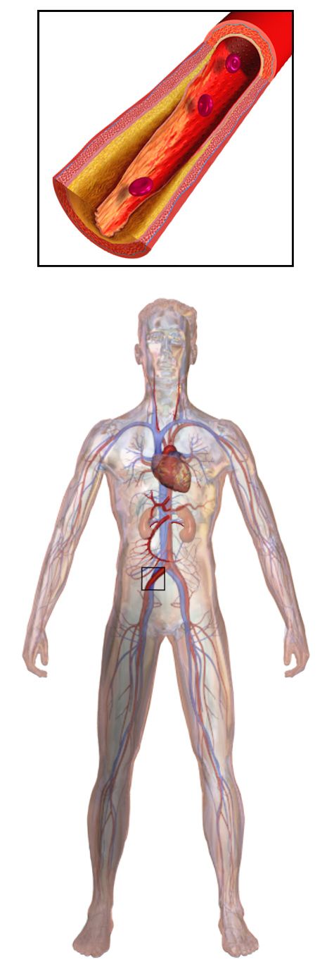
Sickle cell disease
Sickle cell disease is autosomal recessive. A single substitution of valine for glutamic acid at position six of the beta-globin chain produces haemoglobin S, which polymerises when deoxygenated and deforms the red cell into a rigid crescent. Those cells obstruct microvasculature and are cleared early, giving the two halves of the disease: vaso-occlusion and chronic haemolytic anaemia. Symptoms do not appear until fetal haemoglobin declines, usually after four to six months, often first as dactylitis — painful swelling of the hands and feet.
| Event | Recognition | Priority nursing action |
|---|---|---|
| Vaso-occlusive (pain) crisis | Severe deep bone, joint, back or abdominal pain; low-grade fever | Analgesia on a schedule plus intravenous fluids at 1.5 times maintenance; warmth to the area; oxygen only if oxygen saturation is below 95 percent |
| Acute chest syndrome | New pulmonary infiltrate with fever, chest pain, cough, hypoxia, tachypnoea | Leading cause of death — escalate immediately; oxygen, antibiotics, analgesia that permits deep breathing, incentive spirometry every 2 h while awake, possible transfusion |
| Splenic sequestration | Rapidly enlarging spleen, sudden fall in haemoglobin, pallor, hypovolaemic shock; peak risk under 5 years | Life-threatening — volume resuscitation and urgent transfusion; teach caregivers to palpate the spleen daily at home |
| Aplastic crisis | Profound anaemia after parvovirus B19 infection with a low reticulocyte count | Transfusion support; isolate from pregnant staff and visitors |
| Overwhelming infection | Fever in a functionally asplenic child | Fever of 38.5 °C or higher is an emergency — blood culture and parenteral antibiotics without waiting for other results |
| Silent or overt stroke | Focal weakness, aphasia, seizure, declining school performance | Annual transcranial Doppler screening from age 2 to 16; chronic transfusion if velocities are elevated |
Prevention is where nursing changes the trajectory. Penicillin V prophylaxis begins by two months of age at 125 mg orally twice daily and increases to 250 mg twice daily from age three until at least age five. Pneumococcal, meningococcal, Haemophilus influenzae type b and annual influenza immunisation are non-negotiable. Hydroxyurea raises fetal haemoglobin and markedly reduces crises, acute chest syndrome and transfusion need; it requires regular full blood counts because it suppresses the marrow, and it is teratogenic, which matters for adolescents. Families avoid dehydration, extremes of cold, high altitude, unpressurised flight and exhaustion. Meperidine is avoided for sickle pain because its metabolite normeperidine accumulates and lowers the seizure threshold.
Bleeding disorders and iron deficiency
Haemophilia A is an X-linked recessive deficiency of factor VIII, so it is expressed almost entirely in males and carried by females. It presents with prolonged bleeding after circumcision, easy bruising once the child begins to cruise, and haemarthrosis — joint bleeding that a young child reports as stiffness, warmth and a refusal to move the limb before pain is ever described. Treatment is factor replacement, given prophylactically in moderate and severe disease and on demand for bleeds; desmopressin can be used for mild haemophilia A. For an acute bleed, rest, ice, compression and elevation are applied while factor is prepared. Aspirin and non-steroidal anti-inflammatory drugs are avoided, intramuscular injections and rectal temperatures are avoided, and contact sports are replaced with swimming and cycling. The rule that saves lives: after a head injury, the child receives factor first and imaging second — treatment is never delayed for a scan.
Immune thrombocytopenia typically follows a viral illness by one to three weeks and presents in a well-looking child with petechiae, purpura and epistaxis. Most cases resolve spontaneously. Nursing care is protective — soft toothbrush, no rectal temperatures, no intramuscular injections, no contact sports, pressure to venepuncture sites for at least five minutes — with the greatest concern being intracranial haemorrhage when platelets fall below about 20,000/mm³.
Iron deficiency anaemia is the commonest nutritional deficiency of childhood. Risk peaks at 9 to 24 months and again in adolescence. The usual drivers are whole cow milk before 12 months, more than about 700 mL of cow milk daily in a toddler which displaces iron-rich foods and irritates the gut mucosa, and prolonged exclusive breastfeeding without iron supplementation after six months. Oral iron is prescribed as 3 to 6 mg/kg/day of elemental iron in one to three divided doses, given between meals with a source of vitamin C, through a straw or with a syringe toward the back of the mouth to limit dental staining, and the family warned that stools will turn black and tarry. Iron is a leading cause of fatal poisoning in children under six, so the bottle is stored locked and out of reach and the number of tablets in the house is kept small.
Childhood leukaemia and febrile neutropenia
Acute lymphoblastic leukaemia is the most common childhood malignancy, peaking between two and five years. The presentation follows directly from marrow crowding: pallor and fatigue from anaemia, bruising, petechiae and epistaxis from thrombocytopenia, and fever and recurrent infection from neutropenia, with bone and joint pain, lymphadenopathy and hepatosplenomegaly. Diagnosis is by bone marrow aspiration. Treatment runs through induction, consolidation and intensification, and a maintenance phase, totalling roughly two to three years, with intrathecal chemotherapy for central nervous system prophylaxis because systemic drugs do not cross the blood-brain barrier reliably.
Febrile neutropenia is the paediatric oncology emergency. Calculate the absolute neutrophil count: total white cell count multiplied by the sum of the percentage of segmented neutrophils and bands, divided by 100. A white count of 1,200/mm³ with 20 percent segs and 5 percent bands gives 1,200 × 0.25 = 300/mm³, which is severe neutropenia. A single temperature of 38.3 °C or a temperature of 38.0 °C sustained for an hour in a child with an absolute neutrophil count below 500 requires blood cultures and the first dose of broad-spectrum intravenous antibiotics within 60 minutes. The neutropenic child cannot mount classic signs — there may be no purulence, no infiltrate on film, no redness — so fever alone is the signal. No rectal temperatures, no suppositories, no live vaccines, no fresh flowers or standing water in the room, and meticulous hand hygiene by everyone entering.
Tumour lysis syndrome occurs as cells are destroyed early in treatment: potassium, phosphate and uric acid rise while calcium falls, threatening arrhythmia and acute kidney injury. Prevention is aggressive hydration with allopurinol or rasburicase, with close monitoring of electrolytes and urine output. Anticipatory guidance for families covers hair loss, mucositis, nausea, changed taste, and the fact that a child on chemotherapy who develops chickenpox exposure needs to be reported the same day.
Allergy and anaphylaxis
Anaphylaxis is a clinical diagnosis: acute onset of skin or mucosal involvement with either respiratory compromise or hypotension, or two or more organ systems involved after a likely allergen, or hypotension alone after a known allergen. The first action is intramuscular epinephrine into the anterolateral thigh — 0.01 mg/kg of the 1 mg/mL concentration to a maximum of 0.3 mg in a child and 0.5 mg in an adolescent or adult — repeated every 5 to 15 minutes as needed. Autoinjectors are supplied at 0.1 mg for the smallest infants where available, 0.15 mg for children roughly 7.5 to 25 kg, and 0.3 mg at 25 kg and above. Antihistamines relieve the rash and itch only, and corticosteroids do neither quickly; neither treats anaphylaxis and neither prevents a biphasic reaction, so they are adjuncts given after epinephrine and never instead of it — giving them first only delays the drug that works. Lay the child flat with legs elevated unless breathing is easier sitting, and never allow a child to stand or walk after epinephrine because sudden upright posture during hypovolaemia has caused arrest.
In sickle cell disease treat the crisis with hydration, scheduled analgesia and oxygen only when hypoxic, and treat fever and a rapidly enlarging spleen as emergencies; in oncology, fever with an absolute neutrophil count below 500 means cultures and antibiotics within 60 minutes; in anaphylaxis, intramuscular epinephrine comes before every other drug.
Section 4Neurologic, Musculoskeletal and Integumentary Conditions
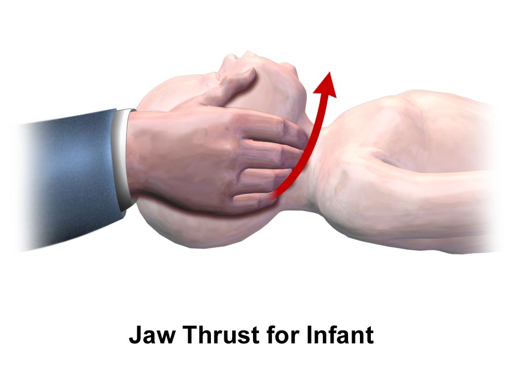
Seizures and status epilepticus
A simple febrile seizure occurs between six months and five years, is generalised, lasts under 15 minutes, and does not recur within 24 hours. It is frightening and usually benign. Nursing care during any seizure is the same: stay with the child, note the time, turn the child to the side, protect the head, loosen tight clothing, clear the surrounding area, and give oxygen if available. Nothing is ever put in the mouth, and the child is never restrained. Afterwards, document the onset, duration, movement pattern, eye deviation, incontinence, colour change and the length and character of the postictal period. Teach parents plainly that antipyretics reduce discomfort but have never been shown to prevent a recurrence, and that a child who has a febrile seizure is not thereby a child with epilepsy.
Status epilepticus is a seizure lasting five minutes or more, or repeated seizures without recovery of consciousness between them. Airway, oxygen, position, glucose check and timing come first, and then the drug ladder is followed on the clock.
| Time | Step | Drug and weight-based dose |
|---|---|---|
| 0–5 min | Stabilise | Airway positioning, oxygen, monitor, capillary glucose, intravenous access, note the time |
| 5–20 min | First-line benzodiazepine | Lorazepam 0.1 mg/kg IV, maximum 4 mg per dose; or midazolam 0.2 mg/kg IM, maximum 10 mg; or rectal diazepam 0.5 mg/kg (ages 2–5), 0.3 mg/kg (6–11), 0.2 mg/kg (12 and over), maximum 20 mg |
| 20–40 min | Second-line agent | Levetiracetam 60 mg/kg IV (maximum 4,500 mg); or fosphenytoin 20 mg phenytoin-equivalents/kg (maximum 1,500 mg PE); or valproate 40 mg/kg (maximum 3,000 mg) |
| Beyond 40 min | Refractory | Repeat second-line or begin anaesthetic infusion with intensive care involvement |
Anticipate respiratory depression after benzodiazepines and have bag-valve-mask and suction at the bedside before the drug is given. For a child on long-term anticonvulsants, teach that medication is never stopped abruptly, that adherence around illness and travel matters, and that helmets are worn for cycling and skating while unsupervised swimming and climbing at height are avoided.
Meningitis and rising intracranial pressure
Bacterial meningitis in a neonate is non-specific: temperature instability, poor feeding, lethargy, a weak or high-pitched cry, a bulging fontanelle and paradoxical irritability, meaning the infant cries more when picked up and comforted rather than less. Nuchal rigidity, the Kernig and Brudzinski signs, photophobia and severe headache belong to older children. Suspicion is enough to act. Droplet precautions begin immediately and continue for 24 hours after effective antibiotics start; blood cultures are drawn and antibiotics are given without waiting for lumbar puncture if the puncture will be delayed. Bacterial cerebrospinal fluid is cloudy with a high white count dominated by neutrophils, high protein and low glucose; viral fluid is clear with lymphocytes, normal glucose and mildly raised protein. Close household and childcare contacts of a child with meningococcal disease require chemoprophylaxis with rifampicin or ceftriaxone.
Rising intracranial pressure declares itself differently by age. In an infant expect a bulging tense fontanelle, separated sutures, an increasing head circumference, a high-pitched cry, irritability, poor feeding and the “setting sun” downward gaze. In an older child expect headache worst on waking, vomiting that may be effortless, diplopia, papilloedema, and a change in personality or school performance. The late signs are the Cushing triad — hypertension with a widening pulse pressure, bradycardia and irregular respiration — and by then herniation is imminent. Nursing management is to elevate the head of the bed about 30 degrees, keep the head midline and the neck unflexed, space nursing activities rather than clustering them so that pressure can return to baseline between interventions, keep the room quiet and dim, avoid suctioning, coughing, straining and hip flexion, and control fever and pain, all of which raise pressure.
A ventriculoperitoneal shunt fails in two ways: obstruction, which reproduces the signs of raised pressure, and infection, which presents with fever, redness along the tract, irritability and poor feeding within the first one to two months after placement. Post-operatively the child is usually positioned flat on the side opposite the valve to prevent too-rapid decompression, and head circumference is measured daily in infants.
Neural tube defects and cerebral palsy
Myelomeningocele is visible at birth as a sac over the spine containing meninges and neural tissue. Before surgical closure, usually within 24 to 72 hours, the infant is positioned prone or side-lying, the sac is covered with a sterile non-adherent dressing moistened with warm sterile normal saline and never allowed to dry, no nappy is placed over the sac, and the infant is handled with strict latex-free precautions from the first moment because these children develop latex allergy at very high rates with repeated exposure. Measure head circumference daily — hydrocephalus develops in the majority. Long-term care involves clean intermittent catheterisation, a bowel programme, and mobility support. Prevention belongs to the preconception period: 400 mcg of folic acid daily for all people who could become pregnant, and 4 mg daily for those with a previously affected pregnancy.
Cerebral palsy is a non-progressive disorder of movement and posture arising from an injury to the developing brain; the lesion does not worsen, though its musculoskeletal consequences do. Spastic forms predominate. Early clues include persistent primitive reflexes, hand preference before 12 months, scissoring of the legs, stiff or floppy tone, and delayed motor milestones. Nursing priorities are safety, communication and nutrition — these children aspirate readily, so upright positioning, thickened textures where indicated and unhurried feeding matter, and growth is monitored closely. Antispasmodics, botulinum toxin injection, intrathecal baclofen, orthoses and orthopaedic surgery all have a place. Speak to the child according to age, not according to motor ability; motor impairment is not intellectual impairment.
Bones, casts and skin
Children’s bones bend before they break, producing greenstick and buckle fractures, and their growth plates are weaker than the surrounding ligament, so a physeal injury occurs where an adult would sprain. Neurovascular assessment distal to any injury, cast or traction is the recurring nursing task: pain, pallor, pulselessness, paraesthesia and paralysis. The earliest and most reliable sign of compartment syndrome, however, is none of the classic five — it is pain out of proportion to the injury, unrelieved by opioids, and worsened by passive stretch of the digits. Report it immediately, loosen or bivalve the cast, and keep the limb at heart level rather than elevated, because elevation above the heart reduces arterial inflow to an already ischaemic compartment.
Some fractures are a safeguarding question. A fracture in a child who is not yet cruising or walking, posterior rib fractures, metaphyseal corner fractures, multiple fractures at different stages of healing, or an explanation that does not match the injury pattern all require escalation and documentation in objective, non-accusatory language. The same is true of a scald with a clean stocking-glove demarcation and no splash marks. The single most useful question is whether the injury fits the child’s developmental stage: an infant who cannot yet roll did not roll off the sofa, and a child too short to reach the tap did not run their own bath. Reporting is a mandatory legal duty triggered by reasonable suspicion, not by proof or certainty — the nurse reports and the investigating agency determines what happened, and no colleague, manager or provider can waive that duty. Keep the child safe, record the caregiver’s account verbatim and your own observations factually, and do not interrogate the family or announce the referral as an accusation.
Developmental dysplasia of the hip is screened with the Ortolani and Barlow manoeuvres in the first weeks of life, and later by asymmetric thigh and gluteal folds, limited hip abduction and a shortened femur on the affected side. The Pavlik harness is worn 23 hours a day for infants under about six months; parents are taught to check skin under the straps, dress the infant in a shirt and knee socks beneath the harness, and never to adjust the straps themselves. In adolescence, slipped capital femoral epiphysis presents in a heavier teenager as hip or referred knee pain with an outwardly rotated limb and limited internal rotation, and the immediate action is strict non-weight-bearing pending surgical fixation. Idiopathic scoliosis is screened with the Adams forward-bend test; bracing is typically considered for curves of 25 to 45 degrees in a growing child and surgical fusion above about 45 to 50 degrees, after which nursing care centres on log-rolling, neurovascular checks and effective analgesia.
Duchenne muscular dystrophy is X-linked, caused by absent dystrophin. Between three and five years the child shows a waddling gait, frequent falls, calf pseudohypertrophy and the Gowers sign of walking the hands up the thighs to rise. Creatine kinase is markedly elevated. Corticosteroids slow the decline; the eventual causes of death are cardiomyopathy and respiratory failure, so cardiac and pulmonary surveillance begin long before symptoms.
Atopic dermatitis is managed by soak-and-seal: a short lukewarm bath, a gentle pat dry, and emollient applied within three minutes, twice or more daily. Topical corticosteroid potency is matched to the site, with only the lowest potencies used on the face, neck and skin folds. Keep nails short, use cotton clothing, avoid fragranced products and overheating, and treat the itch, because scratching is what breaks skin and invites infection.
Treat a seizure lasting five minutes as status epilepticus and give a weight-based benzodiazepine with airway equipment ready; recognise the Cushing triad as a late sign of intracranial pressure rather than a first one; and remember that the earliest sign of compartment syndrome is pain out of proportion and on passive stretch, with the limb kept at heart level, not above it.
Section 5Weight-Based Safety and Living with Chronic Illness
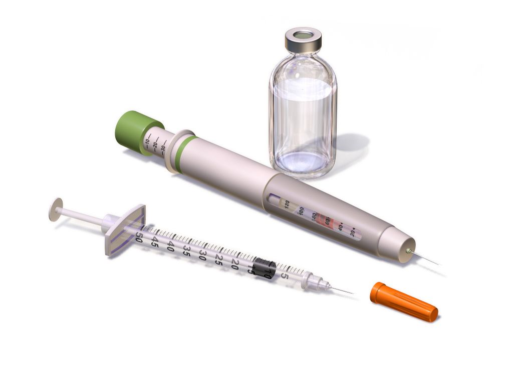
The arithmetic that keeps a child safe
Every paediatric medication order is a calculation, and the nurse is the last person who can catch an error before it reaches the child. Three habits prevent most harm. First, the weight is measured in kilograms on admission and recorded in kilograms only — never pounds, never a weight reported by a parent from memory, never an estimate. Second, every dose is checked against the published safe dose range for that drug per kilogram per dose and per day. Third, every paediatric dose is compared with the maximum adult dose, because a large adolescent multiplied by a per-kilogram figure can easily exceed what any adult would receive.
| Scenario | Arithmetic | Decision |
|---|---|---|
| Amoxicillin 90 mg/kg/day divided every 12 h for an 18 kg child | 90 × 18 = 1,620 mg/day; 1,620 ÷ 2 = 810 mg per dose. Suspension is 400 mg/5 mL, so 810 ÷ 400 × 5 = 10.1 mL | Safe; measure 10.1 mL in an oral syringe |
| Ibuprofen ordered as 400 mg for a 22 kg child | 400 ÷ 22 = 18.2 mg/kg per dose; safe range is 5–10 mg/kg per dose | Hold and clarify — the order is roughly double the upper limit |
| Acetaminophen 15 mg/kg for a 72 kg adolescent | 15 × 72 = 1,080 mg | Exceeds the 1,000 mg maximum single adult dose — give 1,000 mg. The adult ceiling always wins |
| Morphine 0.1 mg/kg IV for a 12 kg toddler | 0.1 × 12 = 1.2 mg | Safe; high-alert drug — independent double check by a second nurse before administration |
| Epinephrine 0.01 mg/kg IM for an 18 kg child in anaphylaxis | 0.01 × 18 = 0.18 mg of the 1 mg/mL solution | Give 0.18 mL, or use the 0.15 mg autoinjector supplied for 7.5–25 kg |
Maintenance fluid volume is calculated by the Holliday-Segar method, expressed hourly as the 4-2-1 rule: 4 mL/kg/h for the first 10 kg of body weight, 2 mL/kg/h for the second 10 kg, and 1 mL/kg/h for each kilogram above 20. Worked for a 26 kg child: the first 10 kg gives 10 × 4 = 40 mL/h; the second 10 kg gives 10 × 2 = 20 mL/h; the remaining 6 kg gives 6 × 1 = 6 mL/h; total 66 mL/h. The daily equivalent confirms it — 100 mL/kg for the first 10 kg is 1,000 mL, 50 mL/kg for the second 10 kg is 500 mL, and 20 mL/kg for the last 6 kg is 120 mL, giving 1,620 mL per day, or 67.5 mL/h. Maintenance fluid for children beyond the neonatal period should be isotonic — 0.9 percent sodium chloride or lactated Ringer solution, usually with 5 percent dextrose — because hypotonic maintenance fluid has repeatedly caused hospital-acquired hyponatraemia and seizures. Potassium is added only once the child is passing urine. Maintenance is added to, not substituted for, deficit replacement and ongoing losses.
Cystic fibrosis as the model chronic condition
Cystic fibrosis is autosomal recessive, caused by mutations in the CFTR chloride channel gene on chromosome 7, producing abnormally viscous secretions in the airway, pancreas, biliary tree, intestine and reproductive tract. It may declare itself at birth as meconium ileus, or later as recurrent respiratory infection, steatorrhoea, growth faltering despite a large appetite, and a salty taste to the skin. Diagnosis is by newborn screening confirmed with a sweat chloride above 60 mmol/L on two occasions, and by genotyping.
Daily care runs on sequence. Airway clearance — chest physiotherapy, high-frequency chest wall oscillation vest, positive expiratory pressure devices, autogenic drainage — is performed before meals rather than after, to avoid vomiting, and bronchodilators precede it while dornase alfa and hypertonic saline are timed to the clearance session. Pancreatic enzyme replacement is taken with every meal and every fat-containing snack, within about 30 minutes of eating, at 500 to 2,500 lipase units/kg per meal and never exceeding 10,000 lipase units/kg/day, a ceiling set because higher doses have been associated with fibrosing colonopathy. Capsules may be opened onto a small amount of acidic soft food such as apple sauce and must not be crushed or chewed. The diet is high in calorie and protein at roughly 110 to 200 percent of the usual requirement, with fat-soluble vitamins A, D, E and K supplemented and additional salt provided in heat and during exercise. CFTR modulator therapy has transformed outcomes for eligible genotypes.
Pain, development and the family as partner
Children feel pain fully and report it differently, so the tool must match the child. Use the FLACC behavioural scale for infants and children up to about three years and for any non-verbal child; the Wong-Baker FACES or Oucher scale from roughly three to seven years; and a 0 to 10 numeric scale from about age eight. Believe the score. Chronic under-treatment of paediatric pain is well documented and it is a nursing failure, not a physiological curiosity. Combine pharmacological management with sucrose and non-nutritive sucking in infants, distraction, guided imagery, warmth, positioning, and above all parental presence.
Development shapes every explanation. A toddler fears separation and needs a parent present, brief warning and a comfort object. A preschooler thinks magically and may believe illness is a punishment, so say plainly that nothing they did or thought caused this. A school-age child wants to know how the body works and to have a job to do — holding the tape, counting to three. An adolescent needs privacy, confidentiality within legal limits, honest information about how treatment affects appearance and social life, and progressive ownership of their own regimen with continuing adult oversight. Perform painful procedures in a treatment room rather than the bed, so the bed remains safe.
Language is clinical care. Describe what is happening rather than assigning blame: an infant is “taking small volumes and tiring at the breast”, not “a difficult feeder”; a family is “managing three medicines across two households and missing evening doses”, not “non-compliant”. That reframing is not politeness — it points at the actual problem, which is usually a solvable one of access, understanding, schedule or cost. Use the family’s words for themselves, whether that is mother, birthing parent, dad, carer or grandparent, and treat the person who does the daily work of the illness as a member of the team.
Escalation and delegation
Escalate with SBAR so that the urgency is unmistakable. Situation: “This is Aisha, the nurse on 4 East, calling about Leo, 4 years old, weight 16 kg, one hour into insulin infusion for DKA.” Background: “He presented with a glucose of 620 mg/dL and pH 7.08, he has had 160 mL of normal saline as a 10 mL/kg bolus, and the insulin infusion is running at 0.1 units/kg/h.” Assessment: “In the last 20 minutes his heart rate has fallen from 148 to 82, blood pressure has risen from 96/58 to 124/78, he is complaining of headache and he has vomited twice. I believe he is developing cerebral oedema.” Recommendation: “I need you at the bedside now, I have raised the head of the bed, I am requesting an order for hypertonic saline or mannitol, and I have activated the rapid response team.”
| Task | Assistive personnel | Licensed practical or vocational nurse | Registered nurse only |
|---|---|---|---|
| Vital signs on a stable child | Yes, reports values to the RN | Yes | Interprets and acts on them |
| Daily weight, intake and output, nappy weighing | Yes | Yes | Evaluates the trend |
| Feeding a stable child, hygiene, positioning, ambulation | Yes | Yes | — |
| Oral rehydration by an established plan in a stable child | May offer fluid and record volume | Yes | Sets and revises the plan |
| Routine oral and intramuscular medication, stable gastrostomy feeds, tracheostomy care on a stable child | No | Yes, within scope and policy | Retains high-alert drugs, titrations and intravenous push |
| Initial and ongoing assessment, neurovascular checks, pain reassessment after intervention | No | Contributes data | Yes |
| Patient and family teaching and evaluation of learning | No | Reinforces established teaching | Yes |
| Any care of an unstable or deteriorating child | No | No | Yes |
Weigh the child in kilograms, calculate every dose per kilogram, verify it against the safe range and then against the maximum adult dose, run maintenance fluid by the 4-2-1 rule with isotonic solution, and keep assessment, teaching, evaluation and the unstable child within registered nurse practice.
The chapter in one breath
Children compensate brilliantly and then fail suddenly. Their larger extracellular water compartment and faster turnover make dehydration the common final pathway of ordinary illness, so grade it by percentage weight lost, rehydrate orally at 50 to 100 mL/kg over four hours when the child can drink, and reserve the 20 mL/kg isotonic bolus for severe loss — remembering that tachycardia, delayed capillary refill and altered alertness arrive long before hypotension, which in a child aged one to ten is anything below 70 plus twice the age in years. Across systems the pattern repeats: mechanism, then assessment, then management, then the complication that kills. Fluids before insulin in ketoacidosis, with cerebral oedema suspected when a treated child develops headache and bradycardia. Hydration, scheduled analgesia and hypoxia-triggered oxygen for sickle crisis, with acute chest syndrome and splenic sequestration as the emergencies. Cultures and antibiotics within 60 minutes for fever with a neutrophil count under 500. A benzodiazepine at five minutes for status epilepticus. Pain on passive stretch as the earliest sign of compartment syndrome. Holding it all together is arithmetic — kilograms, safe dose ranges, the maximum adult dose that always wins, and the 4-2-1 rule — and a family whose daily work with the illness makes them colleagues rather than visitors.
Words worth knowing
| Term | What it means |
|---|---|
| 4-2-1 rule | Hourly maintenance fluid calculation: 4 mL/kg/h for the first 10 kg, 2 mL/kg/h for the second 10 kg, 1 mL/kg/h for every kilogram beyond 20 kg. |
| Absolute neutrophil count | Total white cell count multiplied by the combined percentage of segmented neutrophils and bands, divided by 100; below 500/mm³ is severe neutropenia. |
| Acute chest syndrome | New pulmonary infiltrate with fever, chest pain and hypoxia in sickle cell disease; the leading cause of death and an immediate escalation. |
| Compartment syndrome | Rising pressure within a fascial compartment causing ischaemia; earliest sign is pain out of proportion and on passive stretch, and the limb is kept at heart level. |
| Currant jelly stool | Blood and mucus passed in intussusception; a late finding whose absence does not exclude the diagnosis. |
| Cushing triad | Hypertension with widened pulse pressure, bradycardia and irregular respiration — a late and ominous sign of raised intracranial pressure. |
| Diabetic ketoacidosis | Glucose above 200 mg/dL with pH below 7.3 or bicarbonate below 15 mmol/L plus ketosis; treated with fluids first, then an insulin infusion without a bolus. |
| FLACC scale | Behavioural pain tool scoring face, legs, activity, cry and consolability, used for infants, toddlers and non-verbal children. |
| Gowers sign | Walking the hands up the thighs to rise from the floor, indicating proximal muscle weakness as in Duchenne muscular dystrophy. |
| Haemarthrosis | Bleeding into a joint, the characteristic bleed of haemophilia, first reported by a young child as stiffness and refusal to move the limb. |
| Holliday-Segar method | Daily maintenance fluid calculation: 100 mL/kg for the first 10 kg, 50 mL/kg for the second 10 kg, 20 mL/kg thereafter. |
| Hypochloraemic metabolic alkalosis | The acid-base picture of pyloric stenosis, produced by loss of gastric hydrochloric acid; corrected before surgery. |
| Nephrotic syndrome | Massive proteinuria, hypoalbuminaemia, oedema and hyperlipidaemia; treated with corticosteroids and monitored by daily weight. |
| Oral rehydration solution | Glucose-electrolyte solution at physiological osmolality that treats mild and moderate dehydration, given in small frequent volumes even when the child is vomiting. |
| Paradoxical irritability | An infant who cries more, not less, when picked up and comforted — a meningeal sign in infancy. |
| Pavlik harness | Abduction device for developmental dysplasia of the hip in infants under about six months, worn 23 hours daily and adjusted only by the clinician. |
| Splenic sequestration | Sudden pooling of blood in the spleen in sickle cell disease with rapid anaemia and hypovolaemic shock; caregivers are taught to palpate the spleen. |
| Status epilepticus | A seizure lasting five minutes or longer, or repeated seizures without recovery, treated with a weight-based benzodiazepine on the clock. |
| Tumour lysis syndrome | Rapid cell breakdown producing high potassium, phosphate and uric acid with low calcium; prevented by hydration and urate-lowering therapy. |
| Vaso-occlusive crisis | Painful episode from microvascular obstruction by sickled cells, managed with hydration, scheduled analgesia and oxygen only when hypoxic. |
Check yourself
The Child in Crisis: Emergencies, Safeguarding and the Family
Almost no child arrests without warning. The warning is usually written in the minutes before — a child who has stopped fighting the oxygen mask, a fontanelle that has sunk, a heart rate that has been climbing since the last set of observations, a bruise on an infant who cannot yet roll over. This chapter is about seeing that writing and acting on it in the right order, because in paediatric emergencies the correct first action is frequently not the intuitive one. You will learn to read a child across a room using the Paediatric Assessment Triangle, to grade dehydration by percentage and match it to fluid orders, to calculate every dose per kilogram and check it against an adult ceiling, to run the fixed sequences that save a child in anaphylaxis, status epilepticus, shock or ketoacidosis, and to recognise injuries that do not fit the story or the child’s developmental stage. Then you will learn to stay in the room with the family afterwards — through resuscitation, through bad news, and through the death of a baby or a child.
By the end of this chapter you can
- Apply the Paediatric Assessment Triangle and its TICLS appearance cues to name a child’s physiological state within sixty seconds of entering the room.
- State normal heart rate, respiratory rate and the hypotension threshold for each paediatric age band, and use the formula systolic BP less than 70 + (2 × age in years) for children 1 to 10 years.
- Distinguish compensated from decompensated shock and explain why hypotension and bradycardia are late, ominous findings in a child, and state the compression rate, depth, ratios and defibrillation energies used in paediatric life support.
- Grade dehydration as mild, moderate or severe by percentage weight loss, match each grade to its clinical signs, and calculate the fluid deficit in millilitres per kilogram.
- Calculate maintenance fluids using the 4–2–1 rule and calculate weight-based doses showing the arithmetic, always checking the result against the maximum adult dose.
- Perform the correct first action in anaphylaxis, status epilepticus, paediatric septic shock, diabetic ketoacidosis, poisoning and foreign body airway obstruction, and name the drug, manoeuvre or delay that is contraindicated in each.
- Identify injuries inconsistent with a child’s developmental stage using TEN-4-FACESp and the sentinel injury concept, and describe the nurse’s personal mandatory-reporting duty and documentation standard.
- Escalate deterioration using SBAR, and apply delegation limits that keep assessment, teaching, evaluation and the unstable patient with the registered nurse.
- Support a family through resuscitation, bad news and perinatal or paediatric loss using family-centred, non-blaming language and developmentally appropriate explanations of death, and explain the investigation that follows a sudden unexpected infant death, including the ABCs of safe sleep the investigation asks about.
Section 1The First Sixty Seconds: Reading the Deteriorating Child
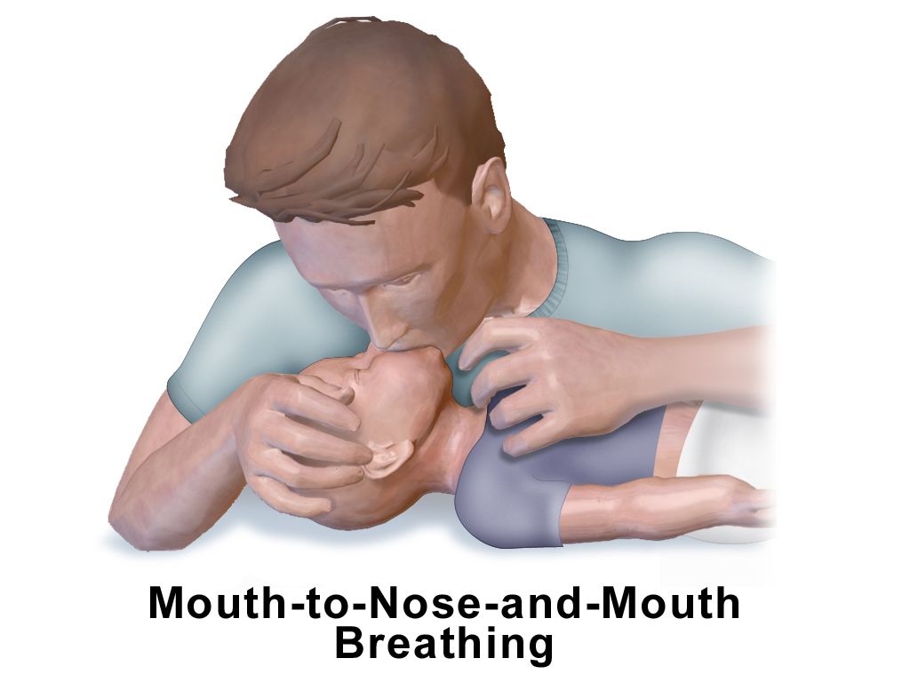
The Paediatric Assessment Triangle
The Paediatric Assessment Triangle (PAT) is a structured way of doing what experienced paediatric nurses do instinctively: forming an impression before touching the child. It uses only what you can see and hear from the end of the bed, takes under a minute, requires no equipment, and works on a child who is asleep, terrified or hostile. Its three legs are appearance, work of breathing and circulation to the skin. It does not replace the primary survey; it tells you how fast the primary survey needs to happen and whether you should be calling for help before you start it.
Appearance is the leg students under-weight, and it is the most important. It is the outward sign of brain perfusion, oxygenation and substrate delivery, and it is summarised by the mnemonic TICLS: Tone, Interactiveness, Consolability, Look or gaze, and Speech or cry. A well child has flexed limbs and resists you; a sick child is limp or has lost the normal flexed posture. A well infant tracks a face and reaches for a bright object; a sick infant stares through you. A well child is comforted by a caregiver; the inconsolable child, and equally the child who has stopped protesting when a stranger approaches, are both worrying. A vigorous angry cry is reassuring. A weak, whimpering or grunting cry is not.
Work of breathing is assessed by listening for abnormal airway sounds and looking for the visible effort of moving air. Stridor is upper airway, wheeze is lower airway, grunting is alveolar — a child grunting is generating their own positive end-expiratory pressure against collapsing alveoli, and grunting is one of the most reliable single markers of serious illness in an infant. Look for nasal flaring, head bobbing, tracheal tug and retractions, naming the level (suprasternal, intercostal, subcostal). Look at the position the child has chosen: the tripod position, the sniffing position, or an infant who will only settle sitting forward. Circulation to skin is the third leg and is judged by colour: pallor, mottling and cyanosis, in that order of progression. Cyanosis in a child is late.
The value of the triangle lies in the combinations. Which legs are abnormal names the physiological problem, and the physiological problem determines the first intervention.
| Appearance | Work of breathing | Circulation to skin | Physiological state | First priority |
|---|---|---|---|---|
| Normal | Normal | Normal | Stable | Complete assessment at a normal pace |
| Normal | Abnormal | Normal | Respiratory distress | Oxygen, position of comfort, keep the child calm |
| Abnormal | Abnormal | Normal | Respiratory failure | Assisted ventilation with bag-mask; call for help now |
| Normal | Normal | Abnormal | Compensated shock | Vascular access and a fluid bolus before the blood pressure falls |
| Abnormal | Normal | Abnormal | Shock that has reached the brain — decompensated once the systolic pressure falls below the age threshold | Immediate bolus, escalate, prepare for deterioration |
| Abnormal | Normal | Normal | Primary brain or metabolic problem | Check glucose, consider seizure, poisoning, sepsis, trauma |
| Abnormal | Abnormal | Abnormal | Cardiopulmonary failure | Resuscitation team; arrest is minutes away |
Check a glucose in every child with an abnormal appearance. Hypoglycaemia mimics sepsis, head injury, poisoning and seizure, and it is the one cause on that list you can correct in ninety seconds. Infants have small glycogen stores and a high metabolic rate; a child who has been vomiting for a day may be profoundly hypoglycaemic with a normal-looking chest and normal skin colour. Treat a blood glucose below about 2.6 mmol/L (47 mg/dL) in a neonate, or below 3.3 mmol/L (60 mg/dL) in an infant or child, and treat any symptomatic child regardless of the number: give dextrose 0.5 to 1 g/kg — in practice 5 to 10 mL/kg of 10% dextrose for an infant, or 2 to 4 mL/kg of 25% dextrose for an older child — and then recheck.
The numbers, by age band
A single set of adult ranges is useless in paediatrics and actively dangerous. A heart rate of 150 is normal in a two-month-old and a resuscitation call in a twelve-year-old. A respiratory rate of 20 is normal in a school-age child and a pre-terminal sign in a neonate. Learn the bands, and learn that heart rate and respiratory rate are the sensitive early markers while blood pressure is the last thing to change.
| Age band | Heart rate, awake (beats/min) | Respiratory rate (breaths/min) | Hypotension if systolic BP is |
|---|---|---|---|
| Neonate, birth to under 1 month | 100–205 | 30–60 | less than 60 mmHg |
| Infant, 1 month to under 1 year | 100–190 | 30–53 | less than 70 mmHg |
| Toddler, 1 year to under 3 years | 98–140 | 22–37 | less than 70 + (2 × age in years) |
| Preschool, 3 years to under 6 years | 80–120 | 20–28 | less than 70 + (2 × age in years) |
| School-age, 6 years to under 12 years | 75–118 | 18–25 | less than 70 + (2 × age in years) up to age 10; less than 90 mmHg at 11 years |
| Adolescent, 12 years and over | 60–100 | 12–20 | less than 90 mmHg |
Work the formula until it is automatic. A four-year-old is hypotensive below 70 + (2 × 4) = 78 mmHg systolic. A seven-year-old is hypotensive below 70 + (2 × 7) = 84 mmHg. A recorded systolic of 82 in that seven-year-old is not “a bit soft”; it is decompensated shock in a child who has already exhausted their compensatory reserve, and it demands an immediate bolus and a call, not a repeat cuff in fifteen minutes. Sleeping heart rates run roughly 20 to 30 beats/min lower than the awake ranges above, so a sleeping infant at 175 is tachycardic even though the number sits comfortably inside the awake band of 100 to 190.
Two other numbers belong here. Temperature: a rectal temperature of 38.0°C or higher in an infant under 28 days is a medical emergency requiring full evaluation for serious bacterial infection and parenteral antibiotics, regardless of how well the baby looks, because neonates mount almost no localising signs. Weight: if the child cannot be weighed, estimate with (age in years × 2) + 8 kg for children 1 to 5 years and (age in years × 3) + 7 kg for children 6 to 12 years — or better, use a length-based resuscitation tape. Every drug and every fluid volume in this chapter depends on that number being right.
From compensation to collapse, and the escalation that prevents it
Children compensate for hypovolaemia and sepsis extremely well and then fail extremely fast. Cardiac output in a small child is largely rate-dependent because stroke volume is relatively fixed, so the first response to any fall in preload is tachycardia, followed by intense peripheral vasoconstriction. That vasoconstriction is what you are seeing in prolonged capillary refill, cool mottled peripheries, a narrowed pulse pressure and weak peripheral pulses with a preserved central pulse — and it is what keeps the blood pressure normal. This is compensated shock, and it is entirely treatable. Once the blood pressure falls, the child has run out of reserve; this is decompensated shock, and mortality climbs steeply.
Bradycardia in a sick child is not a rhythm problem to be observed. It is hypoxia or acidosis reaching the myocardium and it immediately precedes arrest. In paediatric advanced life support, a heart rate under 60 beats/min with signs of poor perfusion despite adequate oxygenation and ventilation is an indication to start chest compressions — you do not wait for pulselessness. Most paediatric arrests are respiratory in origin and hypoxic in mechanism, which is why effective bag-mask ventilation, not defibrillation, is the intervention that saves the majority of these children. Compress at 100 to 120 per minute to a depth of at least one third of the anteroposterior chest diameter (about 4 cm in an infant, 5 cm in a child), at 30:2 for a single rescuer and 15:2 for two rescuers in infants and children. Defibrillation for a shockable rhythm starts at 2 J/kg, then 4 J/kg, then 4 J/kg or more without exceeding 10 J/kg or the adult dose. Adrenaline is 0.01 mg/kg intravenously or intraosseously (0.1 mL/kg of the 0.1 mg/mL concentration), maximum 1 mg, every 3 to 5 minutes.
The purpose of every observation set is escalation, and escalation has a script. SBAR converts a vague worry into a clinical demand. Situation: “I am calling about Maya Okoye, 4 years old, bed 6. Her heart rate is 172 and her capillary refill is 4 seconds.” Background: “She was admitted eight hours ago with gastroenteritis, has had six large watery stools and has not passed urine since arrival.” Assessment: “I think she is in shock. Her blood pressure is 84 systolic, which is still above her hypotension threshold of 78, so by pressure she is compensated — but her appearance has changed and she is now difficult to rouse, so the shock has already reached her brain and she is about to decompensate.” Recommendation: “I need you to review her now and I would like an order for a 20 mL/kg isotonic bolus and a bedside glucose.” Say the words “I need you to come now” when you mean them, and if the response does not match the child, use the chain of command or the rapid response criteria — both of which parents may also activate in many facilities.
Delegation limits tighten as the child destabilises. Unlicensed assistive personnel may weigh a child, record vital signs on a stable patient, apply a pulse oximeter probe, measure intake and output, and stay with a child for comfort. They may not perform the Paediatric Assessment Triangle, triage, interpret a change in condition, or take observations on an unstable child, because those acts are assessment. Teaching a caregiver, evaluating the response to a bolus, and the care of any child whose PAT is abnormal remain with the registered nurse.
| Red flag | Why it matters | Escalate as |
|---|---|---|
| Grunting, head bobbing or a silent chest | Alveolar collapse or exhaustion; imminent respiratory failure | Emergency call, prepare bag-mask |
| Capillary refill over 3 seconds with mottled cool skin | Compensated shock | Urgent review, bolus, glucose |
| Systolic BP below the age threshold | Decompensated shock | Resuscitation team |
| Heart rate under 60 with poor perfusion in an infant or child | Pre-arrest | Start CPR, call arrest team |
| Fever 38.0°C or higher in an infant under 28 days | Possible serious bacterial infection | Immediate medical review, blood cultures, antibiotics |
| A parent saying the child is “not themselves” | Caregiver concern independently predicts deterioration | Reassess in full; document and escalate |
Name the child’s physiological state from the Paediatric Assessment Triangle before you touch anything, and treat tachycardia and delayed capillary refill as shock, because a normal blood pressure in a child is the last reassurance you will get.
Section 2Fluids, Dehydration and the Arithmetic of a Small Body
Grading dehydration by percentage
Children dehydrate faster than adults for structural reasons: a higher proportion of body water is extracellular and therefore easily lost, the surface-area-to-mass ratio is larger so insensible losses are proportionally greater, the metabolic rate is higher, and the immature kidney concentrates urine poorly. An infant losing fluid through vomiting and diarrhoea can lose a tenth of their body mass in a day, which in an adult would be seven litres.
The gold standard for grading dehydration is an acute change in weight, because 1 kg of acute weight loss equals 1 litre of fluid. If a recent well-child weight exists, use it: a child weighing 12 kg who weighed 13.2 kg last week has lost 1.2 kg, which is 1200 mL, which is 9% of 13.2 kg — severe. When no recent weight exists, grade by clinical signs. Note that the same set of signs corresponds to different percentages in infants and older children, because infants have more water to lose. For the table below, use the infant column under 1 year of age and the older-child column from 1 year onwards, and convert percentage to volume with the rule that each 1% of dehydration is 10 mL/kg.
| Severity | Deficit (infant / older child) | Deficit in mL/kg (infant / older child) | Appearance and neurology | Circulation | Mucosa, eyes, fontanelle | Urine output |
|---|---|---|---|---|---|---|
| Mild | 5% / 3% | 50 / 30 mL/kg | Alert, thirsty, normal behaviour | Normal heart rate, capillary refill under 2 s, normal BP | Slightly dry mucosa, tears present, flat fontanelle, normal turgor | Slightly reduced |
| Moderate | 10% / 6% | 100 / 60 mL/kg | Irritable or listless, restless | Tachycardia, capillary refill 2–3 s, normal BP, weak peripheral pulses | Dry mucosa, few tears, sunken eyes and fontanelle, reduced turgor | Oliguria, under 1 mL/kg/h |
| Severe | 15% / 9% | 150 / 90 mL/kg | Lethargic, obtunded, may be unrousable | Marked tachycardia then bradycardia, capillary refill over 3 s, hypotension (late), thready or absent peripheral pulses, cool mottled limbs | Parched mucosa, no tears, deeply sunken eyes and fontanelle, tenting skin | Anuria |
Deep, sighing respirations in a dehydrated child are not respiratory distress — they are respiratory compensation for metabolic acidosis, and they mean the child is sicker than the chest examination suggests. Skin turgor and the fontanelle are useful but imperfect: turgor is falsely reassuring in a chubby infant and in hypernatraemic dehydration, where the skin feels doughy rather than tented. Capillary refill, heart rate, mental status and urine output remain the workhorses.
Maintenance, deficit and boluses: the 4–2–1 rule
Maintenance fluid replaces ongoing obligatory losses — urine, stool and insensible loss — in a child who is not currently depleted. The 4–2–1 rule gives the hourly rate: 4 mL/kg/h for the first 10 kg, plus 2 mL/kg/h for the next 10 kg, plus 1 mL/kg/h for every kilogram above 20 kg. Its daily equivalent is the 100–50–20 rule (100 mL/kg/day for the first 10 kg, 50 for the next 10, 20 thereafter).
Work an example. A child weighs 26 kg. First 10 kg: 10 × 4 = 40 mL/h. Next 10 kg: 10 × 2 = 20 mL/h. Remaining 6 kg: 6 × 1 = 6 mL/h. Total = 40 + 20 + 6 = 66 mL/h. Check it against the daily rule: (10 × 100) + (10 × 50) + (6 × 20) = 1000 + 500 + 120 = 1620 mL/day, which is 67.5 mL/h — close to the hourly answer but not identical. The gap is created in the first 20 kg: there the daily rule allows 1000 + 500 = 1500 mL/day, which is 62.5 mL/h, while the hourly rule allows only 40 + 20 = 60 mL/h, so the daily rule starts 2.5 mL/h ahead of the hourly one. Above 20 kg that reverses and the gap closes, because the hourly rule adds 1 mL/kg/h while the daily rule adds only 20 mL/kg/day, which is 0.83 mL/kg/h — the daily figure loses about 0.17 mL/h for every kilogram above 20. By 26 kg the 6 extra kilograms have clawed back 1 mL/h of the original 2.5, leaving the 1.5 mL/h difference you see. The two rules are therefore furthest apart at exactly 20 kg and converge as the child gets heavier, meeting at 35 kg where both give 75 mL/h. Use one of them consistently rather than mixing them, and treat the second as a sanity check rather than as proof. A second example: a 7 kg infant sits entirely inside the first band, so 7 × 4 = 28 mL/h. A third: a 15 kg toddler is (10 × 4) + (5 × 2) = 40 + 10 = 50 mL/h.
Maintenance is not resuscitation. A child in shock gets a bolus of 20 mL/kg of isotonic crystalloid over 5 to 20 minutes, then is reassessed — heart rate, capillary refill, mental status, urine output — before the next bolus is considered. For a 12 kg child that is 12 × 20 = 240 mL. Several conditions call for a 10 mL/kg bolus given more cautiously with reassessment between each: neonates, diabetic ketoacidosis (where guidelines advise even, unhurried rehydration), severe acute malnutrition, known or suspected cardiac failure, and suspected raised intracranial pressure. The exception is the child who is frankly shocked with hypotension or unrecordable pulses, who still needs 20 mL/kg rapidly — but that exception applies only beyond the newborn period, because three groups keep the cautious volume even in frank shock: neonates (10 mL/kg over 5 to 10 minutes), known cardiac disease and severe acute malnutrition, where 10 mL/kg aliquots with reassessment between each remain the rule. If a child needs more than 40 to 60 mL/kg, escalate for vasoactive support rather than continuing to pour in fluid.
Where dehydration is mild or moderate and the child can drink, oral rehydration solution is as effective as intravenous fluid and safer: give the estimated deficit from the table above over about 4 hours — that is 50 mL/kg for mild and 100 mL/kg for moderate dehydration in an infant under 1 year, and 30 mL/kg and 60 mL/kg respectively from 1 year onwards — given in small frequent volumes (5 to 10 mL by syringe or spoon every 1 to 2 minutes), plus about 10 mL/kg extra for every additional watery stool. Vomiting is not a contraindication — small volumes given slowly are usually tolerated. Plain water, fruit juice and sports drinks are not substitutes; juice and sports drinks are hyperosmolar and worsen diarrhoea, and free water alone risks hyponatraemia. When a deficit is replaced intravenously, the classic scheme gives the bolus first, then replaces the remaining deficit over 24 hours alongside maintenance, with half of the remainder in the first 8 hours. Isotonic maintenance fluid, generally with added dextrose, is now standard for most hospitalised children because hypotonic fluid caused hospital-acquired hyponatraemia.
Weight-based dosing and the adult ceiling
Children are not small adults, and paediatric dosing is not a scaled-down adult order — it is calculated from scratch, per kilogram, every time. Three habits prevent most of the harm. First, weigh in kilograms only and record only kilograms; a pounds-for-kilograms confusion produces a 2.2-fold overdose and it is the single commonest source of paediatric weight errors. Second, show the arithmetic in your own working before you draw anything up. Third, check the calculated dose against the maximum adult dose; a per-kilogram formula applied to a large adolescent can legitimately exceed the highest dose any adult should receive, and when it does, the adult maximum wins.
Work three examples. Paracetamol (acetaminophen) 15 mg/kg per dose for an 18 kg child: 15 × 18 = 270 mg, every 4 to 6 hours — but the interval alone does not finish the calculation, because the daily ceiling is 75 mg/kg, which here is 75 × 18 = 1350 mg. Divide the ceiling by the dose: 1350 ÷ 270 = 5, so this child may have no more than five doses in 24 hours. Six doses at the four-hourly end would be 1620 mg and would breach the ceiling, which is why 15 mg/kg is ordered four- to six-hourly to a maximum of five doses a day rather than around the clock. Now the same formula for a 60 kg adolescent: 15 × 60 = 900 mg per dose, which is under the 1000 mg adult single-dose ceiling and therefore acceptable — but the daily calculation 75 × 60 = 4500 mg exceeds the adult maximum of 4000 mg in 24 hours, so the daily total must be capped at the adult figure. Lorazepam for status epilepticus at 0.1 mg/kg for a 14 kg child: 0.1 × 14 = 1.4 mg; from a 2 mg/mL vial that is (1.4 ÷ 2) × 1 = 0.7 mL. The same formula in a 50 kg adolescent gives 5 mg, which is above the 4 mg maximum single dose, so 4 mg is what is given — the ceiling wins. Intramuscular adrenaline for anaphylaxis at 0.01 mg/kg of the 1 mg/mL concentration for a 22 kg child: 0.01 × 22 = 0.22 mg, which is 0.22 mL into the vastus lateralis, with a maximum single dose of 0.5 mg at any age — 0.3 mg is a widely used convention in small prepubertal children rather than an absolute ceiling. Auto-injector strengths and how to choose between them are set out with the rest of the anaphylaxis sequence in Section 3.
Some errors recur so predictably that they have names. A tenfold error follows a misplaced decimal, which is why you write 0.5 mg and never .5 mg, and 1 mg and never 1.0 mg. A concentration error follows confusing the 1 mg/mL and 0.1 mg/mL adrenaline presentations. A units error follows writing “u” for units in an insulin order. High-alert medicines in paediatrics — insulin, opioids, heparin, concentrated electrolytes, chemotherapy, magnesium sulfate and vasoactive infusions — require an independent double check, in which a second nurse calculates the dose from the original order without seeing your working, rather than simply agreeing with your number.
Three questions before every paediatric dose. Is the weight in kilograms, measured today, and is it this child’s weight? Does my per-kilogram arithmetic reproduce the ordered dose when I calculate it independently? Does the result exceed the maximum adult dose — and if so, have I stopped and queried it? A dose that is mathematically correct per kilogram but above the adult ceiling is still an overdose. Doses may also need adjusting for renal or hepatic impairment and for gestational age in neonates, where dosing intervals lengthen rather than doses shrinking.
Grade dehydration by percentage and match it to signs, calculate maintenance with 4–2–1 and resuscitation with 20 mL/kg (10 mL/kg in neonates, diabetic ketoacidosis, severe acute malnutrition, known or suspected cardiac failure and suspected raised intracranial pressure), and calculate every drug per kilogram while checking the answer against the maximum adult dose.
Section 3Paediatric Emergencies: When the Sequence Saves the Life
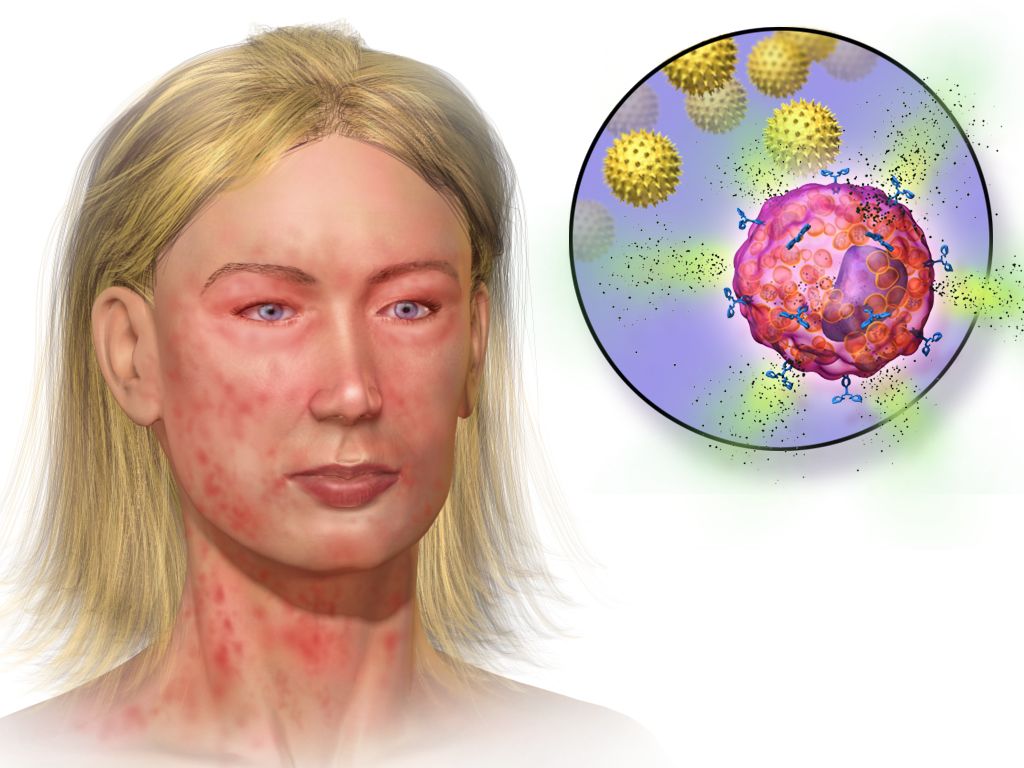
Anaphylaxis: adrenaline into the thigh before anything else
Every emergency in this section has a correct first action, and in most of them the intuitive action is the wrong one. The order is not a matter of tidiness. The order is the treatment.
Anaphylaxis is mast cell and basophil degranulation on a systemic scale, usually immunoglobulin E mediated and in children usually triggered by food — peanut, tree nut, cow’s milk, egg, sesame, shellfish — or by venom, latex or a drug. The mediators released do four things at once: they dilate vessels, open a capillary leak that can shift a third of the circulating plasma volume into the interstitium within minutes, constrict bronchi, and swell the larynx, tongue and pharynx. That combination is why a child can travel from itchy to unrecordable in the time it takes to find a set of observations.
Recognition rests on the speed of onset and the number of systems involved. Suspect anaphylaxis when a child develops, over minutes to a couple of hours, skin or mucosal signs — urticaria, flushing, angio-oedema — together with either respiratory or circulatory compromise; or when two or more systems are involved after exposure to a likely allergen; or when a child becomes hypotensive after exposure to a known allergen. In children the respiratory features dominate, and persistent vomiting or severe abdominal pain shortly after an allergen is a cardinal sign of a systemic reaction rather than a stomach upset. Crucially, something like one child in ten to one in five has no skin signs at all, so the absence of a rash does not exclude anaphylaxis in a child who is stridulous, wheezy, pale and floppy after eating.
The first action is intramuscular adrenaline into the anterolateral thigh, into the vastus lateralis — before an antihistamine, before a steroid, before intravenous access, and before anyone goes to look for the allergy plan. The dose is 0.01 mg/kg of the 1 mg/mL (1:1000) solution, to a maximum single dose of 0.5 mg at any age (0.3 mg is the conventional ceiling used in small prepubertal children), repeated after 5 to 15 minutes if the response is inadequate. The site matters, because intramuscular injection into the thigh produces a higher and faster peak plasma concentration than the deltoid or than any subcutaneous route. Adrenaline is also the only agent that reverses all four mechanisms at once: it constricts vessels, raises blood pressure, shrinks mucosal oedema, dilates bronchi and switches off further mediator release. Delay is what kills. In fatal anaphylaxis the interval from exposure to cardiorespiratory arrest is measured in minutes — shortest with intravenous drugs, then venom, then food — and the finding that runs through fatality reviews is that adrenaline was given late or not at all while an antihistamine was given early. There is no absolute contraindication to adrenaline in anaphylaxis.
Position the child according to what is failing, and change position gently. A child with circulatory compromise lies flat with the legs raised; a child whose breathing is the dominant problem may sit up; a child who is unconscious or vomiting goes into the recovery position. Never make a hypotensive child sit up abruptly, stand, or walk to a trolley. In a vasodilated, volume-depleted circulation venous return is posture-dependent, and suddenly emptying the right ventricle in that state has caused cardiac arrest — deaths are documented in patients who were sat or stood up. Alongside the adrenaline: call for help, remove the trigger where you can (stop the infusion, scrape out a stinger), give high-flow oxygen, obtain access and give a 20 mL/kg isotonic crystalloid bolus if there is any circulatory compromise, and add inhaled salbutamol only for bronchospasm that persists after adrenaline. Observe afterwards for a biphasic reaction, and no child goes home without an adrenaline auto-injector, training in its use, a written action plan and an allergy referral.
| Agent | Dose and route in a child | Role and cautions |
|---|---|---|
| Adrenaline (epinephrine) 1 mg/mL | 0.01 mg/kg intramuscularly into the anterolateral thigh; maximum single dose 0.5 mg at any age, with 0.3 mg the conventional ceiling in small prepubertal children; repeat after 5–15 min if needed | First-line and time-critical. The 1 mg/mL ampoule is the intramuscular concentration; the 0.1 mg/mL presentation is the intravenous strength used in arrest and is not what you draw up here |
| Adrenaline auto-injector | 0.1 mg from about 7.5 kg to under 15 kg where the device is available; 0.15 mg from 15 kg to under 25 kg; 0.3 mg from 25 kg to under 50 kg; 0.5 mg from about 50 kg in larger adolescents and adults; outer thigh, through clothing if necessary | A fixed strength will rarely match the calculated dose exactly; give the nearest appropriate device rather than delaying to draw up an exact volume |
| Oxygen | High flow by non-rebreather mask | For every child with respiratory or circulatory compromise |
| Isotonic crystalloid | 20 mL/kg bolus, repeated as the response dictates | Treats the capillary leak; hypotension persisting after adrenaline is usually unreplaced volume |
| Inhaled salbutamol | By spacer or nebuliser | Adjunct for persistent bronchospasm only; it does nothing for laryngeal oedema or for shock |
| H1 antihistamine | Oral or intravenous | Relieves itch and urticaria only. No effect on the airway, the breathing or the blood pressure, and never given before adrenaline |
| Corticosteroid | Oral or intravenous | Takes hours to act, does not treat the acute event, and current evidence does not support it for preventing biphasic reactions. It must never delay adrenaline |
Two habits protect a child in anaphylaxis more than any refinement of the drug list. The first is treating the reaction you are watching rather than the one described in the notes: a child with a mild history can have a severe reaction, and severity is judged on the airway, the breathing and the circulation in front of you. The second is not waiting for certainty. Adrenaline given to a child who turns out to have had a milder allergic reaction causes transient pallor, tremor and palpitations; adrenaline withheld from a child in anaphylaxis causes deaths.
Status epilepticus: the airway, the clock, then the drug
A convulsive seizure lasting 5 minutes or longer, or repeated seizures without recovery of consciousness between them, is status epilepticus. The five-minute definition is deliberately short: most self-limiting seizures stop within two to three minutes, so one that is still running at five is unlikely to stop by itself and is already doing harm through hypoxia, hyperthermia, acidosis and excitotoxic neuronal injury.
The first action is not the drug. It is the airway, the oxygen and the clock. Open the airway with a jaw thrust if it is needed, turn the child onto their side, suction secretions, give high-flow oxygen, attach monitoring, protect the head, and write down the exact time the seizure started, because every decision from here is timed from that moment. Do not restrain the limbs, and do not put anything in the mouth — a convulsing child cannot swallow their tongue, and objects placed in the mouth break teeth and obstruct airways. Then, in parallel, obtain intravenous or intraosseous access and check a bedside glucose. Hypoglycaemia is a cause of seizure that you can reverse in ninety seconds, and it will not respond to a benzodiazepine.
| Time from seizure onset | Stage | What is done |
|---|---|---|
| 0–5 min | Stabilisation | Airway, lateral position, suction, high-flow oxygen, monitoring, intravenous or intraosseous access, bedside glucose, temperature, and the exact start time written down. No restraint, nothing in the mouth |
| 5 min | First benzodiazepine | Lorazepam 0.1 mg/kg intravenously (maximum 4 mg per dose). Without access: midazolam 0.2 mg/kg intramuscularly or intranasally (maximum 10 mg), buccal midazolam 0.5 mg/kg (maximum 10 mg), or rectal diazepam 0.2–0.5 mg/kg (maximum 20 mg) |
| 10 min | Second and final benzodiazepine dose | Repeat once. Have bag-mask ventilation ready at the bedside — respiratory depression is cumulative across doses |
| 20 min | Second-line agent | Levetiracetam 60 mg/kg (maximum 4500 mg), fosphenytoin 20 mg PE/kg (maximum 1500 mg PE), or sodium valproate 40 mg/kg (maximum 3000 mg). Trial evidence shows the three to be comparably effective, so the choice follows the child’s history and local protocol; valproate is avoided where hepatic or mitochondrial disease is suspected |
| 40 min | Refractory status | Critical care: anaesthetic infusion with intubation and continuous electroencephalography |
While the drugs are being given, someone should be answering the question of why. Check the temperature, the sodium, the calcium and the toxicology; ask about missed anticonvulsant doses, a recent head injury, a ventriculoperitoneal shunt, a possible ingestion, and immunisation and travel history. A febrile seizure in a child between 6 months and 5 years is common and usually benign, but a seizure with fever plus a rash, neck stiffness or an abnormal appearance is meningitis until proved otherwise. And an unexplained seizure in an infant — particularly with vomiting, a tense fontanelle or a falling conscious level and no visible mark — should raise the possibility of abusive head trauma, which is the subject of the next section.
A seizure gets harder to stop the longer it runs. Within minutes of continuous seizure activity, GABA-A receptors are internalised from the synaptic membrane, so the very target the benzodiazepine acts on is disappearing while you watch. That is the pharmacological reason for the clock — for giving the first dose at 5 minutes rather than “seeing how it goes”, and for moving to a second-line agent after two benzodiazepine doses rather than giving a third. A third benzodiazepine at 25 minutes buys respiratory depression, not seizure control.
Shock, ketoacidosis, poison and the obstructed airway
Septic shock in a child is recognised on perfusion and mental state, not on blood pressure. The dysregulated host response produces vasodilatation, capillary leak and myocardial depression, and the child compensates with tachycardia and vasoconstriction — so what you see first is a capillary refill that is prolonged, or in warm shock unnaturally flash-fast; skin that is mottled and cool, or flushed with bounding pulses; a heart rate out of proportion to the fever; a falling urine output; and above all a change in how the child interacts, from irritable, to drowsy, to difficult to rouse. Hypotension is a late and pre-terminal sign, because a child can lose a quarter to a third of their circulating volume before the systolic pressure moves at all. A normal blood pressure is therefore never a reason to wait. The first actions are to say the word sepsis out loud and activate the pathway: high-flow oxygen; vascular access, going to an intraosseous needle after two failed attempts or about 90 seconds; blood cultures if they delay nothing; broad-spectrum antibiotics within one hour of recognition; a lactate; and fluid given as 10 to 20 mL/kg boluses titrated to perfusion, up to 40 to 60 mL/kg in the first hour, with reassessment after every single bolus. Stop the fluid and change strategy if the liver edge becomes palpable, crackles appear or the work of breathing rises, and move to vasoactive support rather than pouring in more. Three groups take the cautious 10 mL/kg bolus from the outset with reassessment between each: the neonate, whose volume expansion is 10 mL/kg over 5 to 10 minutes however shocked they are; the child with known cardiac disease, whose ventricle cannot absorb the preload; and the child with severe acute malnutrition, in whom myocardial and membrane function are impaired and standard-volume resuscitation increases mortality.
Diabetic ketoacidosis inverts the instinct to fix the number you can see. Insulin deficiency with a surge of counter-regulatory hormones produces hyperglycaemia, an osmotic diuresis that empties the circulation, and lipolysis with ketone production that drives the acidosis and the deep sighing Kussmaul breathing. The child is dehydrated, acidotic, depleted of total body potassium whatever the serum potassium happens to read, and hyperosmolar — and the brain has spent hours to days adapting to that osmolality. Fluid comes before insulin. Restore the circulation with isotonic crystalloid: 10 mL/kg over 30 to 60 minutes in the child who is dehydrated but still perfusing, repeated only as perfusion demands — but 20 mL/kg given rapidly, and repeated as needed, in the child who is genuinely shocked with hypotension or unrecordable pulses. Then replace the remaining deficit evenly over 24 to 48 hours. The insulin infusion starts 1 to 2 hours after fluids have begun, at 0.05 to 0.1 units/kg/h, and a child never receives an insulin bolus: a bolus drops glucose and osmolality abruptly, drives potassium into the cells, and is associated with cerebral oedema. Potassium is withheld only for frank hyperkalaemia or anuria. If the potassium is low at presentation, replacement starts with the initial fluid and the insulin infusion is delayed until potassium replacement is running, because insulin given to a hypokalaemic child can precipitate a lethal arrhythmia; if the potassium is normal, replacement starts alongside the insulin once urine output or normal renal function is confirmed. Insulin drives potassium intracellularly and unmasks a total-body deficit that is present whatever the serum level reads. Bicarbonate is not given routinely — it is associated with cerebral oedema and does not improve outcome. The narrow exceptions, on specialist advice, are life-threatening hyperkalaemia and severe acidosis with a pH below 6.9 where cardiac contractility is impaired.
The complication that shapes every one of those decisions is cerebral oedema: uncommon, but the leading cause of death and permanent disability in paediatric ketoacidosis, and characteristically appearing 4 to 12 hours after treatment begins — which is to say, while the laboratory numbers are improving. Its risk tracks how ill the child was on arrival rather than how fast the fluid ran: severe acidosis, a low carbon dioxide, a raised urea, young age and a new diagnosis all predict it, whereas a large randomised trial found that neither the rate of fluid administration nor its sodium content changed neurological outcome. Guidelines therefore still rehydrate evenly over 24 to 48 hours as a precaution, not because speed has been shown to cause the injury. The warning signs are neurological rather than biochemical: headache, recurrent vomiting after treatment has started, agitation giving way to drowsiness, incontinence in a child who was continent, a rising blood pressure with a falling heart rate, cranial nerve palsies, and any fall in conscious level at all. A falling conscious level during ketoacidosis treatment is the emergency, and it is treated before it is imaged: call for help, reduce the intravenous fluid rate by about a third, raise the head of the bed, and give hypertonic 3% saline 2.5 to 5 mL/kg over 10 to 15 minutes or mannitol 0.5 to 1 g/kg over 10 to 15 minutes, repeated after 30 minutes if there is no response. Imaging comes after treatment, never instead of it.
Poisoning and ingestion begins, like everything else, with airway, breathing, circulation and a glucose, then three questions: what, how much, and when. Contact the poisons information service — that call is the single most useful early action — and decontaminate skin or eyes where the exposure was topical. Do not induce vomiting. Syrup of ipecac left paediatric practice because it never improved any outcome: it empties little, it delays or blocks activated charcoal and oral antidotes, and vomiting a corrosive or a hydrocarbon re-injures the oesophagus and risks aspiration pneumonitis. Gastric lavage has gone the same way. Activated charcoal 1 g/kg, to a maximum of about 50 g, may be considered within roughly an hour of a suitable ingestion in an alert child with a protected airway, but it does not bind iron, lithium, alcohols, hydrocarbons or corrosives. Never assume a well-looking child is safe, because paracetamol, sulfonylureas, sustained-release preparations and the toxic alcohols all have silent latent periods. Be especially alert to the agents where a single adult tablet or one mouthful can kill a toddler: calcium channel blockers, tricyclic antidepressants, opioids including methadone, sulfonylureas, clonidine and imidazoline decongestants, theophylline, antimalarials, camphor, oil of wintergreen (methyl salicylate) and concentrated nicotine liquid. A swallowed button battery is a separate emergency — current flowing at the mucosa burns through the oesophageal wall within about two hours — so a child who may have swallowed one needs an immediate radiograph and urgent endoscopic removal, and in a child of 12 months or older who is still swallowing normally, who is within about 12 hours of the ingestion, and in whom there is no suspicion of perforation or sepsis, 10 mL of honey every 10 minutes to a maximum of six doses while transfer is arranged reduces the injury. Beyond that window, or with an unprotected airway, the injury is already established and the honey adds aspiration risk without benefit.
Foreign body airway obstruction turns on a single observation: is the cough effective? A child who can cry, speak or produce a loud forceful cough is moving air, and their own cough generates pressures no manoeuvre of yours can match — so you encourage the coughing, stay with the child, and do nothing else while it remains effective. When the cough turns silent or feeble, when the child cannot vocalise or breathe, when colour deteriorates or consciousness begins to fall, the cough is ineffective and you act at once. In an infant under 1 year, give 5 back blows head-down along your forearm with the jaw supported, then turn the infant and give 5 chest thrusts — two fingers on the sternum at the compression landmark, sharper and slower than compressions — and repeat, checking the mouth between cycles. In a child over 1 year, give 5 back blows then 5 abdominal thrusts, and repeat. Abdominal thrusts are never used in an infant, whose liver is large, low and unprotected by the rib cage. If the child becomes unresponsive, lay them on a firm flat surface, shout for the resuscitation team and begin CPR starting with chest compressions, looking into the mouth before each set of rescue breaths and removing an object only if you can see it. Never perform a blind finger sweep: in a narrow paediatric airway it drives a partially obstructing object down into the larynx, converts a partial obstruction into a complete one, and causes bleeding and swelling that make everything afterwards harder. Every child who received back blows or thrusts needs medical assessment afterwards, and a persistent cough, stridor or unilateral wheeze after a choking episode means a retained foreign body until bronchoscopy says otherwise.
| Emergency | Do this FIRST | Never |
|---|---|---|
| Anaphylaxis | Adrenaline 0.01 mg/kg of the 1 mg/mL solution intramuscularly into the anterolateral thigh | Give an antihistamine or a steroid first, or sit or stand a hypotensive child up |
| Status epilepticus | Airway and lateral position, oxygen, note the exact time, check the glucose — then a benzodiazepine at 5 minutes | Restrain the child, put anything in the mouth, or give a third benzodiazepine instead of a second-line agent |
| Septic shock | Oxygen, access, cultures, antibiotics within the hour, and fluid titrated to perfusion | Wait for the blood pressure to fall before calling it shock |
| Diabetic ketoacidosis | Restore the circulation with isotonic fluid; the insulin infusion follows 1 to 2 hours later | Give an insulin bolus, give bicarbonate routinely, or rehydrate quickly in a child who is not shocked — the shocked child with hypotension or unrecordable pulses still gets 20 mL/kg rapidly |
| Falling conscious level during ketoacidosis treatment | Treat as cerebral oedema: slow the fluid, head up, hypertonic saline or mannitol, call for help | Send the child for imaging before treating |
| Poisoning or ingestion | Airway, breathing, circulation and a glucose; establish what, how much and when; call the poisons service | Induce vomiting, or give a home remedy to neutralise it |
| Choking with an effective cough | Encourage the coughing and stay with the child | Interrupt an effective cough with blows or thrusts |
| Choking with an ineffective cough | Infant: 5 back blows then 5 chest thrusts. Child over 1 year: 5 back blows then 5 abdominal thrusts | Perform a blind finger sweep, or use abdominal thrusts on an infant |
The words you choose decide what the family tells you. In poisoning, choking and unexplained collapse the history is the diagnostic test, and a caregiver who feels accused gives a shorter and less accurate one. So ask “what was within reach?” rather than “how did they get hold of it?”, and “tell me exactly what you saw” rather than “were you watching?” Say “this happens in ordinary homes” and mean it. None of this softens the safeguarding thinking of the next section — a blame-free manner and an alert mind are entirely compatible — but it is how you find out what was swallowed, how much, and when.
In paediatric emergencies the order of actions is the treatment: intramuscular adrenaline into the thigh before any other drug in anaphylaxis, the airway and the clock before the benzodiazepine in status epilepticus, perfusion and mental state rather than blood pressure in septic shock, fluid before insulin in ketoacidosis with a falling conscious level treated as cerebral oedema, and back blows and thrusts — never a blind finger sweep — once a cough becomes ineffective.
Section 4Safeguarding: Injuries That Do Not Fit the Story or the Stage
Development is the yardstick
The single most useful safeguarding question is not “could this have happened?” but “could this child have done this?” An injury is concerning when it is inconsistent with the mechanism offered, and it is equally concerning when the mechanism is plausible in principle but impossible for a child at that developmental stage. A four-month-old cannot roll off a sofa if they have never rolled. A five-month-old cannot pull a mug of tea from a table if they cannot sit unsupported or pincer-grasp. A non-ambulatory infant cannot sustain a spiral femur fracture from “twisting in the cot”. Knowing the milestones from Chapter 11 is therefore a safeguarding skill: rolling at about 4 to 6 months, sitting unsupported at 6 to 8 months, crawling at 7 to 10 months, cruising at 9 to 12 months, walking at 12 to 15 months.
This is the basis of the aphorism “those who don’t cruise rarely bruise”. Bruising in a pre-mobile infant is rare enough that it is treated as a sentinel injury — a minor-looking injury in a young infant that frequently precedes severe or fatal abuse, and that represents the opportunity to intervene before the catastrophic event. Sentinel injuries include any bruise, any intraoral injury such as a torn frenulum, and any burn in an infant who is not yet mobile. They are easy to dismiss because they are small. They should never be dismissed.
The TEN-4-FACESp rule gives a validated screen for bruising in children under 4 years, flagging bruises that warrant evaluation for abuse.
| Letter | Region or feature | Why it is concerning |
|---|---|---|
| T | Torso (chest, abdomen, back, buttocks, genitourinary) | Accidental bruising clusters over bony prominences of the limbs and forehead, not soft protected areas |
| E | Ear | The ear is shielded by the skull and shoulder; bruising implies a direct blow or pinch |
| N | Neck | Suggests grabbing or strangulation |
| 4 | Any bruise anywhere in an infant under 4.99 months | Pre-mobile infants do not self-bruise |
| F | Frenulum (labial or lingual) | Forced feeding or a blow to the mouth |
| A | Angle of the jaw | Protected site |
| C | Cheek, fleshy part | Protected by the zygoma in accidental falls |
| E | Eyelid | Isolated eyelid bruising without a forehead injury is atypical |
| S | Subconjunctival haemorrhage | Associated with strangulation and direct trauma |
| p | Patterned bruising | Reproduces the object — a hand, belt, cord, buckle |
Injuries and histories that raise concern
Certain injuries carry a high specificity for inflicted trauma because of the forces required to produce them. Others are concerning mainly because of the context. Both belong in your assessment, and neither constitutes a diagnosis — the diagnosis of abuse is a multidisciplinary process, and there are important medical mimics such as bleeding disorders, osteogenesis imperfecta, metabolic bone disease, Mongolian blue spots or congenital dermal melanocytosis, and cultural practices such as cupping and coining, which must be explored respectfully rather than assumed.
| Finding | Why it raises concern |
|---|---|
| Posterior or posteromedial rib fractures | Require anteroposterior squeezing of the chest, so they are highly specific for inflicted injury; they are rarely but genuinely reported after two-thumb encircling-hands infant CPR, so a documented resuscitation is recorded and considered rather than excluded by rule |
| Classic metaphyseal lesion (bucket-handle or corner fracture) | Produced by torsion or traction on a limb |
| Spiral or transverse femur fracture in a non-ambulatory infant | Requires rotational or high force the child cannot generate |
| Fractures at multiple different stages of healing | Implies repeated events over time |
| Scapular, sternal or spinous process fracture | High-force injuries in protected sites |
| Immersion burn with a sharp line of demarcation, stocking-and-glove distribution, symmetry, sparing of flexural creases | Consistent with being held in hot liquid; a child who falls in has splash marks and irregular edges |
| Small, round, deep burns about 8 mm across | Cigarette contact |
| Retinal haemorrhages with subdural haemorrhage and few or no external marks | Abusive head trauma from rotational acceleration and deceleration |
| Any anogenital injury, or sexually transmitted infection in a prepubertal child | Requires urgent specialist evaluation |
| Poor growth, untreated dental disease, missed critical appointments, developmental regression, inadequately treated chronic illness | Possible neglect, which is the most common form of maltreatment |
Histories matter as much as injuries. Be concerned when there is no history offered for a significant injury; when the history changes between tellings or between caregivers; when it does not fit the injury or the child’s developmental stage; when there was an unexplained delay in seeking care; when the injury is blamed on a young sibling or a pet; when the child is unusually watchful, flinches, or shows no expectation of comfort; or when the caregiver is hostile to examination or focused on something other than the child. Be concerned too about medical child abuse — illness fabricated or induced by a caregiver — when symptoms occur only in one person’s presence, when the history is dramatic but investigations are persistently normal, or when a caregiver pushes for invasive procedures.
The duty to report, and how to document
Nurses are mandated reporters in every United States jurisdiction and hold equivalent safeguarding duties in most other systems. Four features of that duty are commonly misunderstood by students. First, the threshold is reasonable suspicion, not proof: you are not required to know that abuse occurred, only to have cause to suspect it, and investigation is the child protection agency’s job, not yours. Second, the duty is personal and non-delegable; it is not discharged by telling the physician or the charge nurse, and “the doctor said he would handle it” is not a defence. Third, reporting in good faith carries statutory immunity from civil and criminal liability, whereas failure to report is itself an offence in most jurisdictions and a licensure matter in all of them. Fourth, you generally report first and, depending on institutional policy and risk to the child, the family is informed by the designated team member — you do not need the caregiver’s permission and you do not warn them in a way that endangers the child. Reports are typically made by telephone immediately and in writing within 24 to 48 hours.
Your interviewing role is deliberately limited. Ask open, non-leading questions once — “tell me what happened”, “can you tell me more about that?” — and then stop. Repeated interviews contaminate evidence and re-traumatise children; formal forensic interviewing is done by trained specialists. Never promise a child that you will keep what they tell you secret; say instead that you will tell the people whose job it is to keep them safe. Where sexual assault is disclosed, preserve evidence per policy, note that in a prepubertal child evidence is rarely recovered from the body beyond about 24 hours while clothing and bedding remain productive far longer — the 72 to 120 hour kit windows apply mainly to adolescents, and acute examination is indicated regardless for injury, infection and safety assessment — discourage washing or changing clothes, and place clothing in paper rather than plastic bags.
Documentation in safeguarding is a legal document you may be asked to defend years later. Record what you observed, not what you concluded. Quote the child and the caregiver verbatim, in quotation marks, attributed to the speaker. Describe each injury by site, size in centimetres, shape, colour and any pattern, and map it on a body diagram; photograph only per institutional policy with a scale in frame. Do not attempt to date a bruise by its colour — colour-based ageing has been shown to be unreliable. Avoid the words “alleged”, “claims” and “denies”, which editorialise; write “the caregiver states”. Record who was present, the time of each event, and every notification you made with the name and time.
Poverty is not neglect. A family without transport, without a working refrigerator for insulin, or without the money for a prescription has an unmet need, not a moral failing, and the intervention is a social work referral, a resource, or a delivery arrangement — not a maltreatment report. Neglect is failure to provide when the caregiver has the means and the capacity to do so. Confusing the two harms families who are already disadvantaged and diverts scarce child protection resources. Report the child who is unsafe; resource the family who is struggling; and be honest with yourself about which one you are looking at.
Test every injury against the child’s developmental stage as well as the story, treat any bruise in a pre-mobile infant as a sentinel injury, and report on reasonable suspicion — the duty is yours personally, it does not require proof, and good-faith reporting is protected.
Section 5The Family in Crisis: Presence, Bad News and Loss

Family presence and the conversation during the emergency
Families should be offered the option of being present during invasive procedures and resuscitation. The evidence, and the position statements of emergency and critical care nursing organisations, support this: presence does not increase interference or litigation, and families who witness a resuscitation show lower rates of prolonged grief, intrusive imagery and unanswered questions afterwards. Offering is the operative word — presence is invited, never required, and a family who declines is not failing their child. The practical requirement is that a dedicated support person, not a team member with a resuscitation role, stays with the family throughout to narrate what is happening, answer questions and escort them out if needed.
During the crisis, communication has two audiences and two structures. To the team, use SBAR, which was scripted in Section 1. To the family, speak in short sentences, use the child’s name, avoid the abbreviations that fill the room, and check understanding rather than asking “does that make sense?” Give a warning shot before serious news: “I need to tell you something difficult.” The SPIKES framework structures the fuller conversation — Setting (private, seated, phones handed over, interpreter arranged, the right people present), Perception (“what have you been told so far?”), Invitation (“how much detail would you like?”), Knowledge (plain language, small amounts, then pause), Emotions (acknowledge and stay silent rather than filling the silence), and Strategy and summary (what happens next, who to contact, written information).
Use the word died. “Passed away”, “we lost her” and “gone” are ambiguous, and to a young child they are literally confusing — children have searched for a sibling who was “lost” and refused to sleep after being told a baby “went to sleep”. Avoid the phrases that sound like comfort and land as dismissal: “at least you have other children”, “at least it was early”, “everything happens for a reason”, “you can have another”, “you are young”. What helps is simple, honest presence: “I am so sorry. We did everything we could. Would you like me to stay?”
Perinatal, neonatal and paediatric loss
Perinatal loss covers miscarriage, stillbirth (in the United States, fetal death at 20 weeks’ gestation or later), termination for fetal anomaly, and neonatal death. The nursing task is to make choices available and to follow the family’s lead, because the interventions that support long-term grief are almost entirely about agency and memory. Ask the baby’s name and then use it, every time. Offer — without pressure and with the option to change their mind later — seeing and holding the baby, bathing and dressing the baby, hand and footprints, a lock of hair with permission, photographs, a blanket or hat, and religious or cultural rites. Offer time, including the option of a cooling cot where available. Document what was offered, what was chosen and what was declined so the family is not asked the same painful questions repeatedly, and flag the record so that staff do not walk in with routine cheerfulness.
Do not neglect the physiology of loss. After a birth at 16 weeks or beyond, the birthing parent will experience lactogenesis: milk usually comes in on day 2 to 4 whether or not there is a baby to feed, and being unprepared for that is described by bereaved parents as one of the cruellest surprises of the experience. The crisis-care task here is to warn the family before it happens, so that it is expected rather than ambushing them at home, and to offer — where the family wishes it — the option of donating expressed milk, which some parents find deeply meaningful. The suppression regimen itself is postpartum management and is set out in Chapter 7. Hand the family back to routine postpartum care rather than letting bereavement displace it, and make sure the screening for perinatal mood and anxiety disorders described in Chapter 8 actually happens, because the risk after loss is substantially raised.
Where a child dies suddenly and unexpectedly, families face investigation on top of grief. In sudden unexpected infant death, a scene investigation, a death scene re-enactment and an autopsy are routine and legally required in most jurisdictions; explain in advance that this happens for every such death and is not an accusation, and that the questions about the sleep surface and who was in the bed are asked of every family. Risk peaks between 1 and 4 months. The sleep environment comes up in that conversation because the investigation asks about it, so you need the ABCs of safe sleep at hand: Alone (room-sharing without bed-sharing), on the Back for every sleep, in a Crib or cot with a firm flat surface and nothing else in it — no bumpers, pillows, quilts, positioners or soft toys. Deliver it without blame, since the death has already happened and the parents will be searching for what they did wrong; the wider anticipatory guidance for well infants belongs to the well-child chapters, not here.
Children, grief, and the team that carries it
Siblings grieve, and they grieve according to where they are developmentally. Under about 3 years, a child registers separation and disruption rather than death, and responds to routine, familiar caregivers and physical comfort. From about 3 to 5 years, thinking is magical and death is understood as reversible or temporary; these children commonly believe they caused the death by a thought or a bad behaviour and must be told explicitly and repeatedly that nothing they did or thought caused it. From about 6 to 9 years, understanding becomes concrete and often personified; the mature concept of death — that it is permanent, universal, irreversible, and involves the ending of bodily functions — usually consolidates by about 9 to 10 years. Adolescents hold an adult concept but may express grief through withdrawal, anger, somatic complaints or risk-taking, and need honest information and peer connection. Across all ages: use concrete language, answer the same question as many times as it is asked, keep routines, and let children attend rituals if they choose — having been prepared for exactly what they will see and accompanied by an adult whose only job is them.
The family in crisis needs practical scaffolding as much as emotional support: an interpreter rather than a relative translating, a chaplain or the family’s own faith leader, social work, bereavement follow-up with named contacts and dates, written information they can read later because almost nothing said in the first hour is retained, and where relevant referral to palliative care, which belongs alongside active treatment and not only at the end. Requests about organ and tissue donation should be decoupled from the death notification and made by a trained requestor from the organ procurement organisation, not by the nurse who has just told the family their child died.
Finally, the team. Caring for a dying child, a resuscitation that failed or a safeguarding case generates moral distress and second-victim reactions that are normal responses to abnormal work. Structured debriefing after critical events, peer support, and knowing how to access employee assistance are professional competencies, not weaknesses. And even in the aftermath, delegation limits hold: unlicensed personnel can sit with a family, fetch what is needed, prepare a memory box and provide physical care, but the assessment of a grieving parent’s risk, the teaching, the evaluation of coping and the care of the unstable patient remain with the registered nurse.
What families remember. Bereaved parents in follow-up studies rarely recall the drugs, the diagnosis or the equipment. They recall whether the staff used their baby’s name, whether anyone cried with them, whether they were rushed, whether someone said the child’s death was not their fault, and whether anyone came back. You cannot change the outcome in these moments. You can change, permanently, what the family carries out of the room — and that is a clinical intervention with an evidence base, not merely kindness.
Offer family presence rather than imposing it, deliver news with a warning shot and the word “died”, make memory-making and the physiology of loss part of the care plan, and match every explanation of death to the child’s developmental stage.
The chapter in one breath
Crisis care in paediatric and perinatal nursing rewards pattern recognition and punishes improvisation. Start at the doorway with the Paediatric Assessment Triangle — appearance judged by TICLS, work of breathing, circulation to skin — because which legs are abnormal names the physiological state and the state names the first intervention. Anchor every observation to an age band, remembering that heart rate and capillary refill change early while blood pressure falls last, that a child 1 to 10 years is hypotensive below 70 + (2 × age in years), and that bradycardia with poor perfusion in an infant or child means starting compressions. Grade dehydration by percentage and match it to signs, calculate maintenance with the 4–2–1 rule, resuscitate with 20 mL/kg (10 mL/kg in neonates, diabetic ketoacidosis, severe acute malnutrition, known or suspected cardiac failure and suspected raised intracranial pressure), and calculate every dose per kilogram while checking the answer against the adult ceiling. In paediatric emergencies the order is the treatment: intramuscular adrenaline into the thigh before antihistamine or steroid in anaphylaxis, the airway and the clock before the benzodiazepine in status epilepticus, perfusion and mental state rather than blood pressure in septic shock, fluid before insulin in ketoacidosis with any falling conscious level treated as cerebral oedema before it is imaged, no induced vomiting in poisoning, and back blows and thrusts — never a blind finger sweep — once a cough becomes ineffective. Test every injury against the child’s developmental stage, treat bruising in a pre-mobile infant as a sentinel injury, and report reasonable suspicion yourself. Then stay in the room, use the child’s name, and say “died”. After a stillbirth or a death, ask the baby’s name and use it, offer memory-making without pressure, warn the birthing parent that lactogenesis is coming before handing that care back to the postpartum chapters, and explain the investigation and the ABCs of safe sleep it asks about after a sudden unexpected infant death.
Words worth knowing
| Term | What it means |
|---|---|
| Paediatric Assessment Triangle (PAT) | A 60-second no-touch impression using appearance, work of breathing and circulation to skin; the pattern of abnormal legs names the physiological state. |
| TICLS | Tone, Interactiveness, Consolability, Look or gaze, Speech or cry — the components of the appearance leg of the PAT. |
| Compensated shock | Inadequate perfusion with a still-normal blood pressure, maintained by tachycardia and vasoconstriction; signalled by delayed capillary refill and cool mottled skin. |
| Decompensated shock | Shock with hypotension — systolic BP below 60 (neonate), 70 (infant), 70 + (2 × age) for 1 to 10 years, or 90 over 10 years; a late and ominous finding. |
| 4–2–1 rule | Hourly maintenance fluid: 4 mL/kg/h for the first 10 kg, 2 mL/kg/h for the next 10 kg, 1 mL/kg/h for each kilogram above 20 kg. |
| Fluid deficit | The volume already lost, estimated as 50/100/150 mL/kg for mild, moderate and severe dehydration in an infant and 30/60/90 mL/kg in an older child, or measured directly as acute weight loss (1 kg equals 1000 mL). |
| Independent double check | A second nurse calculates a high-alert dose from the original order without seeing the first nurse’s working, then the two results are compared. |
| Anaphylaxis | A rapid-onset multisystem allergic reaction with respiratory or circulatory compromise; treated with intramuscular adrenaline before any other drug, and not excluded by the absence of a rash. |
| Intramuscular adrenaline | 0.01 mg/kg of the 1 mg/mL solution into the anterolateral thigh — maximum single dose 0.5 mg at any age, 0.3 mg being the conventional ceiling in small prepubertal children — repeatable after 5 to 15 minutes; there is no absolute contraindication in anaphylaxis. |
| Status epilepticus | A convulsive seizure lasting 5 minutes or longer, or repeated seizures without recovery between them; airway, oxygen, timing and glucose come first, a benzodiazepine at 5 minutes, and a second-line agent at about 20 minutes. |
| Paediatric septic shock | Sepsis with inadequate perfusion, recognised on capillary refill, skin and mental state rather than blood pressure, because hypotension in a child is a late and pre-terminal sign. |
| Cerebral oedema in ketoacidosis | The leading cause of death in paediatric diabetic ketoacidosis, typically 4 to 12 hours into treatment: headache, recurrent vomiting, rising blood pressure with a falling heart rate, and a falling conscious level. Treated before it is imaged. |
| Activated charcoal | 1 g/kg, up to about 50 g, considered within roughly an hour of a suitable ingestion in an alert child with a protected airway; it does not bind metals, alcohols, hydrocarbons or corrosives. |
| Ineffective cough | A silent or feeble cough in a choking child who cannot speak, cry or breathe — the point at which back blows and thrusts begin. An effective cough is left entirely alone. |
| Blind finger sweep | Sweeping a finger through a choking child’s mouth without seeing the object; forbidden, because it drives the object deeper and injures a narrow airway. |
| Sentinel injury | A minor-looking injury — typically a bruise, intraoral injury or burn — in a pre-mobile infant, frequently preceding severe or fatal abuse. |
| TEN-4-FACESp | A bruising screen for children under 4: Torso, Ear, Neck, any bruise under 4.99 months, plus Frenulum, Angle of jaw, Cheek, Eyelid, Subconjunctival haemorrhage and patterned bruising. |
| Mandated reporter | A professional legally obliged to report reasonable suspicion of child maltreatment personally and promptly, protected by good-faith immunity and liable for failure to report. |
| SPIKES | Setting, Perception, Invitation, Knowledge, Emotions, Strategy and summary — a structure for delivering serious news. |
| ABCs of safe sleep | Alone, on the Back, in a Crib on a firm flat surface with nothing else in it — the core counselling to reduce sudden unexpected infant death. |